
Apuntes
Topic 3b: circulatory system
Contenido del apunte (11)
Página 1
Function:
pumps blood around the body with the use of 4 chambered heart and vessels
circulation of oxygen and CO2 (respiratory system), nutrients (small intestine), hormones (endocrine glands), ions and fluids (urinary system)
removal of nitrogenous waste (urinary system)
Closely linked to multiple organ systems
Can be divided into 3 sections:
coronary division: vessels that serve the heart
pulmonary division: vessels that serve the heart and lungs
systemic division: vessels that serve the rest of the body
Página 2
Blood: connective tissue that delivers oxygen, nutrients, water, hormones, and ions to all of the cells in the body. Also picks up CO2 and cellular waste, has a role in thermoregulation and fighting off infection.
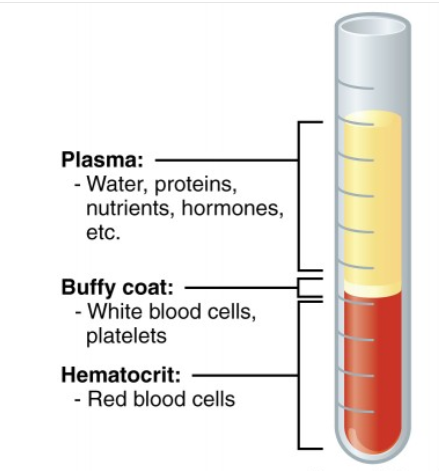
plasma: the liquid matrix, occupies 55% of the total volume of blood.
Primary component is water, which allows the volume of blood to be altered by changing the water levels in plasma; this is accomplished by the kidneys.
Plasma volume impacts blood pressure (increased volume results in increased pressure and vice versa), and is regulated by the kidneys
albumin: major osmoregulatory protein in the blood, increased [] leads to increase in BP
Also contains nutrients, cell waste, respiratory gases, ions, hormones, and proteins.
Plasma proteins are produced by the liver:
immunoglobulins: immune response
albumins: molecular transport within the blood
fibrinogen: inactive form of a clotting protein
hematocrit value: comparison of cell volume to plasma volume; The percentage of blood occupied by cells. Hovers around 45%. HCT value is primarily influenced by RBCs (because they are the most abundant blood cell type).
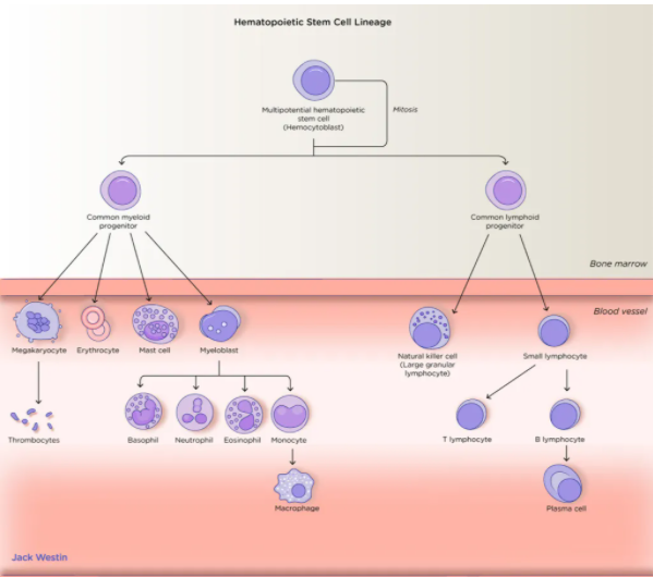
formed elements: aka blood cells, derived from hematopoietic stem cells
erythrocytes: aka RBCs, deliver oxygen
most abundant type of blood cell
mature in the bone marrow under the influence of erythropoietin (EPO, a hormone)
Erythropoiesis: continuous production of red blood cells in the marrow of large bones
do not have organelles, therefore life span is limited to 120 days, after which they are degraded by the liver and spleen via hemolysis
bilirubin: end product of breakdown of hemoglobin from RBCS, ultimately excreted into the small intestine via bile from the liver
have a biconcave disc shape allowing them to be flexible as they move through small vessels as well as increasing surface area
Hemoglobin: the critical component of erythrocytes, allowing the transport of gases
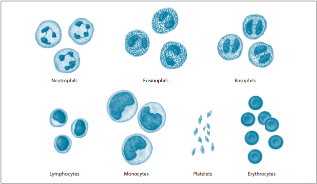
leukocytes: aka WBCS, primarily involved in immune response
diverse collection of cells, derived from stem cells in the red bone marrow, generally present in lower levels however levels increase during infections
Granulocytes: cytoplasm with granular appearance, includes neutrophils, basophils, and eosinophils.
Agranulocytes: cytoplasm that does not have a grainy appearance. Include monocytes (which mature into macrophages), and lymphocytes (T cells and B cells).
thrombocytes: aka platelets, fragments of bone marrow cells (megakaryocytes)
replaced often as they live 10-12 days only after maturing.
Important to blood clotting
humans have 4-6 liters of blood, the entirety of which can be circulated in <1 minute
pH: hovers around 7.4
temperature is slightly warmer than body temperature allowing changing patterns of circulation (via vasoconstriction and vasodilation) to distribute heat where needed
blood type:
determined by blood cell antigens (AB and Rh)
Although the immune system does not make antibodies against self- antigens (i.e., your blood type), it will make antibodies against foreign antigens (i.e., incompatible blood types).
Type O blood is the universal donor, because it has no surface antigens.
Type AB blood is the universal recipient because they do not make antibodies.
The Rh factor is controlled by a separate gene than ABO blood type. Anti-Rh antibodies are made when the Rh factor is absent (i.e., you have Rh- blood). Rh- individuals can receive only Rh- blood.
Rh factor also has a role in pregnancy; when the mother is Rh-, and the fetus has Rh+ blood, the mother's immune system will produce anti-Rh antibodies, which will cross the placenta and attack the fetus's blood cells.
Página 3
Coagulation:
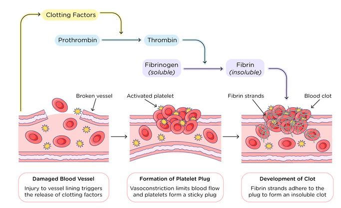
process involving a cascade leading to blood clotting to reduce blood loss after damage to a blood vessel
platelets release thromboplastin
thromboplastin converts prothrombin (inactive plasma protein) to thrombin (active form).
Thrombin converts fibrinogen to fibrin (insoluble threads of the thrombin protein).
Fibrin forms a meshwork around the platelet plug (platelets stuck to one another at the site of injury), holding necessary molecules tight to the injury site, forming the initial clot.
Clotting also requires other plasma proteins, calcium, and Vitamin K.
Página 4
Blood vessels
arteries and veins are the main ones, branch further into narrower vessels
Endothelial cells: specialized cells allowing for selective permeability in the walls of blood vessels
help in the movement of chemicals that facilitate thermoregulation (epinephrine).
allow movement of leukocytes, release of molecules from the blood to the site of injury to help with clotting
Vessel Structure:
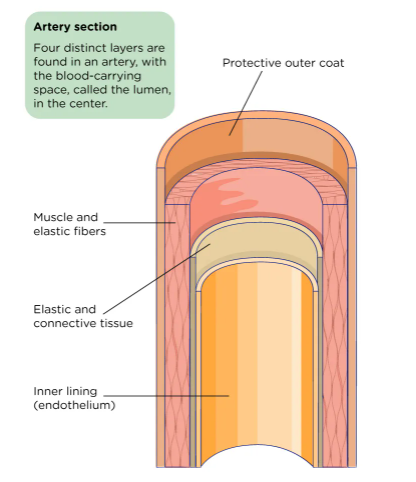
3 tunics (layers) form the walls
tunica intima: inner layer, smooth inner lining of endothelial cells in contact with red blood cells.
Tunica media: middle layer composed of smooth muscle surrounding endothelium; helps regulate blood flow.
in arteries, helps facilitate vasoconstriction and vasodilation of arteries regulates blood flow, blood pressure, and temperature.
also found in veins
Tunica externa: outermost layer made of connective tissue (collagen and elastic fibers) surrounding the middle layer; stretches and supports blood vessels.
found in both veins and arteries
Types of vessels:
Arteries: large vessels carrying blood away from the heart, have thick walls, very elastic to accommodate blood pressure.
main artery is the aorta
Arteries leaving the heart branch into --> arterioles which narrow further --> capillaries (smallest vessel type).
Capillary beds: a collection of (10-100) capillaries branching off a single arteriole, carry blood away from the body and exchange nutrients, oxygen
So small that RBCS line up single file to pass through them.
The blood entering the systemic capillary blood is also high in nutrients, and after exchange the blood carries CO2 and wastes.
In pulmonary circulation, deoxygenated blood enters the pulmonary capillary bed, and blood is oxygenated.
Precapillary sphincters regulate entry of blood into the capillary beds.
Excess water that is not taken in by osmosis is returned to circulation via the lymphatic system. Some materials also enter the capillaries via endocytosis or exit via exocytosis.
connect arterioles and venules, enable exchange of gas, water, nutrients and waste between blood and surrounding tissues
3 types: continuous, fenestrated, and sinusoidal
Mechanism of gas exchange: bulk flow (mass movement of fluids into and out of capillary beds) involving 2 pressure-driven movements
filtration: movement of fluids along their pressure gradient (high to low) from capillary bed to tissues; driven by hydrostatic pressure
reabsorption: movement of fluids along their pressure gradient (high to low) from the tissues to the capillaries; driven by osmotic pressure
[Solute] in the capillaries is > [solute] of surrounding fluids, resulting in osmosis drawing water back into the venule end of the capillary bed.
blood loses heat via radiation at the capillary beds when near the skin, because capillary beds have a large surface area
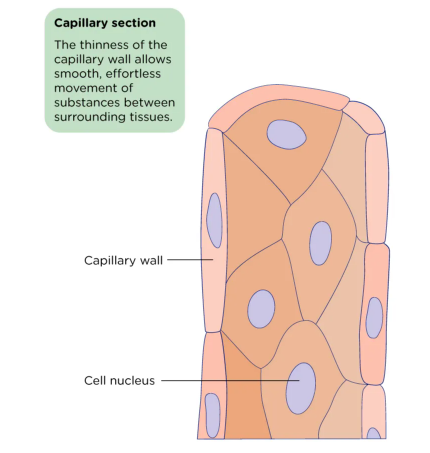
After the capillary beds, capillaries widen in diameter --> become venules.
The venules, carrying deoxygenated blood, move back towards the heart --> become veins, which merge into the heart.
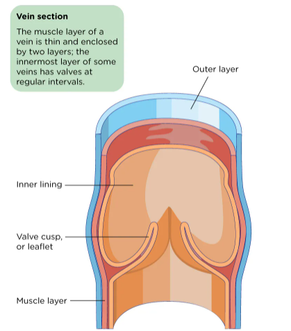
Veins: less thick walled than arteries, bring blood back to the heart.
Movement of blood in venules and veins is facilitated by the contraction of smooth muscles; they also contain valves that close to prevent backflow.
Contraction of skeletal muscle helps veins work against gravity to get blood back to the heart.
Vascular resistance must be overcome for blood to flow
vessel length: resistance increases with increased length
vessel radius: resistance increases with decreased radius
arterioles can alter their radius via vasodilation/vasoconstriction
blood viscosity: resistance increases with increased viscosity
veins tend to have lower resistance and lower pressure, rely on valves to prevent backflow
Constant flow is required, so pressure and resistance must be adjusted to maintain it
Página 5
Structure of the heart:
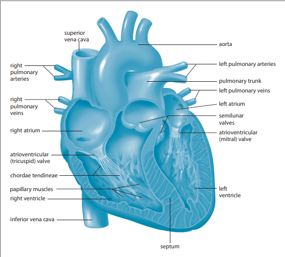
in humans, has 4 chambers (2 atria, 2 ventricles)
atria: upper chambers, R and L, receive blood
ventricles: lower chambers, R and L, pump blood
Semilunar valves regulate the flow of blood out of ventricles
aortic valve: prevents backflow at the aorta
pulmonary valve: prevents backflow at the pulmonary artery
Each atrium and ventricle are separated by atrioventricular (AV) valves.
mitral valve: separate the L atrium and L ventricle
tricuspid valve: separate the R atrium and R ventricle
right and left sides have distinct functions and are separated by the septum (thick barrier)
Right side has the pulmonary circuit, left side has the systemic circuit
R/L being the side of the person, not YOUR R/L side as you look, but THEIR R/L side
3 layers:
epicardium: outer layer of cells, made of 2 layers
pericardium: fluid filled sac surrounding the entire heart, helps reduce friction
myometrium: middle layer made of cardiac muscle, bulk of the heart wall
valves and chamber linings are made of tissues other than muscle
endocardium: lining of the inner wall of the heart
Flow of blood through the heart:
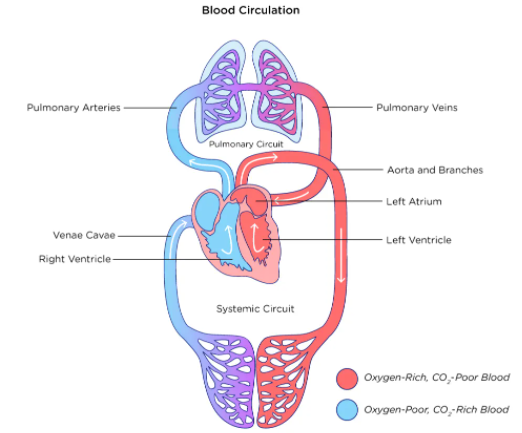
pulmonary circuit: deoxygenated blood flows through the R side of the heart; moves deoxygenated blood from the heart to the lungs and oxygenated blood back to the heart.
Pulmonary arteries branch off the right side of the heart and carry deoxygenated blood towards the lungs.
The pulmonary arteries are the only arteries in the body that DO NOT carry oxygenated blood
Gas exchange with the alveoli and the pulmonary capillaries oxygenates the blood, which returns to the left side of the heart via pulmonary veins, into the systemic circuit.
Pulmonary veins are the only veins carrying oxygenated blood.
systemic circuit: oxygenated blood flows through the L side of the heart; oxygenated blood from the lungs goes through the heart, throughout the body, and brings deoxygenated blood back to the heart.
Arteries of this circuit branch off the left side of the heart and carry oxygenated blood to the capillaries in the rest of the body.
Deoxygenated blood returns to the right side of the heart through systemic veins.
The aorta is the largest artery in the body; located next to the inferior vena cava. Branches into arteries, arterioles, and capillaries.
blood circulation in humans is known as double circulation as blood reaches the heart 2x
Página 6
Types of circuits/Blood Flow:
regulated by size of blood vessels, action of smooth muscle, uni-directional valves, and blood pressure
Coronary arteries: the 1st branches off the aorta, provide circulation to the surface of the heart. Heart attack is caused by blockage of the coronary arteries leading to death of cardiac muscle secondary to loss of blood flow to the muscle. After blood flows through the coronary arteries, deoxygenated blood is returned to the pulmonary circuit by coronary veins.
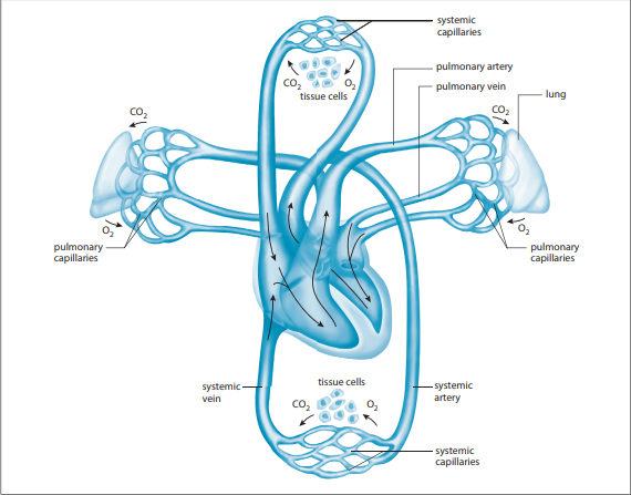
Systemic circuit: pulmonary veins --> L atrium--> bicuspid valve --> L ventricle --> aorta--> organs, muscles, rest of body
oxygenated blood from the lungs carried by the pulmonary veins enters the heart through the L atrium.
The L atrium contracts in sync with the right atrium, pushing oxygenated blood through the bicuspid valve into the L ventricle.
L ventricle contracts In sync with the right ventricle, pushing oxygenated blood into the aorta via the aortic semi lunar valve.
Aorta carries oxygenated blood to the rest of the body.
pulmonary circuit: vena cavae--> R atrium --> tricuspid valve--> R ventricle --> pulmonary arteries --> lungs
The R atrium receives deoxygenated blood from the superior vena cava (which has come from the upper organs and arms) and the inferior vena cava (which has come from the lower organs and the legs)
Deoxygenated blood moves to the R ventricle through the tricuspid valve
R ventricle pumps deoxygenated blood through the pulmonary arteries, which carry the blood to the lungs for re-oxygenation.
Página 7
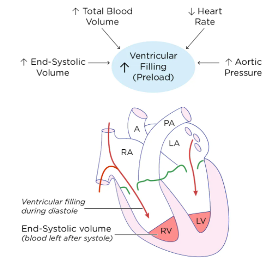
Cardiac cycle: the events behind a single heartbeat.
The two atria contract simultaneously, pushing blood into the ventricles
systole: the two ventricles contract pushing blood out of the heart
diastole: a brief resting period during which both atria refill with blood. The cycle begins again.
EKGs are used to visualize the electrical currents generated by the heart during the cardiac cycle.
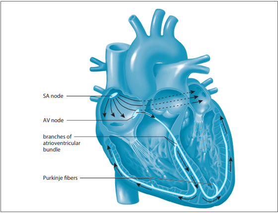
Conducting system of the heart: impulses that generate heart contraction; cardiac muscle is involuntary and is maintained by this system unless stimulated by the nervous system.
SA node: sinoatrial node, AKA pacemaker, a bundle of conducting cells in the top of the right atrium that initiates contractions. It sends electrical impulses through the two atria causing them to contract. Generates its own rate of contraction at an average of 70 contractions/minute.
AV node: atrioventricular node, impulse from the SA node arrives here and then is forwarded to the ventricles
Bundle of His: impulse from AV node is spread through here
Purkinje fibers: in the walls of ventricles, impulse from bundle of His spreads here causing ventricle contraction
ANS can adjust the rate of contraction. The sympathetic branch can increase the rate of contraction, while the parasympathetic branch can decrease the rate of contraction. The medulla oblongata monitors conditions (such as blood pH level, which indicates CO2 and O2 levels) and signals adjustment prn.
Página 8
Blood pressure: measurement of the force that blood exerts on the walls of a blood vessel, typically measured within arteries. Helps push blood through the vessels.
Expressed with two values—systolic and diastolic pressure.
Systolic pressure should be higher and indicates the peak pressure exerted on arteries when the heart is beating.
artery walls stretch to accommodate increased pressure
Diastolic pressure is lower and is a measurement of the lowest pressure in the vessels between heartbeats.
elasticity of vessels allows them to relax back to their regular diameter
these values are reported in mmHg
Factors affecting BP;
hormones
stress
exercise vs rest
position of the body
eating/diet
Blood pressure is produced by hydrostatic pressure
fluid moves from areas of high hydrostatic pressure to low hydrostatic pressure
in the arteries, pressure near the heart is very high, so blood flows to the arterioles, which slows the rate of flow because they are more narrow
BP should be enough to overcome peripheral resistance of the arteries/arterioles.
Primarily regulated by renal regulation of blood volume. There is a direct relationship between blood volume and blood pressure.
Página 9
Oxygen transport by hemoglobin:
hemoglobin structure: 4 protein chains wrapped around a heme (iron) core
unique structure allows 1 hemoglobin molecule is able to carry 4 molecules of O2
Each erythrocyte contains ~250 million hemoglobin molecules--> 1 erythrocyte carries ~1 billion oxygen molecules
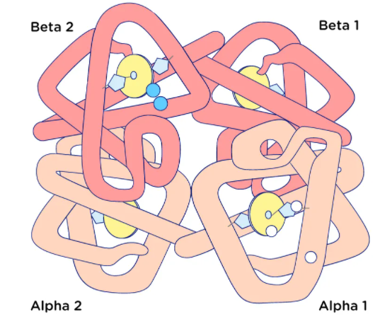
fetal hemoglobin has greater affinity for oxygen than adult hemoglobin and carbon monoxide has a much greater affinity for hemoglobin then oxygen which is why CO is poisonous
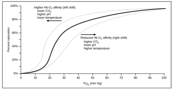
Bohr effect: increased [H+] (aka low blood pH) and increased [CO2] will decrease hemoglobin’s affinity for O2 (most common in areas of high altitude)
When hemoglobin binds 1 oxygen molecule, conformational change in the shape of hemoglobin allows the remaining 3 molecules to load more easily. The same process for unloading.
This effect encourages the unloading of oxygen from hemoglobin when cells are in need
The opposite is true as well (high [O2] in the lungs encourages the dissociation of H+ ions (which combine with bicarbonate ions to form CO2 and H2O) from hemoglobin, so that CO2 can be exhaled).
example: myoglobin in muscle cells during exercise
CO2 also bind to hemoglobin and forms carbaminohemoglobin
Increased temperature and increased [CO2] also cause hemoglobin's affinity for oxygen to be reduced
Página 10
Transport of Carbon Dioxide:
3 methods
dissolution directly into the blood (5-7%)
binding to hemoglobin (10%)
carried as bicarbonate ion (85%)
carbonic anhydrase converts CO2 to carbonic acid (H2CO3) and vice versa
Carbonic acid is an unstable intermediate that dissociates quickly into H+ ions and bicarbonate ions (HCO3-)
this allows for continued uptake of CO2 into the blood down its [] gradient, as it does not build up
However, this can also alter blood pH quickly if not regulated by the bicarbonate buffer system
chloride shift: bicarbonate ion transported out of RBC into the plasma, in exchange for Cl- ion; at the lungs, these ions are exchanged once again so CO2 can be reformed and exhaled
CO2 is more soluble in blood than oxygen
Página 11
Nervous and Endocrine control:
Cardiovascular center in the ANS is a cluster of neurons responsible for regulation of cardiac output
cardioaccelerator center: receives sympathetic stimulation from cardiac accelerator nerve, to regulate heart rate and stroke volume
cardioinhibitor center: receives parasympathetic stimulation from the vagus nerve and slows cardiac function by decreasing HR and stroke volume
Norepinephrine and epinephrine, signaling from chemoreceptors (detect acidification) are common stimuli
baroreceptors: specialized stretch receptors located in blood vessels and heart chambers that respond to degree of stretch caused by the presence of blood, send signals to regulate BP
vasoconstriction can be used locally to reduce blood flow in a specific area, however when it occurs in a generalized way, it causes an increase in systemic BP
Renin-angiotensin-aldosterone system: hormone system regulating BP and water balance
when blood volume is low, kidneys secrete renin
renin converts angiotensinogen (released by liver) to angiotensin I
angiotensin I is converted to angiotensin II by ACE (angiotensin-converting enzyme) found in the lungs
angiotensin II is a vasoactive peptide that causes blood vessels to constrict, resulting in increased BP
this is why ACE-I (ACE inhibitors) can be used as anti-hypertensives
Información
- Descripción
- Apunte sobre Topic 3b: circulatory system, creado por 000 000 el 13/01/2022.
- Creado por
- 000 000
- Visitas
- 2
- Valoración