
Notes
Topic 3b: respiratory system
Content of the notes (8)
Page 1
Primary function: gas exchange (providing the body with oxygen and eliminating CO2), thermoregulation (loss of heat in the form of moist air during exhalation, reducing body temperature), and filtration (protection from infectious particles.
Also functions in pH regulation, vocal communication, sense of smell.
Pulmonary arteries: low in oxygen and high in CO2, emerge from the right side of the heart and carry deoxygenated blood to the lungs.
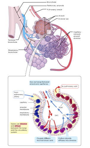
Branch into pulmonary capillaries which surround the alveoli in the lungs. CO2 diffuses from the pulmonary capillaries into the alveoli for exhalation.
Oxygen from the lungs is taken up by hemoglobin in the erythrocytes within the capillaries. This oxygenated blood travels back to left side of the heart through the pulmonary veins, and then is distributed throughout the body via the systemic circuit.
Page 2
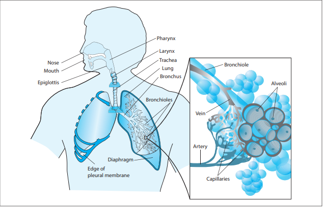
Structure: a series of tubes that bring air to the alveoli. Because the system is open, it is vulnerable to infection. Air enters the Nose or mouth--> pharynx + larynx--> trachea--> lungs --> bronchi + bronchioles+ alveoli
Air is inhaled through the nose or mouth. Then in the nose and pharynx, air is warmed to body temperature, moisturized to facilitate gas exchange, and then filtered.
Both the nose and the pharynx are covered with mucous membranes to prevent desiccation of the tissues, and to collect particles or microbes that may have entered with air.
The nose also has the function of olfaction.
the pharynx has two passageways: esophagus and larynx
The larynx is the passageway for air, opening is covered by the glottis.
The larynx is also the voice box, is made of cartilage and has vocal cords that vibrate to produce sound.
Typically the esophagus is closed off and the glottis is open, unless the person is swallowing. When a person is swallowing, the epiglottis—small piece of cartilage covering the glottis—stops food from entering the larynx.
As air flows through the larynx, it makes its way into the trachea and the lower respiratory tract.
The trachea is supported by C-shaped rings of cartilage. The internal surface is lined with mucus-producing goblet cells and ciliated epithelia that propel foreign particles trapped in mucus forward.
The trachea branches off into the lungs
Lungs: made of resilient tissue, encompass the bronchioles and alveoli.
R lung has 3 lobes and L lung has 2.
Each lung is surrounded by pleural membrane (Fluid filled).
As air passes to the lungs, it is diverted through bronchi
There is 1 bronchi for each lung; these are the 2 primary bronchi. The primary bronchi then ultimately branch into the bronchioles, after first branching into secondary and tertiary bronchi.
Smooth muscle surrounding the bronchioles adjust their diameter depending on oxygen demands.
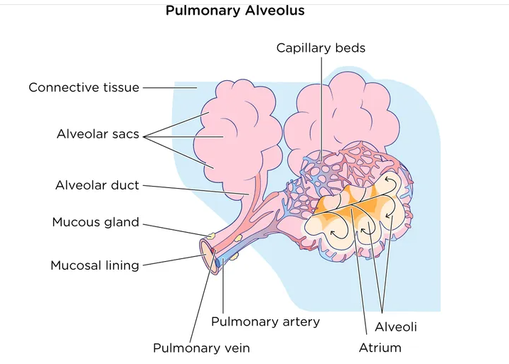
Bronchioles after more divisions, terminate in alveolar ducts, which are surrounded by the alveolar ducts containing multiple alveoli (tiny air sacs).
Alveolar ducts are attached to the end of each bronchiole, with each duct ending in ~100 alveolar sacs. Each sac contains 20-30 alveoli
Alveoli are made of thin-walled parenchymal cells (simple squamous epithelium), and are in contact with capillaries.
Alveoli are small and numerous, providing a large amount of surface area for gas exchange.
Page 3
Breathing mechanisms:
Dependent on pressure gradients and thoracic cavity muscles
Boyle's Law: in a closed space, pressure and volume are inversely related
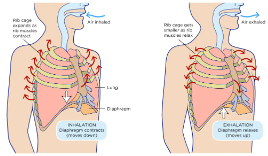
Inhalation:
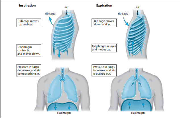
lung volume increases as a result of contraction of diaphragm and intercostal muscles.
pressure decreases (as per Boyle's law), resulting in pressure gradient (pressure in thoracic cavity > pressure in atmosphere/externally)
Inhalation occurs as air rushes in
There is a resulting increase in volume as air comes in; this is attributed to an increase in alveolar space as the bronchi/bronchioles are stiff and do not change in size
Exhalation:
a passive process, no muscle contraction involved
Elastic recoil: when air fills the lungs, the tissues of the lung exert pressure back towards the interior of the lungs
competing inward and outward forces--> inward forces try to inflate the lungs, as outward forces try to deflate the lungs
The intercoastal muscles relax, returning the chest wall to its original position
the diaphragm relaxes, moving higher into the thoracic cavity, increasing pressure internally in comparison to externally--> causing air to rush outwards
Diaphragm: thin muscle separating the thoracic cavity from abdominal cavity.
The diaphragm is innervated and neurally connected to the part of the medulla that controls breathing.
Inspiratory neurons cause contraction of the diaphragm, which is followed by a period of inactivity allowing for relaxation of the diaphragm and exhalation.
The diaphragm is stimulated 12-15x/minute in its relaxed state. This rate increases during times of increased O2 demand and excessive CO2 production.
Intercostal muscles: contract to move the rib cage up and outwards to assist inhalation.
For inhalation, the chest cavity volume must increase (diaphragm contracts and pushes down), to decrease pressure (i.e. inverse relationship of volume and pressure) so air flows from an area of greater pressure (outside the body) to an area of low pressure (inside the chest cavity).
When these muscles relax, the volume of the chest cavity decreases resulting in higher pressure inside than outside, forcing exhalation.
Page 4
Surface tension in the lungs:
force exerted by water molecules on the surface of lung tissue as they pull together
inward force (from hydrogen bonding between water molecules) on the surfaces of lungs lowers surface area as the tissue is pulled together
there is a considerable amount of surface tension in the lungs because the air is moist
alveoli are not able to resist surface tension on their own, and they deflate as air is forced out by contraction of the pleural cavity during exhalation
without something to reduce the surface tension, the airways would collapse after exhalation and re-inflation would be much more difficult during inhalation
Surfactant fluid secreted by the alveoli decreases surface tension, keeps alveoli inflated, preventing them from collapsing. Without this fluid, the lungs do not function
surfactant is a lipoprotein, works similar to detergent by reducing surface tension
Page 5
Alveolar Gas Exchange:
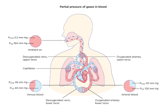
After inhalation, the partial pressure of oxygen in the alveoli is > partial pressure of oxygen in the capillaries surrounding the alveoli.
The gases will flow from an area of high concentration to low concentration, so after inhalation, oxygen moves from the alveoli into the capillaries and binds to hemoglobin.
Concentration gradients cause CO2 to diffuse into the alveoli (low concentration) from the capillaries (high concentration), where it can be exhaled.
The lungs do not fully deflate after exhalation, resulting in inhaled air mixing with residual air from the previous respiration, creating a lower partial pressure of oxygen within the alveoli
Respiratory quotient (RQ): ratio of CO2 production to oxygen consumption, varies between 0.7 to 1.0.
key factor used to calculate partial pressure of O2 in the alveolar spaces within the lung
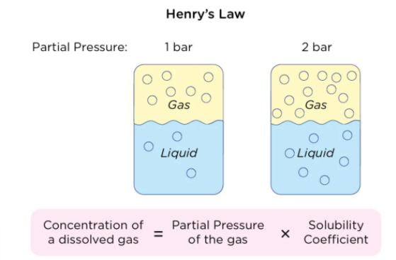
Henry's Law: [gas in a liquid] is directly proportional to solubility and partial pressure of that gas
the higher the partial pressure of the gas, the greater amount of # of gas molecules that will dissolve in the liquid
[gas in a liquid] is also dependent on solubility of that gas in the liquid
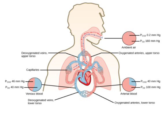
Partial pressure of oxygen in the systemic capillaries is 100 mmHg however in the surrounding tissues the partial pressure of oxygen is 40 mmHg.
This pressure gradient pushes oxygen out of the capillaries and into the tissues.
Carbon dioxide and oxygen move independently of each other; there is a carbon dioxide pressure gradient between the systemic capillaries and the surrounding tissues, pushing carbon dioxide out of the tissues into the capillaries.
Page 6
Other functions of the lung other than respiration --->
Thermoregulation:
majority of thermoregulation happens via evaporation of heat in the form of sweat
some happens via vasodilation (cools body by increasing heat loss from the blood vessels in the nasal and tracheal capillary beds) and vasoconstriction (warms the body by reducing heat loss)
panting: in dogs, the main form of thermoregulation
Particulate Filtration:
The nose is particularly structured for filtration because of the presence of cilia and hair to trap particulate matter (pollutants and pathogens).
nasal hairs push foreign particles back outwards to prevent entry into trachea
lining of respiratory tract has goblet cells secreting mucus to trap particles, and the cilia push the mucus outwards
Page 7
Acid-Base Balance:
Respiratory system maintains blood pH via bicarbonate buffer system
bicarbonate buffer system: an acid-base homeostatic mechanism involving the balance of carbonic acid, bicarbonate ions, and carbon dioxide to maintain blood pH
This is the result of the formation of carbonic acid when CO2 interacts with water.
Carbonic acid breaks down into bicarbonate ions and hydrogen ions; bicarbonate ions help buffer pH in the body.
This is a reversible reaction, and the bicarbonate and hydrogen ions join together again to produce carbonic acid when the pH is too acidic. The reformed CO2 is then exhaled in order to adjust pH.
Carbonic anhydrase: catalyzes the formation and dissociation of carbonic acid.

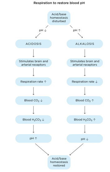
When blood pH decreases, [H+] increases or [bicarbonate ion] decreases, so more carbon dioxide is produced (the equation moves towards the left) to maintain equilibrium.
Increased [H+] is a trigger for signals to the brain to increase the respiratory rate (to exhale the increased carbon dioxide and to help pH move back to its more neutral set point).
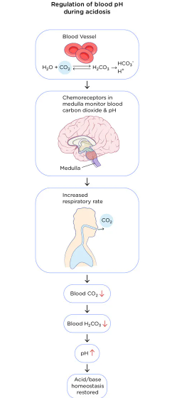
When blood pH increases, [H+] decreases or [bicarbonate ion] increases, less carbon dioxide is exhaled, because the respiratory rate is decreased. Ultimately, this results in hydrogen ion concentration in the blood increasing, decreasing the pH towards its more neutral setpoint.
Page 8
Control of Ventilation by the Nervous System:
The primary influence on ventilation rate is the CO2 level, which is monitored by chemoreceptors in the brain and in certain large blood vessels.
As CO2 levels increase, the pH decreases due to buildup of carbonic acid, resulting in an increase in the breathing rate to eliminate excess CO2. This results in increase in oxygen levels.
Involuntary respiration: controlled by the brainstem (medulla oblongata and the pons)
The brainstem contains central chemoreceptors to detect blood pH levels and sends signals to the respiratory centers of the brain to adjust ventilation rate to maintain blood pH (bicarbonate buffer system).
the medulla oblongata is the primary respiratory control center; sends signals to the muscles that control respiration.
Also controls the reflexes for non-respiratory air movements (coughing, sneezing, Swallowing, and vomiting reflexes).
Ventral respiratory group: stimulates expiratory movements.
Dorsal respiratory group: stimulates inspiratory movements.
the pons is located under the medulla; its primary function is controlling the rate of involuntary ventilation.
Apneustic center: sends signals for long and deep breaths; controls the intensity of breathing, increases tidal volume.
Inhibited by the stretch receptors of the pulmonary muscles when inspiration is at its maximum depth or when there is signaling from the pneumotaxic center.
Pneumotaxic Center: allows fine control of respiratory rate by sending inhibitory signals. These signals limit phrenic nerve activity, and inhibit the signals of the apneustic center. It decreases tidal volume.
These two centers work antagonistically to control respiratory rate.
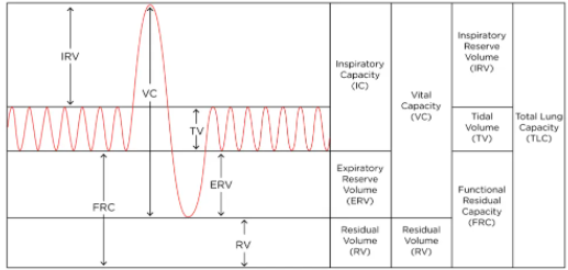
tidal volume: the amount of air that moves in or out of the lungs with each respiratory cycle
Voluntary respiration: controlled by the cerebral cortex
primary motor cortex: the neuro center responsible for initiating any voluntary muscular movement, including voluntary respiration.
Contraction and relaxation of the internal and external intercostal muscles is controlled by the superior portion of the primary motor cortex.
Can be overridden by chemoreceptor stimulus, hypothalamus stress response
Required for higher functions such as voice control
Important respiratory nerves:
the phrenic nerves: stimulate diaphragm activity.
The vagus nerve: innervates the diaphragm, controls movements in the larynx and pharynx.
the posterior thoracic nerves: stimulate the intercostal muscles located around the pleura.
Information
- Description
- Note on Topic 3b: respiratory system, created by 000 000 on 15/01/2022.
- Created by
- 000 000
- Views
- 0
- Rating