10193033
Description
Flashcards by Lexi Crosbie, updated more than 1 year ago
|
|
Created by Lexi Crosbie
over 8 years ago
|
|
| Question | Answer |
| Clay Shoveler's Fracture | Definition: Avulsion fracture(s) on the spinous processes of C6-T1. Causes: hyperflexion |
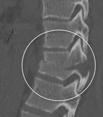
| Compression Fracture | Definition: the collapse of a vertebral body most often in the thoracic/lumbar regions. The anterior edge collapses, changing the shape of the verebral body into a wedge instead of a block. Causes: often associated with osteoporosis. Results from flexion / axial loading |
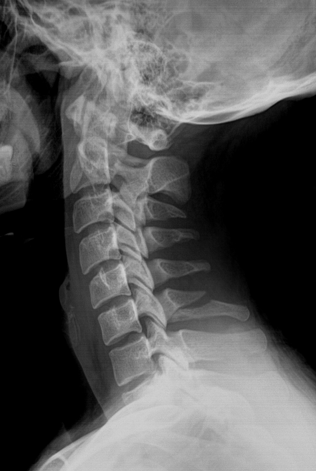
| Hangman's Fracture | Definition: extends through the pedicles of C2 with out without subluxation of C2 on C3. Unstable if alive. Causes: occurs when neck is subjected to extreme hypertension. |
| Jefferson's Fracture | Definition: comminuted fracture where the anterior and posterior arches of C1 are fractured as the skull slams onto the ring. Causes: axial loading |
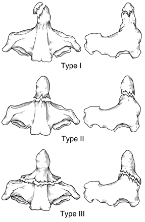
| Odontoid Fracture | Definition: involves the dens and can extend into the lateral masses or arches of C1. - Type I: avulsion fracture and involves only the tip. Mostly stable - Type II: most common, fracture through the base/waist of C2 - Type III: fracture extends into the body of the C2 vertebrae |
| Teardrop Burst Fracture | Definition: vertebral body is comminuted with triangular fragments avulsed from the anteroinferior border and fragments from the posterior vertebral body displaced into the spinal canal. Most commonly have neurological damage. Cause: compression with hyperflexion |
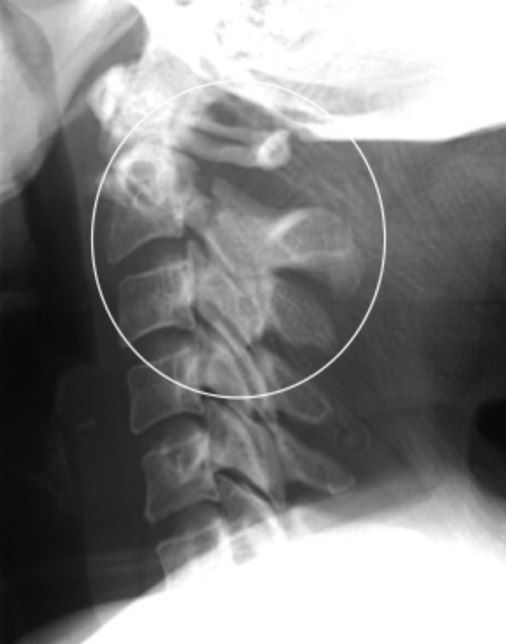
| Rotational Injury | Definition: injury involving complex forces causing injury onto the head of neck.When they involve hyperflexion, complex fractures to the interarticularis and pedicles may occur. A 'jumped facet(s)' phenomenon may occur unilaterally or bilaterally. |
| Unilateral Locked Facet | Definition: the intervertebral foramina is visible above but not below on a true lateral image. Cause: flexion and rotation injury |
| Bilateral Locked Facets | Definition: the upper cervical spine is anteriorly displaced over the lower spine. There is a widening of the spinous processes at the site of injury. Results in a complete dislocation of the spine. Cause: hyperflexion injury |
| Herniated nucleus pulposis | Definition: i.e. slipped disc. When the soft inner part of the intervertebral disk protrudes through the fibrous cartilage outer layer and into the spinal canal, causing severe pain and possible numbness that radiates into the extremities. Often L4/5 level causing sciatica. |
| Muscle Spasm | Looks like: loss of normal lordotic curve on a lateral view or torticollis on the AP view. |
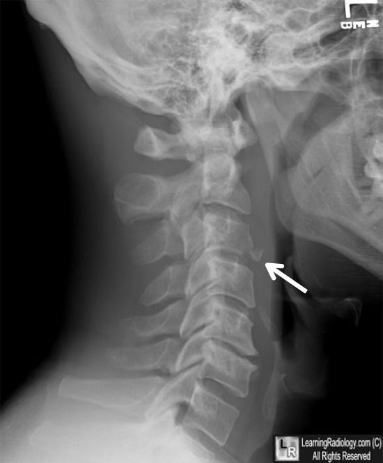
| Cervical Ribs | Definition: rudimentary rib that projects laterally from C7 but does not reach the sternum |
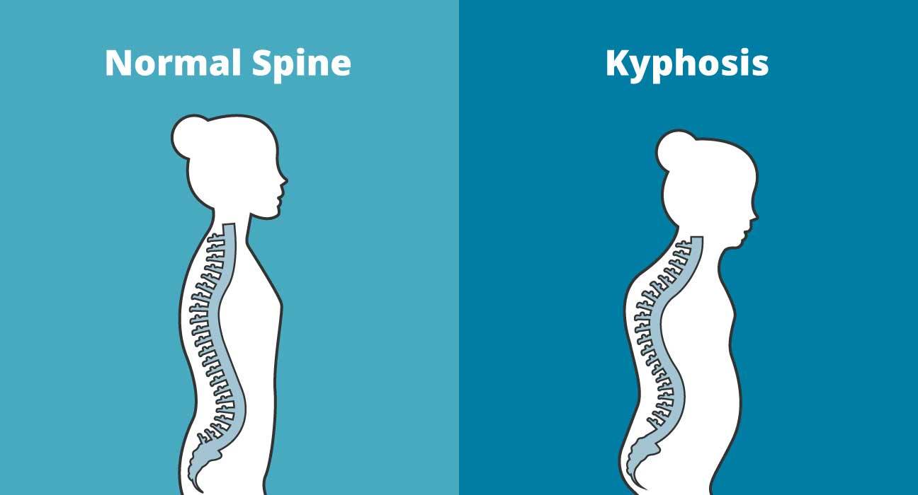
| Kyphosis | Definition: abnormal or exaggerated convex curvature of the thoracic spine that results in a stooped posture and reduced height. Cause: possibly compression fractures, poor posture, rickets or other spinal diseases. |
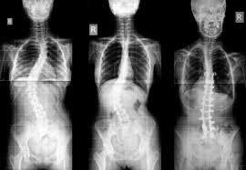
| Scoliosis | Definition: an abnormal or exaggerated lateral curvature of the spine |
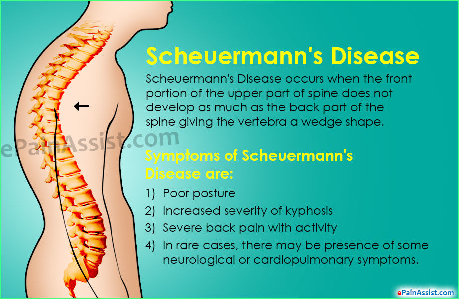
| Scheuermann's Disease | |
| Spondylitis | Definition: inflammation of the vertebra. Can go on to become ankylosing spondylitis |
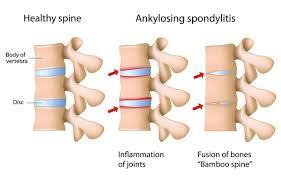
| Ankylosing Spondylitis | Definition: inflammation of the sacroiliac, intervertebral and costovertebral joints leading to pain and stiffness. Continues until ankylosis occurs where the bones form a union. AKA Bamboo Spine |
| Spondylosis | Definition: age related degeneration of the intervertebral discs. Can contribute to arthritis changes that may affect the zygapophyseal joints and intervertebral foramen. |
| Chance Fracture | Definition: anterior wedge fracture of the vertebral body with horizontal fracture through posterior elements or distraction of facets joints and spinous processes. Cause: flexion and distraction injury of the spine e.g. MVA with a lap belt. |
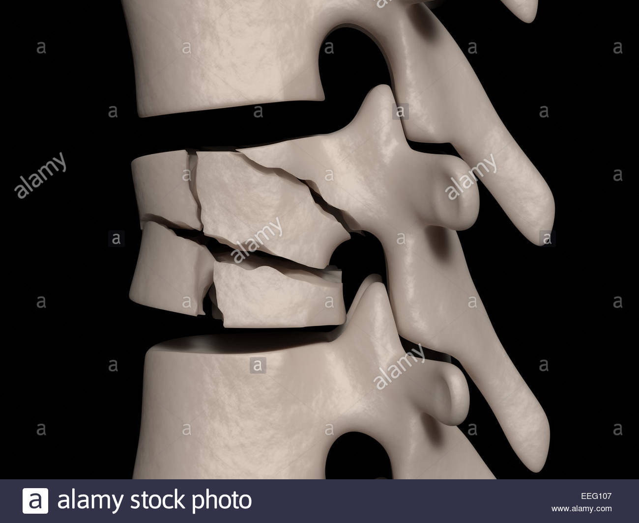
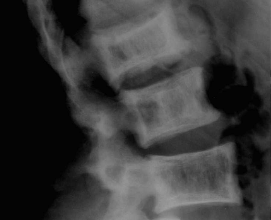
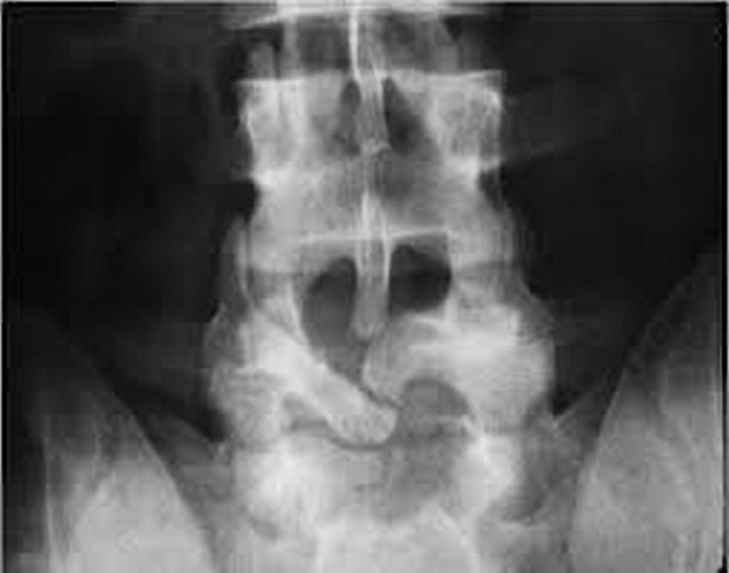
| Burst Fracture | Definition: vertebra is crushed by forces however unlike usual compression fractures, they are fractured in more than one place and direction. Can potentially injury spinal cord and tissues. On x-ray there is a loss of vertebral height and the anterior portion is often compressed more than the rear, interpedicular widening. Cause: severe trauma i.e. MVA, fall from a height |
| Paget's Disease | Definition: increased remodelling of the affected bones, leading to thickening and deformation. Most commonly found in the L spine but is seen throughout spinal column. Cause: hyperactive osteoclasts and osteoblasts. |
| Lordosis | Definition: the normal concave curvature of the L=spine and also an abnormal or exaggerated concave lumbar curvature. Causes: pregnancy, obesity, poor posture, rickets or tuberculosis of the spine. |
| Spina bifida | Definition: congenital condition in which the posterior aspects of the vertebrae fail to develop and therefore expose part of the spinal cord. Varies in severity and most often occurs around L5. |
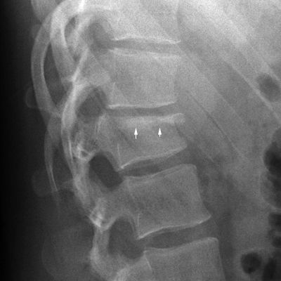
| Spondylolisthesis | Definition: involves the forward movement of one vertebrae in relation to another. Cause: often due to a developmental defect in the pars interarticularis or may happen due to spondylolysis or severe osteoarthritis. Most common at L5/S1. |
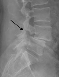
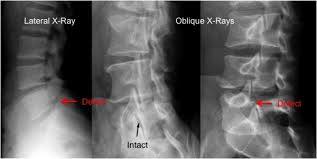
| Spondylolysis | Definition: dissolution of a vertebrae, such as from a lack of development of the vertebral arch and separation of the pars interarticularis of the vertebra. On the oblique x-ray, Scottie Dog appears broken. |
Want to create your own Flashcards for free with GoConqr? Learn more.