
Flashcards
Liver pathology
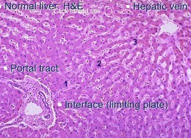
Cards in this deck (45)
- 1liver vasculatureIncoming portal vein and hepatic artery
Outgoing hepatic vein - 2—
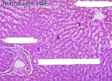
- 3broad causes of injury to the liverDrugs or toxins incl. alcohol
Abnormal nutrition/metabolism
Infection
Obstruction to bile or blood flow
Autoimmune liver disease
Genetic/deposition disease
Neoplasia
Others - 4acute and chronic inflammation in liver diseaseAcute inflammation = agent causes injury but is then removed (Days to weeks)
Chronic inflammation = agent causes injury and persists (Months to years)
“Acute-on-chronic”
Chronic liver disease often presents with acute exacerbation plus evidence of underlying chronicity e.g. fibrosis - 5what is fulminant inflammation in the liver?“Fulminant” = severe acute, rapidly progressing towards liver failure
- 6sites of damage in liver due to injurious agentsLiver injury often mainly to parenchyma (hepatocytes); but bile ducts or rarely blood vessels can be the main target
Parenchyma, bile ducts, blood vessels and connective tissue are inter-dependent, so damage to one damages the others - 7Definition of cirrhosis= End-stage liver disease
Definition is three-fold:
- Diffuse process with
- Fibrosis &
- Nodule formation - 8difference between acute hepatitis and acute cholestasis/cholestatic hepatitisboth with inflammation but acute bile stasis is more marked in cholestatic hepatitis
- 9histology of acute hepatitisDiffuse hepatocyte injury seen as swelling. A few have died, described as “spotty necrosis” . There is an inflammatory cell infiltrate in all areas: portal tracts, interface and parenchyma.
dying hepatocytes and plasma cells - 10Cholestatic hepatitis/acute cholestasis - causes - histologyCauses:
Extrahepatic biliary obstruction
Drug injury e.g. antibiotics,
Histology: brown bile (bilirubin) pigment +/- acute hepatitis - 11histology of chronic hepatitisinflammation near portal tract and interface
portal fibrosis and formation of fibrous septum (band) - 12histology of hepatitis Bground glass cytoplasm in hepatocytes = accumultion of 'surface antigen', one of three main HB viral antigens
- 13Chronic biliary/cholestatic disease - causes - histologyCauses:
- Primary biliary cirrhosis (PBC)
- Primary sclerosing cholangitis (PSC)
Histology: Focal, portal-predominant inflammation and fibrosis with bile duct injury; granulomas (arrow) in PBC - 14examples of genetic/deposition liver disease (3)Haemochromatosis (iron)
(excess iron is shown by (blue) Perl’s stain: normally no blue stain present)
Wilson’s disease (copper)
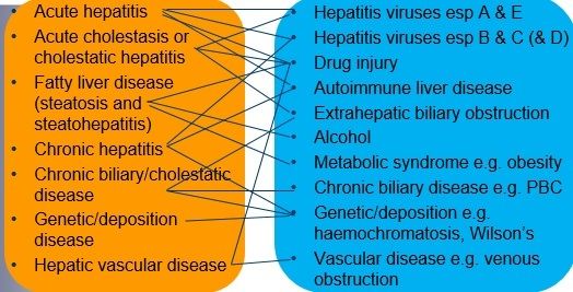
Alpha-1-antitrypsin deficiency - 15morphology and causes of diffuse liver disease
- 16aims of investigation in diffuse liver disease- diagnosis
- cause
- grade
- stage - 17aims of management of diffuse liver diseaseManagement aims to reduce symptoms, reduce inflammation and prevent or slow progression of fibrosis
Treatment may include:
- Specific treatment against cause e.g. removal of alcohol or drug, weight loss, optimal diabetic control, specific antivirals or immunosuppression, and/or…
- Supportive treatment e.g. for severe acute hepatitis or for cirrhosis in general - 18drugs that predictably cause liver damageparacetamol and methotrexate
(also herbal)
drug-induced liver disease is common and mimics other conditions - 19categories of focal liver diseasefocal liver lesions = space-occupying lesions
-->
non-neoplastic
- developmental/degenerative eg. cysts
- inflammatory eg. abscess
OR
neoplastic
- benign
- malignant - 20Liver cysts - why - commonestUsually developmental or degenerative in origin
Commonest = Von Meyenberg complex (= simple biliary hamartoma)
- resemble metastases by naked eye at operation; often submitted for pathology including urgent intra-operative frozen section
No treatment required - 21liver neoplasms (benign and malignant)Benign 5%
eg. Hepatocellular adenoma
Haemangioma
Malignant
eg. Heppatocellular carcinoma
Cholangiocarcinoma - 22importance of haemangioma and hepatic adenomaImportance because of differential diagnosis with metastases
Haemangioma
Benign blood vessel tumour
Biopsy avoided because of risk of bleeding
Hepatic adenoma
Rare
Mainly young women, often associated with hormonal therapy
Risk of bleeding and rupture so excision if large - 23Hepatocellular carcinoma - what - associated withMost common primary liver tumour
Usually arises in cirrhosis and associated with elevated serum AFP (alpha feto-protein)
Screening available - 24normal liver functions- Protein, carbohydrate and fat metabolism
- Plasma protein and enzyme synthesis
- Production of bile
- Detoxification
- Storage of proteins, glycogen, vitamins and metals
- Immune functions - 25aims of histopathologyEstablish disease diagnosis and severity
Predict outcome and guide treatment - 26% liver disease due to infection and fatty liver?15% infectious
Fatty liver due to alcohol or obesity etc makes up at least 70% of liver disease in West - 27symptoms and signs of liver disease1. From liver itself e.g. hepatomegaly
2. From portal hypertension e.g. ascites
3. From chronic liver dysfunction e.g. pruritis, spider naevi
4. Non-specific e.g. nausea, falls, tremor - 28symptoms and signs related to abnormal liver structure and functionAbnormal biliary system
- Accumulation of bilirubin (esp. acute cholestasis): jaundice
- Accumulation of bile acids (esp. chronic cholestasis): pruritis
Abnormal parenchyma
e.g. RUQ pain
In chronic disease e.g. hormonal changes
Liver failure only once <25% function
Abnormal vasculature
Abnormal connective tissue matrix
- Together, e.g. portal hypertension - 29Liver investigations: bloodLiver function tests (biochemistry)
- Transaminases: AST, ALT
- Alkaline phosphatase (Alk P)
- Gamma glutamyl transferase (GGT)
- Bilirubin (Bil)
- Albumin (Alb)
Liver-related haematology tests
- Prothrombin time (PTT)
Synthetic function esp. PTT and Alb - 30other liver testsViral serology
Autoimmune serology
Tests for liver metabolic/genetic disease
Iron, copper, alpha-1-antitrypsin
Radiology (imaging): especially masses
- Ultrasound of abdomen
- CT scan of abdomen
- ERCP/MRCP
Biopsy (in only few cases) - 31Acute hepatitis - presentation - causes - managementPresentation:
- Short hx of RUQ tenderness, malaise etc
- Elevated AST/ALT (and often Bil)
Causes:
- Viral hepatitis, autoimmune, drug injury (identify through serology and drug history)
- Paracetamol overdose causes severe acute hepatitis with massive necrosis
Management:
- Remove or treat cause
- Liver support if severe dysfunction e.g. confluent necrosis; a few may need transplant - 32Fatty liver disease (steatosis and steatohepatitis) - presentation - causesCommonest cause of liver disease in West
Clinical presentation:
- Acute OR chronic “hepatitis”, or
- Asymptomatic abnormal LFTs
Causes:
- Alcohol
- Non-alcoholic (NA: so NAFLD, comprising NAS & NASH) or so-called metabolic syndrome, including Type II diabetes, obesity and hyperlipidaemia
- Drugs e.g. methotrexate, amiodarone, steroids - 33Hepatitis C liver pathologymainly in portal tracts, as chronic inflammation with lymphoid aggregates
- 34Chronic hepatitis - clinical definition - presentation - causesClinical definition: Liver inflammation (abnormal LFTs) for at least 6 months
Presentation:
Chronic hepatitis or acute exacerbation
Causes:
Viral hepatitis B or C, autoimmune, rarely drug injury (identify through serology and history) - 35chronic hepatitis pathology reportingLiver biopsy assessed for:
1. Activity (grade): degree of inflammation
- Portal, interface & parenchymal inflammation
- Guides treatment
2. Stage: amount of fibrosis
- Prognosis (& guides treatment)
Yields histological summary and numerical score - 36Chronic biliary/cholestatic disease - presentation - causesClinical presentation:
- Chronic liver disease e.g. itch (symptoms more of excess bile acids than excess bilirubin) , or
- Abnormal LFTs: mainly AlkP and GGT, often relatively mild, persisting for >6 months
Causes:
- Primary biliary cirrhosis (PBC)
- Primary sclerosing cholangitis (PSC) - 37Histology of chronic biliary/cholestatic diseaseFocal, portal-predominant inflammation and fibrosis with bile duct injury;
granulomas in PBC - 38Primary biliary cirrhosis - who - markers - managementMainly middle-aged women
Auto-immune disease with serum anti-mitochondrial antibodies (AMA) and high IgM
Despite the name, PBC is NOT cirrhotic from the outset, but usually progresses to fibrosis then cirrhosis over years.
No cure but ursodeoxycholic acid eases symptoms and slows progression; liver transplantation, if available, at end-stage - 39Primary sclerosing choldangitis - association - riskRare, associated with ulcerative colitis
Risk of progression to cholangiocarcinoma - 40Effect of Alpha-1-antitrypsin deficiency on the liverA-1-AT deficiency due to lack of secretion from and thus accumulation in hepatocytes
- 41Hepatic vein outflow obstructionMajor form is due to e.g. hepatic vein thrombosis (Budd-Chiari syndrome) which is often fatal; pro-thrombotic tendency predisposes and early identification permits anti-coagulation
Lesser degrees are more common and milder e.g. nodular regenerative hyperplasia - 42What might cause a liver abscess?may arise from ascending cholangitis
also from hydatid and other parasitic disease - 43Cholangiocarcinoma - what - diagnosis - prognosis - treatmentCholangiocarcinoma = adenocarcinoma of bile ducts, either intra- or extra-hepatic i.e. also primary liver tumour
Diagnosis: imaging and cytology
(N.B. Looks like standard adenocarcinoma on microscopy)
Prognosis: poor
Treatment: curative surgery or palliation - 44Liver metastasescommon site of mets
mainly metastatic carcinoma, esp. adenocarcinoma
esp. from GIT (portal blood supply) - 45Treatment of liver metschemo
single mets may be resected
Information
- Description
- GI/Liver Flashcards on Liver pathology, created by Ellie Britt on 04/01/2018.
- Created by
- Ellie Britt
- Views
- 4
- Rating