11876439
Description
Flashcards by Anna Hogarth, updated more than 1 year ago
|
|
Created by Anna Hogarth
almost 8 years ago
|
|
| Question | Answer |
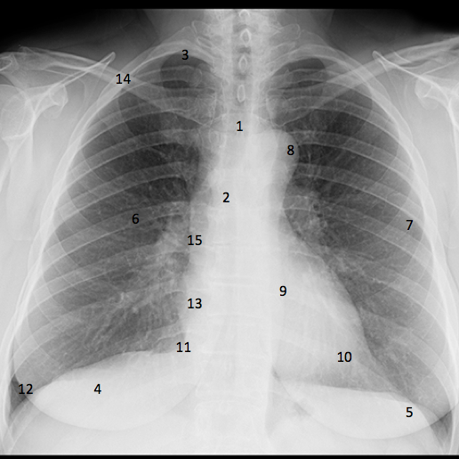
| 1) Trachea 2) Carina 3) Lung apex 4) Right hemidiaphragm 5) Left hemidiaphragm 6) Posterior rib 7) Anterior rib 8) Aortic knuckle/notch 9) Heart 10) Apex of heart 11) Cardiophrenic angle 12) Cardiophrenic recess 13) Right atrium 14) Clavicle 15) Right hilum | |
| What five things can an chest radiograph be used to diagnose? | 1) Infection 2) Fluid overload 3) Fracture 4) Foreign object (inhaled - usually in children) 5) Misplaced object |
| How many days of background radiation is chest X-ray worth? | 3 |
| How is the quality of an x-ray assessed? | R - rotation I - Inspiration P- projection E- Exposure |
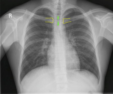
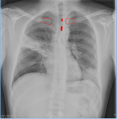
| How is rotation assessed? | By checking whether the clavicles and spiny processes are equidistant |
| What are the two problems with a rotated film? | 1) Distorts heart size 2) Trachea looks displaced |
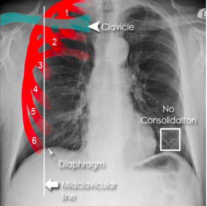
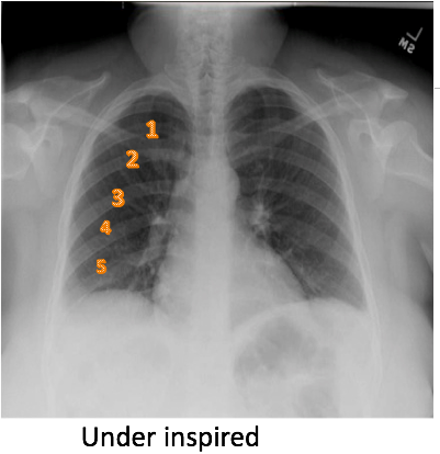
| How can you tell if inspiration has been sufficient? | Should be able to see the entire thoracic cavity with 5-7 anterior ribs at the midclavicular line |
| How do you tell if a film is PA or AP? | 1) Position of the scapula (should always be retracted by asking patient to cross arms) 2) Cardiothoracic ratio should be less than 50% 3) Should say on the film.. |
| In which position is the heart enlarged? | AP |
| AP position | |
| When would you take an AP radiograph? What structure will be in the lung field? What can't you comment on? | 1) If the patient is too ill to stand 2) Scapula 3) The heart size |
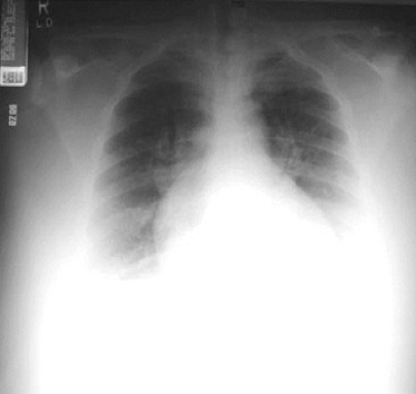
| How can you tell if a film is over or under exposed? | By looking at the cardiophrenic angle to the vertebral column. |
| Underexposed | Overexposed |
| What are the five parts of CXR interpretation? | A - Airway B - Breathing C - Circulation D - Diaphragm E - Everything else |
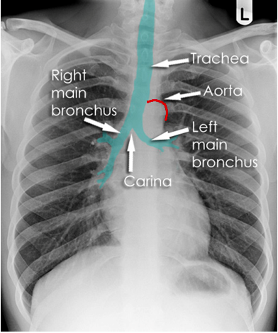
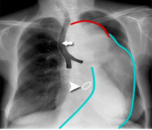
| What components of the airway are being examined in an airway? | 1) Trachea 2) Carina 3) Main bronchi |
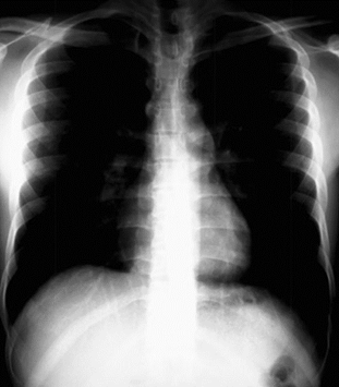
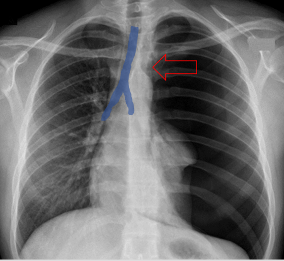
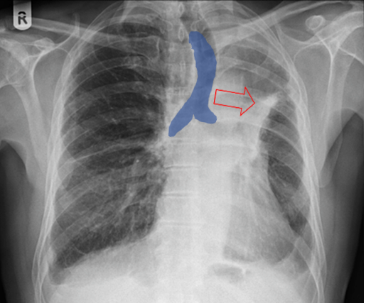
| Describe what you can see here. What two things may be causing this? | 1) Trachea is being pushed 2) Tension pneumothorax, masses |
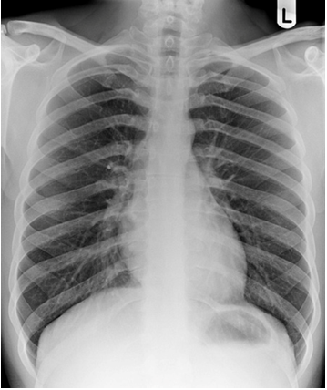
| What can be seen here? What might cause this? (4) | 1) Trachea is being pulled Collapse, loss of lung volume, consolidation (build up of fluid), fibrosis. Anything that causes an increase in negative pressure can cause mediastinal structures to be pulled over, damaging them. |
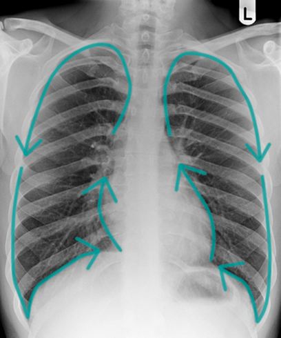
| When looking at breathing what are the divisions called? | Upper, middles and lower zones (along with hilar regions). Can't tell from a chest x-ray exactly what you are looking at. |
| How can you tell that the lungs are filling the pleural cavity? | Lung markings should reach the chest wall. |
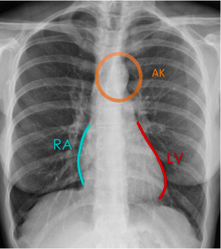
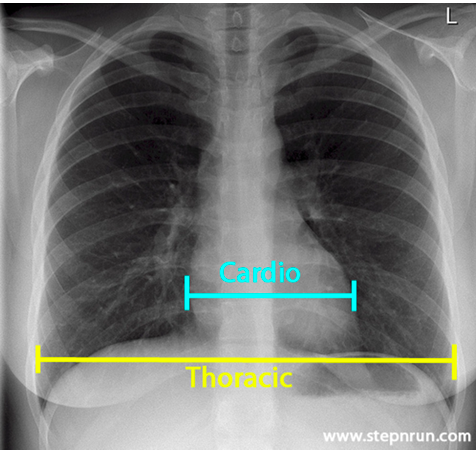
| What are the three areas of focus when examining circulation from a chest radiograph? | 1) Midline structures (inferior mediastinum) - aortic knuckle, widening of mediastinum 2) Heart contours (adjacent lung pathology) 3) Cardiothoracic ratio - greater than 50% is abnormal, an enlarged heart be a sign of lung disease. |
| Note that the aortic knuckle should be 2-3cm in diameter | |
| Describe | The black markings are sternotomy wires. Note the enlarged knuckle and mediastinum - patient had a cardiothoracic aneurysm. |
| Describe. | The cardiothoracic ratio is greater than 50% - cardiomegaly. |
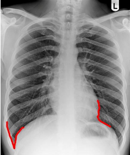
| Note the costophrenic and cardiophrenic angles. The right hemidiaphragm should be higher. You can usually see a gastric bubble on the LHS. In pathological conditions liquid may gather in the recesses. | |
| What pathologies can you look for in the diaphragm? | 1) Flattened hemidiaphragms (common in COPD) 2) Blunted angles 3) Gastric bubbles |
Want to create your own Flashcards for free with GoConqr? Learn more.