
Flashcards
Anatomy practicals week 3
Cards in this deck (46)
- 1Describe the thoracic cage-Encloses: sternum, ribs (12), costal cartilages, 12 thoracic vertebrae
-Protects the heart, lungs, liver and spleen - 2Borders of the thoracic cavity?Superior: upper surface of first rib, their costal cartilages, the manubrium of the sternum and the body of T1
-Inferior: diaphragm - 3What is the mediastinum?Structures lying within the central region of the thoracic cavity including the heart, great vessels, oesophagus and trachea
Lungs lie on either side - 4What nerve supplies the diaphragm? Nerve roots? What do the intercostal nerves supply?Phrenic, C3-5
Each supplies 3 intercostal muscles, the parietal pleura deep to them and lateral/anterior aspects of the overlying skin via a main branch under the rib and a collateral branch on the rib beneath - 5Describe the trachea-Extends from C6 to T4/5
-Can be palpated at the suprasternal/manubrial notch
-Held open by C-shaped rings of elastic cartilage
-Splits at the carina (T4) - 6Describe the lungs-Left has 2 lobes: upper/lower lobe divided by an oblique fissure
-Right has 3 lobes: upper/middle/lower lobes divided by oblique and horizontal fissures
-Left is smaller since the heart is mostly on the LHS - 7Describe stages of lung development-Begins in 4th week as mediam laryngotracheal tube in floor of primitive pharynx
-Separated from developing oesophagus by the growing tracheo-oesophageal septum
-Laryngotracheal tube develops a lung bud at its distal end which divides into 2 bronchial bugs by 5th week
-These grow laterally into primitive pleural cavities and divide to form terminal bronchioles - 8Describe the pseudoglandular period (weeks 5-17) Describe the canalicular period (weeks 16-25)-Lung has a glandular appearance
-Major elements divide as far as terminal bronchioles
-Lumina of bronchi and terminal bronchioles enlarge
-Respiratory bronchioles and alveolar ducts develop
-Lung tissue becomes highly vascularised - 9Describe the terminal sac period (weeks 24-birth)-Alveolar ducts develop terminal sacs (primitive alveoli)
-Matures into flatter, more squamous epithelium at~26 weeks
-Lung are developed enough for foetus to survive if born premature - 10Describe the alveolar period (late foetal- 8 years)-Epithelium of terminal sacs continues to thin so that adjacent capillaries bulge into them forming immature alveoli
-Each respiratory bronchiole terminates into a cluster of thin-walled sacs (future alveoli) separated by loose connective tissue
-95% of alveoli develop after birth - 11Is the trachea a narrow structure?-Can narrow to allow oesophagus to expand
-Loose connection of adventitia allow it to move within the neck/thorax - 12Bony attachments of the diaphragmLumbar vertebrae and arcuate ligaments
Costal cartilages of ribs 7-10 (attach directly to ribs 11-12)
Xiphoid process of the sternum - 13What are the different openings of the diaphragm? Vertebral levels? What structures pass through?-Oesophageal opening (T10): oesophagus, vagus nerves, and oesophageal branches of the left gastric vessels
-Aortic opening (T12): aorta, thoracic duct (lymphatic vessel) and azygous vein
-Caval opening (T8): IVC - 14Where is the apex of the lung located? Is there a difference between the left/right bronchus?Above first rib into floor of the neck
Right bronchus is wider, shorter and more vertical than the left one - 15What are bronchopulmonary segments?-Anatomically separate sections of the lung, each with its own bronchus and artery
-Can be surgically removed without affecting the rest of the lung
Right: 10 segments
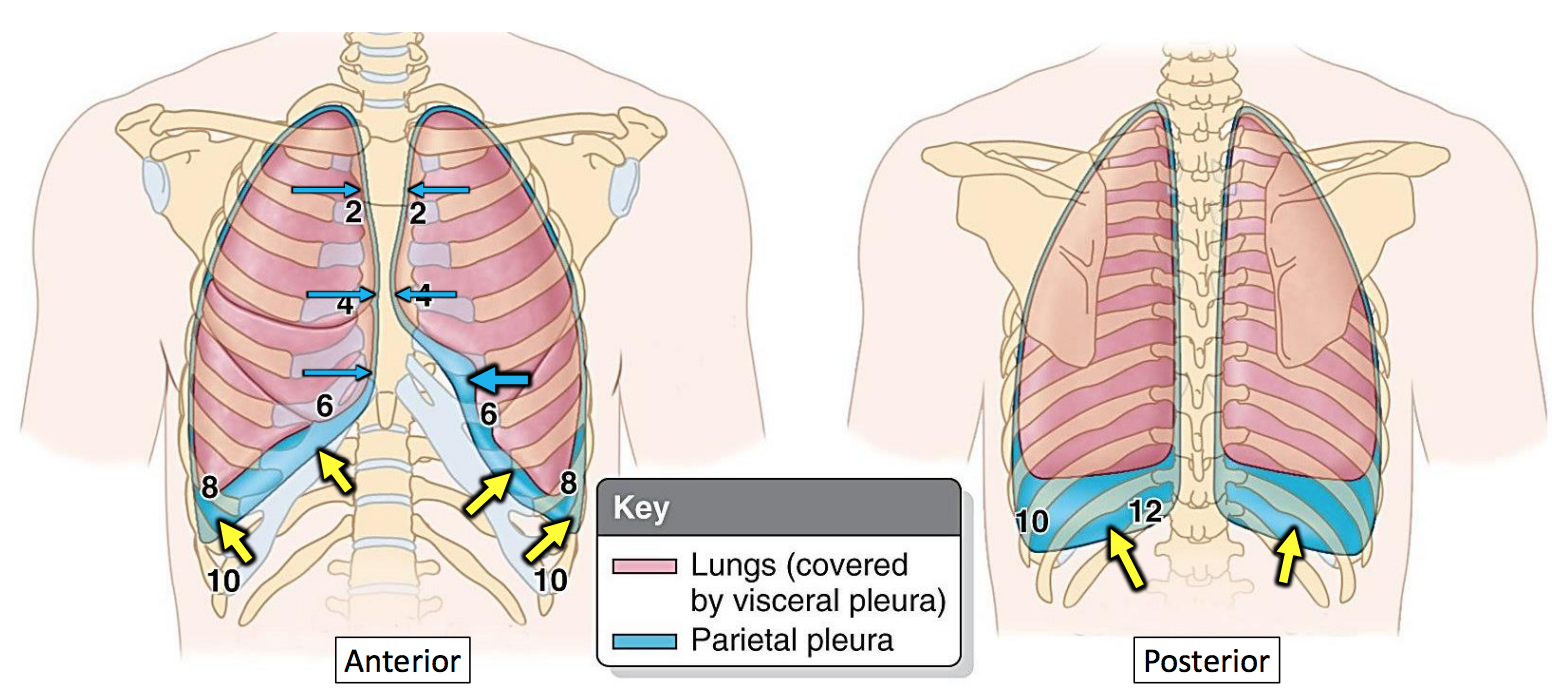
Left: 8 segments - 16What is the pleural cavity?Potential space between the visceral/parietal pleura/ Has a small volume of serous fluid
- 17What is the costophrenic angle? Clinical sig.?Angle (~30 degrees) at the junction of costal pleura and diaphragmatic pleura (ie. right in the corner between the pleura and the diaphragm).
Thoracocentesis (pleural tap)is performed here when patient is in full expiration (less risk of puncturing lungs/pneumothorax) - 18Describe the... of the external intercostals -insertion -action -nerveAttachments: Originate at the lower border of the rib, inserting into the superior border of the rib below
Actions: Elevates the ribs, increasing the thoracic volume
Innervation: Intercostal nerves (T1-T11). - 19Describe the... of the internal intercostals -insertion -action -nerveAttachments: Originates from the lateral edge of the costal groove and inserts into the superior surface of the rib below
Actions: The interosseous part reduces the thoracic volume by depressing the ribcage, and the interchondral part elevates the ribs
Innervation: Intercostal nerves (T1-T11) - 20Describe the... of the innermost intercostals -insertion -action -nerveAttachments: Originates from the medial edge of the costal groove and inserts into the superior surface of the rib below
Actions: The interosseous part reduces the thoracic volume by depressing the ribcage, and the interchondral part elevates the ribs
Innervation: Intercostal nerves (T1-T11) - 21Describe the... of the transverse thoracis -insertion -action -nerveAttachments: From the posterior surface of the inferior sternum to the internal surface of costal cartilages 2-6
Actions: Weakly depress the ribs
Innervation: Intercostal nerves (T1-T11) - 22Describe the... of the subcostals -insertion -action -nerveAttachments: originate from the inferior surface of the lower ribs, near the angle of the rib. They then attach to the superior border of the rib 2 or 3 below
Actions: Share the action of the internal intercostals
Innervation: Intercostal nerves - 23Due to disease or injury, a haemothorax, pleural effusion or pneumothorax may occur and must be removed using a needle. Where should the needle be inserted?5th intercostal space at the midaxillary line
Cannula in 2nd intercostal space in midclavicular line (releases pressure) - 24Why would cutting the visceral pleura not cause pain? Would cutting the parietal pleura cause pain?-Sensory fibres only detect stretch
-Parietal pain would sense localised pain as it is innervated by phrenic/intercostal nerves - 25An apical lung tumour may damage the sympathetic chain above the region of the second thoracic ganglion, resulting in ‘Horner’s syndrome’. Describe Horner’s syndrome and use your knowledge of the autonomic nervous system to explain this response.Symptoms: miosis of eye, partial ptosis, enophthalmos, anhidrosis
Occurs on same side as lesion in sympathetic nerve - 26A patient aspirates some filling material whilst in the dentist’s chair. Where is the material likely to be found? Give a reason for your answer.Right lung. Left bronchus is at an angle
- 27f a similar thing occurs in a recumbent patient, the apical segment of the right lower lobe may become affected by pneumonia or abscess formation. How would you position a patient to allow natural drainage of an infection in the apical segment of the lower lobe of his right lung?Lie on left side
- 28A patient complains of severe pain in the abdominal region, but the doctor asks for a radiograph of both the chest and abdominal regions. Why?-Pain is not localised but referred
-Could be a bowel perforation
-Pocket of air under diaphragm - 29What is paradoxical respiratory movement and when might it occur?-Abnormal movement of chest wall and diaphragm during inhalation and exhalation
-Movement is the opposite of what you would expect eg. chest wall moves in and diaphragm stays relaxed during inhalation
-Can occur due to trauma of ribs, weakened inspiratory muscles - 30Define pneumoniaBacterial infection causing inflammation of lung. Affects alveoli
- 31Define pulmonary thromboembolismSudden blockage in a lung artery. Caused by deep vein thrombrosis that breaks loose and travels up from leg to the lung via blood
- 32Define emphysemaLong-term progressive disease due to over-inflation of alveoli. Shortness of breath as respiratory lung tissue are impaired/destroyed
- 33Define bronchiectasisDisease where there is permanent enlargement of parts of the airway leading to build up of excess mucous (more vulnerable to infection)
- 34Cystic fibrosis is a human congenital disease which usually presents at less than a year of age. How does the defective gene product influence the respiratory tract? Why do patients with cystic fibrosis require physiotherapy ‐ how does it help?-CFTR protein is absent/not functioning
-Causes buildup of thick mucous lining the airway
-Reduces SA available for gaseous exchange
-Bacteria colonise leading to repeated infections
-Physiotherapy can clear the mucous by stimulating a cough, chest percussion, postural drainage, shaking and vibrations - 35Describe the vertebral levels of the pleura and the lungs at various significant anatomical points
- 36What are the 3 major functions of the respiratory system?-Bulk movement of air between lungs/external environment
-Modification of air
-Efficient exchange of gases between the alveolar lumen/capillaries - 37What are Clara cells?Non-ciliated bronchiolar cells that secrete glycosaminoglycans which protect the bronchiole lining
- 38Function of olfactory sensory neurones? Supporting cells?Bipolar neurones which congregate to form the olfactory nerve
Provides metabolic and physical support for the olfactory epithelium - 39Function of basal cells? Brush cells?Stem cells that are capable to differentiating into supporting/olfactory cells
Microvilli-bearing columnar cell that transducts general sensation - 40Types of epithelium that lies: nasal cavity nasopharynx vocal cordsNasal cavity: ciliated pseudostratified epithelium
Nasopharynx: respiratory epithelium (ciliated pseudostratified columnar epithelium)
Vocal cords: stratified squamous epithelium - 41What types of cells are present in the walls of the alveolar sacs to prevent aspirated microorganisms from entering the bloodstream?Macrophages
- 42How does interstitial fibrosis impair gaseous exchange?Scarring of interstitium (tissue between pulmonary alveoli/bloodstream) thickens the diffusion path.
There is excess collagen, making the lung stiff - 43What is the response of the alveoli to bacterial infection as might occur during pneumonia?Pneumonia: fluid inside lungs is visible on CXR
-Macrophages engulf/digest bacteria
-Cytokines and fluid is released into alveolar cavity reducing SA for gas exchange - 44How will pulmonary oedema affect the blood‐gas barrier, and therefore what would be its impact on function?-Increases barrier as fluid is pushed into alveoli
-In older people, back flow of blood causes weakening of heart - 45How would pleural adhesions affect the function of the lungs – and what are they?Pleural adhesions: formation of fibrotic bands in the pleural space
There is less intrapleural space+ more friction. Lung can't expand fully - 46When a patient has a sore throat, which tissue is inflamed?Tonsils
Information
- Description
- Basic topography of the lower respiratory tract
- Created by
- Dolu Falowo
- Views
- 70
- Rating