
Flashcards
Vision 1
Cards in this deck (20)
- 1What is the general anatomy of the eye?Pupil: Opening where light enters the eye
Sclera: White of the eye
Iris: Gives color to eyes
Cornea: Glassy transparent external surface of the eye
Optic nerve: Bundle of axons from the retina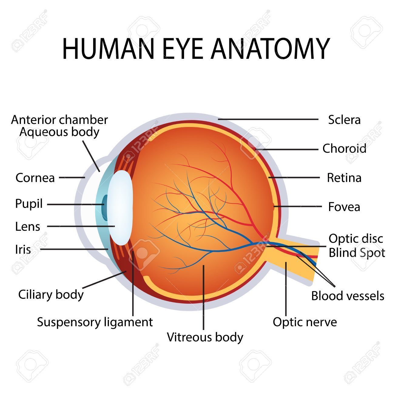
- 2How does the eye form an image?Eye collects light, focuses on retina, forms images (inverted)
The ability to focus an image on the retina depends on the refractive power of both the cornea and the lens as well as on the shape of the eye globe
Cornea has refractive index of 42 diopters (parallel light rays will be focused 2.4 cm behind it)
Cornea has refractive index f 42 diopters (parallel light rays will be focused 2.4 cm behind it) - 3How does the lens accommodate light?Changing shape of lens allows extra focusing power
Most of the focusing performed by the cornea but lens is important for enhancing the refraction of near objects (additional 12 diopters) - 4Describe errors of refractionEmmetropic = normal eye
Presbyopia = Our lens hardens with age and ciliary muscles weaken. This causes a decreased ability in accommodation.
Hyperopia = farsightedness; refractive power insufficient for close objects
Myopia = nearsightedness; refractive power too strong for distant objects - 5How does the pupil help to form an image?The Pupillary Light Reflex
Connections between retina and brain stem neurons that control muscle around pupil
Continuously adjusting to different ambient light levels
Consensual (both pupils react similarly and simultaneously)
Pupil similar to the aperture of a camera
Circular (constrictor) muscles act to decrease the pupil size under parasympathetic control
Radial (dilator) muscles act to increase the pupil size under sympathetic control - 6What is the visual field?Amount of space viewed by the retina when the eye is fixated straight ahead
Image is inverted - 7What is visual acuity?Ability to distinguish two nearby points
Determined largely by photoreceptor spacing and refractive power
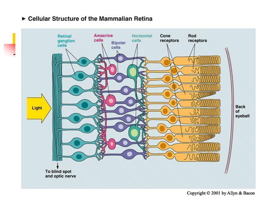
Visual Angle: Distances across the retina described in degrees - 8What is the cellular structure of the retina?Direct (vertical) pathway for signal transmission:
Ganglion cells
Bipolar cells
Photoreceptors
Lateral connections influence signal processing :
Horizontal cells, Receive input from photoreceptors and project to other photoreceptors and bipolar cells
Amacrine cells, Receive input from bipolar cells and project to ganglion cells, bipolar cells, and other amacrine cells - 9Give an overview of photoreceptors
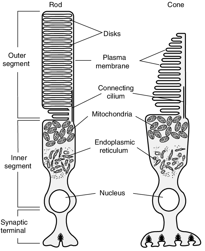
- 10What is the basis of phototransduction/vision?1) Vertebrate Photoreceptors have a depolarized rmp (Vm) Compared to other neurons, resting Vm is more positive (~ 20mV)
2) With light exposure, Vm hyperpolarizes
This positive Vm is because of the “dark current”. A cGMP-gated Na+ channel that is open in the dark and closes in the light
This change in Na+ with light is the signal that enables the brain to perceive objects in the visual field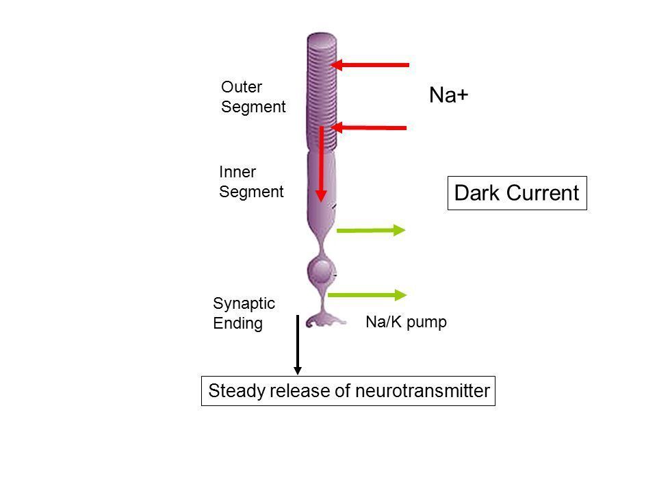
- 11Describe dark currentIn the dark,
PNa ~ PK (Na channels in the outer segment)
Vm therefore between ENa and EK - 12What molecule in Rod cells responds to light?Visual pigment molecules called Rhodopsin are composed of Retinal (Vitamin A derivative) and Opsin (GPCR) are present in membrane folds (called disks in the outer segment)
Rhodopsin converts 11-cis-Retinal to all-trans-Retinal (the active form) - 13What is the molecular pathway in response to light?all-trans-Retinal activates transducin molecular cascade decreases cGMP
This leads to closure of cGMP-gated Na+ channel
Lowered Na entry results in hyperpolarization
High Gain Mechanism…… 1 opsin>>>>1000 trasducin 1 PDE>>>>>1000 cGMP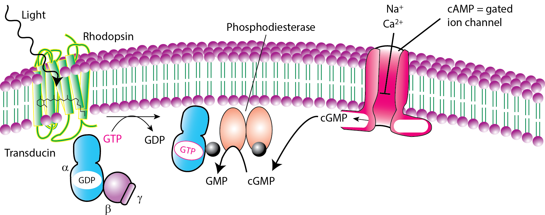
- 14How is the dark current channel modulated?Open in the dark
Closes in response to light
Nucleotide-gated channel (opened by cGMP)
Permeable to Na+
Keeps photoreceptor Vm more positive than most neurons
Steady release of neurotransmitter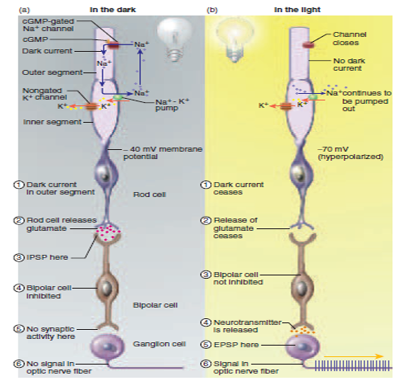
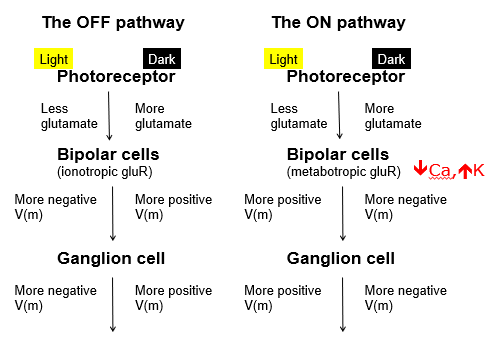
- 15Describe the on and off pathways in light and dark
- 16Describe signal transmission to ON and OFF cellsOnly ganglion (and some amacrine cells) produce action potentials, all other cells produce graded changes in membrane potential
The hyperpolarization of photoreceptors elicits both hyperpolarization and depolarization within bipolar and ganglion cells
These graded potentials modulate the discharge rate of ganglion cells
ON and OFF bipolar and ganglion cells respectively detect increases and decreases in luminance - 17What facilitates visual acuity?Visual Acuity Ability to distinguish two nearby points. Determined largely by photoreceptor spacing and refractive power
- 18What is the distribution of rods and cones in relation to function?Rods – seeing in dim light
Cones – seeing in normal daylight
More convergence in rod system, increasing sensitivity while decreasing acuity
Peripheral rods have large spacing (lower density) and high convergence to a large ganglion cell whereas foveal cones are high density and have low convergence to a small ganglion cell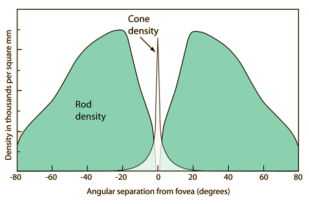
- 19What is the basis for colour vision?Photoreceptors are only activated by a small portion of the spectrum of electromagnetic waves……..we do not see in the infra red or the UV range
The electromagnetic range perceived comprises distinct wavelengths
Different opsins activate at specific wavelengths, e.g. short-wave for blue, long wave for red and middle-wave for green - 20What is the duplex theory of vision?Rods are achromatic in the peripheral retina with high convergence, high light sensitivity and low visual acuity
Cones are chromatic in the central retina (fovea) with low convergence, low light sensitivity and high visual acuity
Information
- Description
- Sensory and motor neuroscience Flashcards on Vision 1, created by Megan MacLeod on 28/04/2019.
- Created by
- Megan MacLeod
- Views
- 0
- Rating