
Flashcards
C&B [047] Heart Development
Cards in this deck (23)
- 1Dorsal Surface Ventral Surface Rostal CaudalDorsal surface closest to developing nervous system
Ventral Surface closest to bell button
Rostal - Head
Caudal - Tail - 2Fetal Blood Circuits: Viteline Umbilical Embryonic- Viteline: yolk sac to embryo
- Umbilical: between embryo + placenta
- Embryonic Circuit: w/in embryo-heart + blood vessels - 3When does embryo develop circulatory systems? Why?- Once tissue layers have developed
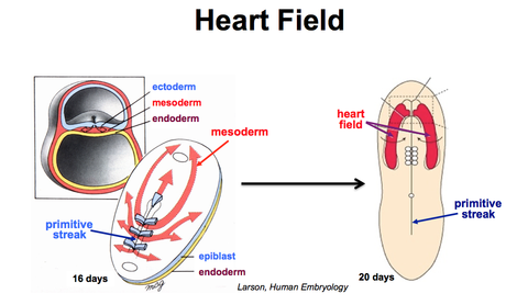
- To carry Nutrients + gases around body for exchange @ placenta - 4Heart Field- Mesoderm cells that'll form heart ingress through primitive streak
- During Gastrulation
- Moves towards embryo rostral end
- Forming crescent shaped "heart field"
- these cells made by lateral plate mesoderm's splanchnic layer - 5Where do all cardiac cell types arise through?- The lineage diversification of 3 embryonic precursor pools:
i) Cardiogenic Mesoderm
= Endocardial cells -> cushion cells
Atrial + Ventricular myocytes -> conduction cells
ii) Proepicardium
= Smooth Muscle of Coronary Artery, Endothelium, Fibroblast
iii) Neural Crest
= aortic smooth muscle + ANS - 6From staining embryo for gene-specific mRNA in whole mount in situ hybridisation we have seen what? What does mutations in this gene cause?- Distribution of Nkx2-5 mRNA in mouse embryo
- only transcribed in heart
- 1st found in cardiac crescent
= then continue to express as they transform to 4 chambered heart
- NKX2.5 mutation cause atrial septal defects - 7Describe the location of the heart field during development Head folding What surrounds yolk sac- Rostral to brain
- Head folding brings it into thorax
+ makes foregut
- Mesoderm w. blood islands surround yolk sac:
i) contains stem cells that make embryonic blood + vasculature
in chick - 8Ventral Closue Endocardial Tubres Ventral Folding- Cardiac Descents forms endocardial Tubes
- Endocardial Tubes lie on either side of developing foregut
- at 20 days: developing forges still open to yolk sac
- Ventral Folding:
i) brings foregut edges together => a tube
ii) Brings endocardial tubes tog. + surrounds them in myogenic mesoderm - 9Arms of Endocardial Tubes- Endocardial tubes' arms divided into
i) atrial (w. venous inlet) + ventricular regions
ii) rostral arterial outlet end: where 2 tubes meet - 10When does human embryonic heart begin to beat? When does the heart fully form?- Human Embryos: heart beats 22-23 days after fertilisation
- Heart fully forms in 8-9th week - 11Where do embryonic veins drain into? Where do arteries arise from? What is the placent's o2ed blood directed through? What are the blood circuits reorganised to?- Embryonic veins drain into inlet sinus go heart
- Arteries arise from outlet end
- Placenta O2ed blood directed through pharyngeal arches:
i) to developing brain + body
- Circuits completely reorganised to the heart's vessels - 12What does cardiac crescent form? What does the heart tube looping achieve? What do the cardiac neural crest cells invade?- Cardiac Crescent: segments of linear heart tube
- Heart Tube loops to bring RA + LA
(rostral to ventricle)
- Cardiac Neural Crest Cells invade aortic arch - 13What do cardiac neural crest cells remodel?- Cardiac Neural Crest Cells drive remodelling of:
i. pharyngeal arteries (3,4,6)
ii. Aortic Sac
= producing mature aortic arch - 14What divides RA +LA and RV + LV? What forms cardiac valves? (i.e. what valve segments are there?)- Septa
- Conotruncal (CT) and Atrioventricular valve segments
=> cardiac valves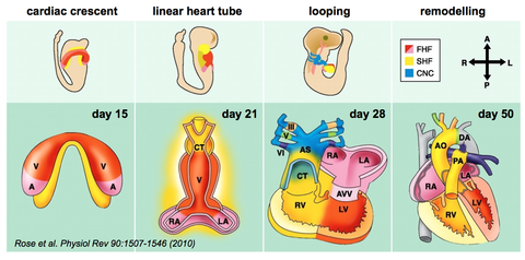
- 15What are endocardial cushions made of? Where do endocardial cushions flow? What is formed when opposing cushions fuse?- Endocardial Cushions:
i) made of ECM
ii) and mesenchyme
- Form in atrioventricular region + outflow tract region
- Opposing cushions fuse =>
i) atrioventricular canals
ii) Outflow Vessels
iii) Membranous part of interatrial / ventricular septa - 16What forms from the roof of atrial chamber? What is foramen primum?- Spina Vestibuli + Septum primum
- as crescent shaped wedge toward AV canal
- Diminishing connection between R + L atria - 17What happens to close off the foramen primum? How does foramen secundum form? How does septum secundum form?- Septum primum + AV septum fuse
- Cell death makes foramen secundum
- Septum Secundum grows from RA roof to AV septum - 18What do foramen oval and secundum allow? Trabecular Layer How are the ventricles divided into R + L? Foramen Ovale- For blood entering RA to pass into LA
- Trabecular Layer: myocardium thickens = outer compact muscle layer forms too
- Muscular Interventricular septum grows towards AV septum
= dividing ventricles to R + L halves
- Foramen Ovale: opening left by septum secundum no closing properly - 19What forms the atrial valve? What direction does the blood in atria flow in? and why? How does the foramen oval close?- Septum primum + secundum => valve allowing blood to flow from:
RA high pressure -> LA low press
- BUT not from LA -> RA
bc blood can't flow against RA press
- @ Birth: press increases in LA
i) Pushes septum primum against secundum
ii) Closes Foramen Ovale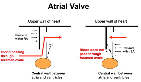
- 20What is sculpted from the muscular walls of the ventricles?- AV valves
- Papillary Muscles
- Chordae Tendinae
- Cusps - 21Describe Path of oxygenated blood.- From Placenta
- into RA
- from IVC
- through LA, LV, Aorta and Arteries to developing brain - 22Describe path of deoxygenated blood- From upper body
- Through RA from SVC
- into RV + pulmonary Artery - 23What is the pathway of blood to be deoxygenated ?- Ductus arteriosus bypasses lungs
- Carrying deo2ed blood to aorta
+ placenta
Information
- Description
- Flashcards on C&B [047] Heart Development, created by jyghowes on 05/02/2015.
- Created by
- jyghowes
- Views
- 0
- Rating