22085585
Description
| Question | Answer |
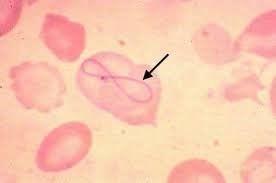
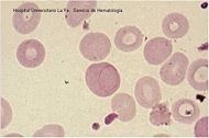
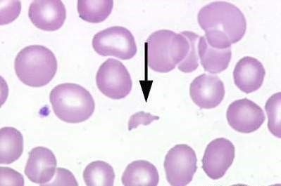
| Cabot rings are purple-staining, threadlike filaments in the shape of a ring or as a figure of eight (8) appearing in the RBC. They are thought to be nuclear remnants and more specifically derived from the microtubules of the mitotic spindle. Cabot rings are seen in conditions where we have disturbed erythropoiesis and most frequently in thalassemia and megaloblastic anemia, basically in similar conditions to Howell-Jolly bodies | |
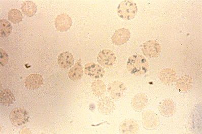
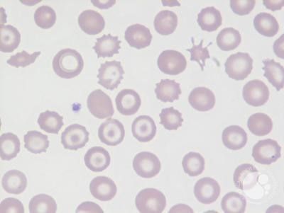
| Acanthocytes are RBC(s) with irregularly spaced projections. These cells have a decreased survival time (condition associated with hemolysis) and are predominantly found in a condition known as acanthocytosis or abetalipoproteinemia (absence of -lipoprotein from the RBC membrane). This condition is normally inherited; however, acanthocytosis may be acquired in association with a number of conditions such as alcoholic cirrhosis and malabsorption states. In this condition, the RBC membrane undergoes distortion to give finger - like projections. At most, there would be 5 projections | |
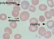
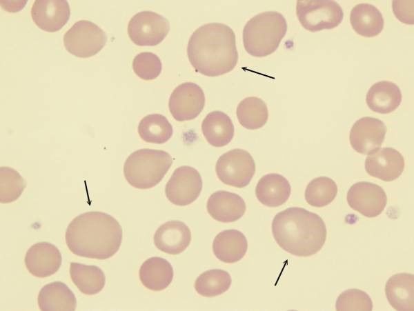
| Dimorphic picture/appearance describes heterogeneity in the size of red blood cells, usually with two distinct populations. It can be found in partially treated iron deficiency, mixed deficiency anemias (e.g. folate/B12 and iron together), following red cell transfusion, or in cases of sideroblastic anaemia. | |
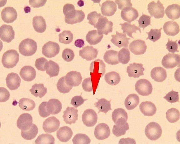
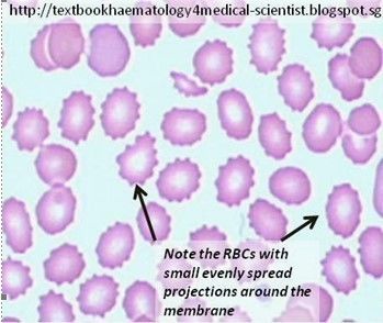
| Echinocytes (burr cells) have multiple short pointed projections uniformly spaced over the cell surface. The central pallor is retained. Echinocytes are often confused with acanthocytes (spur cells). However, the projections of echinocytes are smaller, more numerous (6-10/RBC), and more regular than those of acanthocytes. Echinocytes are characteristic of end stage liver disease or kidney failure and should be differentiated from acanthocytes or spur cells. They can also be seen as an artifact of slide preparation or prolonged specimen storage | |
| Hb H These are bluish green granules that appear with Supravital Stains only. They are precipitates of beta globin chains and are associated with Hb H disease (a form of α thalassemia). | |
| Anisocytosis: Refers to variation in the size of RBC(s). Normally, there is minimal variation in the size of RBC(s). When the variation is significant, we call it anisocytosis. Anisocytosis is not characteristic of any one disease, but it is present in a great majority of erythroid disorders and in virtually all anemias. | |
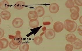
| Hb C Cystals These are elongated crystals with blunt ends that appear darkly stained in color and are associated with HbC disease. | |
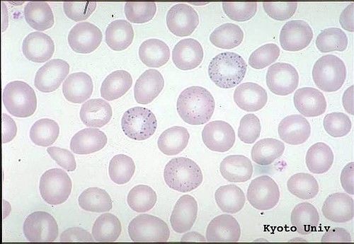
| Basophilic stippling is the fine or coarse deep blue to purple staining inclusion that appears in immature RBC(s) stained with Wright and Giemsa. Stippling represents aggregates of ribosomes and is associated with chronic hemolytic anemias. The three disorders particularly associated with coarse basophilic stippling are lead poisoning, sideroblastic anemia, and thalassemia. | |
| Cabot rings are purple-staining, threadlike filaments in the shape of a ring or as a figure of eight (8) appearing in the RBC. They are thought to be nuclear remnants and more specifically derived from the microtubules of the mitotic spindle. Cabot rings are seen in conditions where we have disturbed erythropoiesis and most frequently in thalassemia and megaloblastic anemia, basically in similar conditions to Howell-Jolly bodies. | |
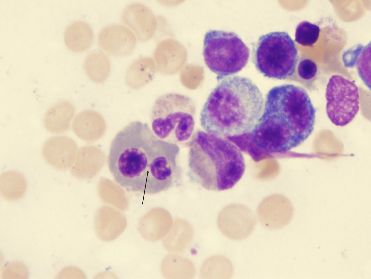
| Diserythropoiesis This refers to an abnormality in the nuclear membrane resulting in bilobed or multilobulated erythroblasts | |
| HbC crystals These are elongated crystals with blunt ends that appear darkly stained in color and are associated with HbC disease. | |
| Howell-Jolly bodies Howell-Jolly bodies are round, purple staining nuclear fragments seen in the RBC. They usually appear in conditions where we have chromosomal breakage. As this RBC passes through the spleen, the spleen will take out the HJB and therefore the cell will pass out without it. So, when we see HJB(s) in a peripheral blood smear, it means that the spleen is not present or if present its function is impaired. HJB are common: 1) In some hemolytic anemias (i.e. Thalassemia and SCA) 2) In megaloblastic anemia Resemble nucleus Certain abnormalities Inherited disorders Spleen is present: supposed to clean Aspleenia Slpeneectomy | |
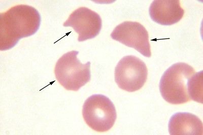
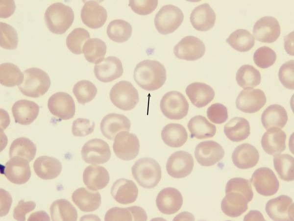
| Heinz bodies Heinz bodies which are clumps of denatured Hb are RBC inclusions which are stained with a supravital stain. With Romanowsky stains, they appear as pale focal areas within the RBC (Peripheral hemoglobinization). The RBC(s) here are also known as bite cells. In man, the finding of Heinz bodies in the blood is associated with G6PD deficiency, following exposure to certain drugs or oxidants. Heinz bodies (arrow) do not appear with the regular stains.(Wright’s-Giemsa stain with 100x objective). They appear as a pale focal area. Photograph courtesy of Dr. Perry Bain. | |
| Heinz bodies are more prominent with the use of a vital stain (New methylene blue stain with 100x objective). Photograph courtesy of Dr.Perry Bain. | |
| Hypochromic When the amount of hemoglobin per red cell is less than normal --> the central pallor becomes larger and the RBC is said to be hypochromic. Hypochromic RBC(s) are seen in Fe-deficiency anemia, Thalassemia as well as Hereditary Sideroblastic Anemia. | |
| Helmet or triangular cells There is no special appearance for schistocytes but two specific forms are important in diagnosis. The fragmented RBC may appear triangular or may have the appearance of a helmet cell (like Napoleon’s hat). A triangular cell or a helmet cell, characterizes a type of anemia in which the RBC(s) are fragmented within the blood vessels, these latter containing fibrin threads. This anemia is a hemolytic anemia that occurs secondary to intravascular coagulation. Intravascular destruction Particular Clots within Dissemination Coagulation within vessels Pathognomonic | |
| Microcytosis When the MCV < 80fl, the RBC (s) are said to be microcytic. There are many types of anemias where the average cell is smaller than normal. These include: • Fe-deficiency anemia • Thalassemia • Hereditary Sideroblastic Anemia | |
| Macrocytosis When the MCV > 100 fl, the RBC (s) are said to be macrocytic. Macrocytosis is encountered in many instances: • Megaloblastic anemia • New born babies • Active erythropoiesis • Liver disease (chronic) i.e. Alcoholism. (Altered lipid content of the red cell membrane) | |
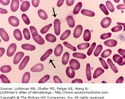
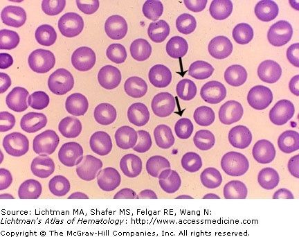
| Ovalocyte, elliptocyte or cigar shaped RBC The commonest abnormal shape is the ovalocyte, elliptocyte or cigar shaped RBC, characteristic of the following disorders: • Fe-deficiency • Thalassemia • Megaloblastic anemia (Macroovalocytes) However note that these cells may be normally present in the blood smear in small numbers. N.B: The cigar shaped RBC is very similar to the ovalocyte or elliptocyte with slight modifications, i.e. it is a little bit longer and thinner. When the great majority of RBC(s) (~ 95%) are ovalocytes, elliptocytes or cigar shaped we call the condition congenital ovalocytosis or elliptocytosis. This condition is inherited as an autosomal dominant gene. In congenital elliptocytosis, we have one of two cases: * 95% of subjects with elliptocytosis are benign and have no disease. Here we do not know what makes the RBC deformed to become elliptical but these cells are known to function normally. * Only 5% show a condition of hemolytic anemia (abnormality or defect in spectrin, a skeletal protein present in the RBC membrane). | |
| Schistocytes Schistocytes or fragmented RBC(s) are encountered in a great majority of hemolytic anemias especially intravascular hemolytic anemias. They are also seen in conditions in which we have ineffective erythropoiesis (destruction of RBC(s) in the bone marrow). | |
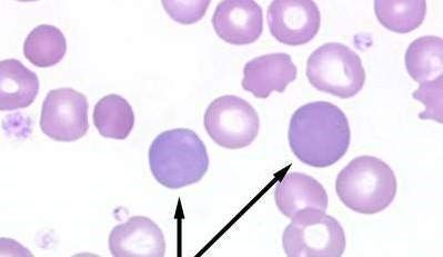
| polychromatophilic RBC is an immature non nucleated RBC which appears diffusely bluish in color when stained with a regular Romanousky stain. This is an early or immature reticulocyte with a significant number of ribosomes and RNA still present in the cytoplasm. Because RNA is acidic in nature, it takes up methylene blue. Since the wright stain contains alcohol we have fixation of the cells the remaining RNA and ribosomes diffuse in the cell to give it a diffuse grayish-bluish color. This cell is known as a reticulocyte when stained with a supravital stain. So if we do not fix the RBC(s), but subject them to a potent oxidizing agent such as Brilliant Cresyl Blue --> we will precipitate the remaining RNA and ribosomes so that these give a deep blue precipitate instead of showing a diffuse grayish color. | |
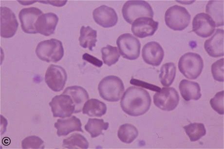
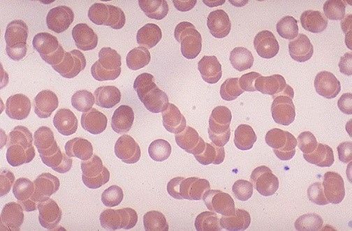
| Rouleaux are stacked/clumped groups of red cells caused by the presence of high levels of circulating acute-phase proteins which increase red cell 'stickiness'. They are often an indicator that a patient has a high ESR and are seen in infections, inflammations and Multiple Myeloma. | |
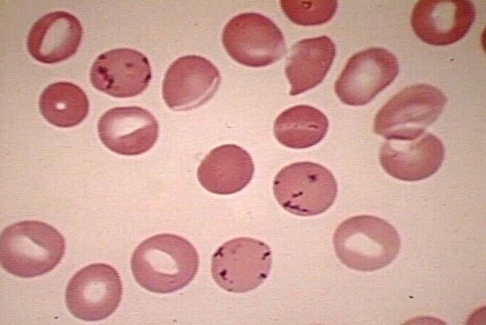
| Pappenheimer bodies Pappenheimer bodies (siderotic granules) are small, irregular, dark staining granules that appear near the periphery of a RBC in a film stained with Wright and Giemsa. With Perls’ Prussian Blue stain (special stain for iron), these bodies stain positively, indicating their iron content. The spleen normally removes these inclusions without destroying the cell. However, after splenectomy, pappenheimer bodies are visible on the blood film. Normally, no more than three small iron particles are noted in developing RBC(s) in the B.M. An erythrocyte that is positive for siderotic (iron) granules in a Prussian Blue stain is designated a siderocyte. A normoblast (nucleated erythrocyte) with siderotic granules is called a sideroblast. With severe disturbance of Hb synthesis, pathologic sideroblasts and siderocytes are present in the B.M. and P.B. Siderotic granules may be present in sideroblastic anemia and refractory anemias. | |
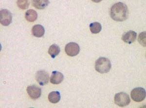
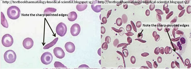
| Sickle cells are deformed RBC(s) in the shape of a sickle or crescent. To be considered as sickle cells, they must come to a point at one end. These cells are associated with HbS and are found in sickle cell anemia. | |
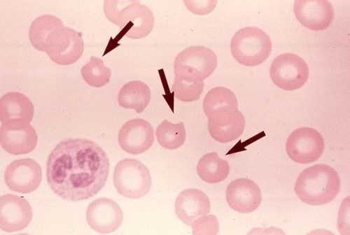
| spherocyte is a small red blood cell with no central pallor that appears a little bit darker in color. Spherocytosis could be inherited or acquired (in association with ABO/ Rh incompatibility or autoimmune hemolytic anemias) and it is always associated with hemolysis due to the rigidity of the membrane | |
| Target cell The normal RBC shape is governed by a wide surface area/volume ratio for adequate exchange of gases. If you decrease the internal volume of the cell, or you increase the surface area keeping the volume constant, the RBC membrane will undergo folding and will look like a target. • Thus any condition which removes Hb from inside the cell will wrinkle the RBC. This is a mark of volume compared to the surface area. Under this, we have anemias due to in heme synthesis (namely Fe-deficiency anemia) or due to in globin synthesis (Hemoglobinopathies such as thalassemia or sickle cell anemia). | |
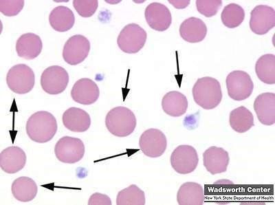
| Stomatocytes are erythrocytes with an elongated (mouth-like) area of central pallor. As many as 3% of RBC(s) may be stomatocytes in a normal smear. The presence of few stomatocytes in a peripheral blood smear is entirely harmless, but when they are increased in number we get a case of congenital stomatocytosis. As is the case with congenital elliptocytosis, almost all cases are harmless and only ~ 5% present a hemolytic type of anemia. Stomatocytes might be seen as a non-specific finding in a variety of situations, such as liver disease (cirrhosis), alcoholism or may even be considered an artifact of preparation | |
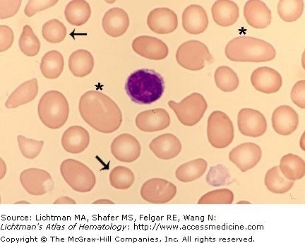
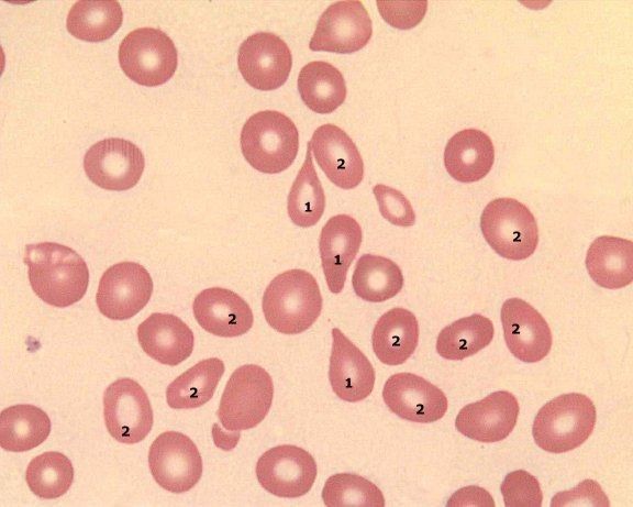
| Tear-drop cells (Pear shaped) Tear-drop cells may be encountered in a variety of anemias. However, when, these cells are numerous, it is very suggestive of myelofibrosis (a condition where the BM becomes full of fibrous tissue) that would lead to extramedullary hematopoiesis or blood cell production outside the bone marrow. | |
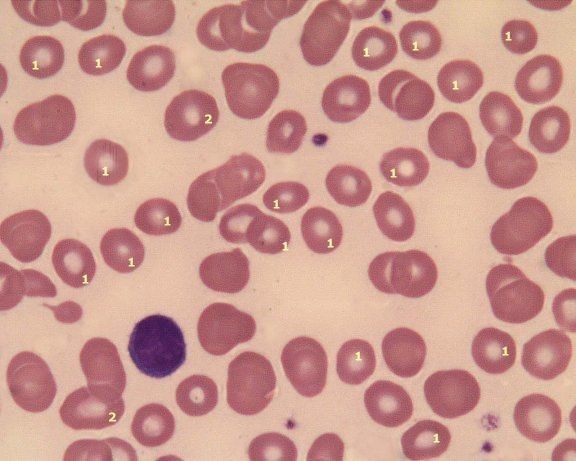
| Target, Spur and burr cells in liver disease | |
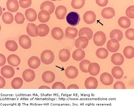
| Normochromic RBC (s) The normal RBC is saturated with Hb and as such the central pallor constitutes around one third of the cell size. This cell is known as a normochromic RBC. | |
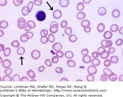
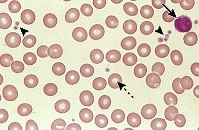
| normal peripheral blood smear High-power view of a normal peripheral blood smear. Several platelets and a normal lymphocyte can also be seen. The red cells are of relatively uniform size and shape. The diameter of the normal red cell should approximate that of the nucleus of the small lymphocyte; central pallor should equal one-third of its diameter. Courtesy of Carola von Kapff, SH (ASCP). |
Want to create your own Flashcards for free with GoConqr? Learn more.