23595021
| Question | Answer |
|
Image:
Image (binary/octet-stream)
|
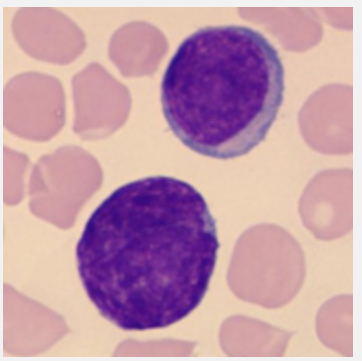
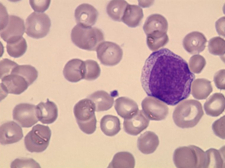
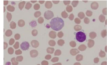
Lymphoblast 10-20 microns Round to oval nucleus with fine, evenely stained chromatin structure, 1-2 nucleoles Blue cytoplasm with clear zone closest to the nucleus No granules |
| Prolymphocyte 10-20 microns Round nucleus, somewhat looser structure than the lymphoblast. One (1) distinct nucleol Blue cytoplasmc No granules | |
|
Image:
Image (binary/octet-stream)
|
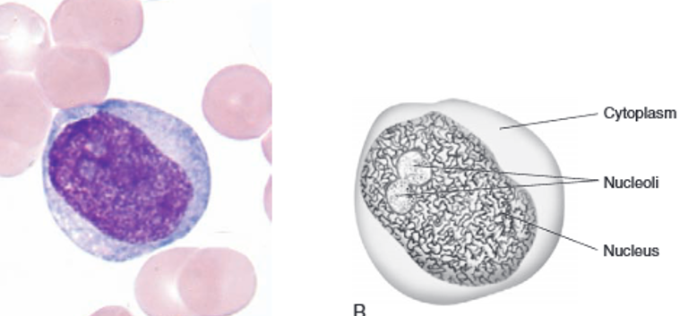
Prolymphocyte 10-20 microns Round nucleus, somewhat looser structure than the lymphoblast. One (1) distinct nucleol Blue cytoplasmc No granules |
| large Lymphocyte 9-15 microns Oval/bulging nucleus, somewhat looser chromatin structure than small lymphocyte. No nucleoles Plentiful colorless to light blue cytoplasm Few bright violet granules | |
| Plasma Cell 7-15 microns Round nucleus, eccentrically placed. Compact (radial) chromatin structure Dark blue cytoplasm with perinuclear zone (sometimes vacuoles) | |
| Megakaryoblast The cell that gives rise to the megakaryocyte is the megakaryoblast, which is difficult to identify morphologically | |
| Megakaryocyte 25-90 microns Round, irregular cell wal Nucleus with 2-16 lobes. The size of the cell varies according to number of lobes. Finely dispersed chromatin mesh in the nucleus. Small nucleoles may be present. Blue to pink cytoplasm | |
|
Image:
Image (binary/octet-stream)
|
monoblast |
|
Image:
Image (binary/octet-stream)
|
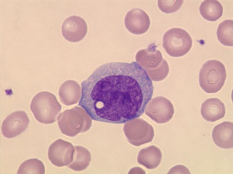
Promonocyte 15-20 microns Irregularly shaped nucleus without lobes. Fine chromatin structure with nucleoles Gray-blue cytoplasm A few granules can occur |
|
Image:
Image (binary/octet-stream)
|
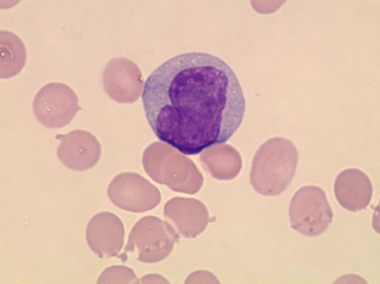
Monocyte 12-24 microns Kidney-shaped, bulging or segmented nucleus, very loose chromatin structure Powder-fine, violet granules can occur Vacuoles often appear in the cytoplasm |
| Myeloblast 15-20 microns Large round nucleus in the middle of the cell, thin, fine chromatin structure, 2-5 nucleoles Relatively small amount of blue cytoplasm, sometimes with a clear zone No granules | |
| Promyelocyt 12-24 microns Oval, possibly slightly bulging nucleus, often with eccentric position. Slightly coarser chromatin structure than myeloblast. 3-4 visible nucleoles Plentiful pale blue cytoplasm Moderate to plentiful unevenly sized violet primary granules | |
|
Image:
Image (binary/octet-stream)
|
Myelocyte 10-18 microns Round to oval nucleus, often eccentrically placed. Coarse and more condensed chromatin structure. No nucleoles Plentiful pink to colorless cytoplasm (more immature, bluish) Very fine moderate to dense granulation (secondary granules) Neutrophilic myelocyte: Violet granules Eosinophilic myelocyte: Bright pink granules Basophilic myelocyte: Blue-violet (black) granules |
| Metamyelocyte 10-16 microns Indented nucleus, coarse clumped chromatin structure. No nucleoles. Pale pink to colorless cytoplasm Many secondary granules Neutrophilic metamyelocyte: Violet granules Eosinophilic metamyelocyte: Pink granules Basophilic metamyelocyte: Blue-violet (black) granules | |
| Band Neutrophil Granulocyte 10-15 microns Band shaped nucleus with coarse, clumped chromatin structure. The narrowest part is at least 1/3 of the widest. No nucleoles Pale pink to colorless cytoplasm Plentiful very fine violet granules | |
|
Image:
Image (binary/octet-stream)
|
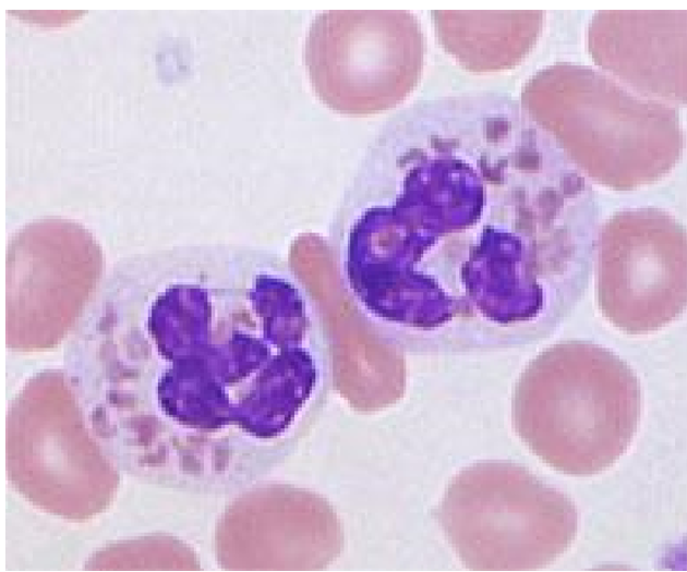
Segmented Neutrophil Granulocyte 10-15 microns Coarse, clumped chromatin structure. 3-5 lobes. No nucleoles. Pale pink to colorless cytoplasm Abundant secondary violet granules |
| Eosinophil Granulocyte 10-15 microns Coarse, clumped chromatin structure.2-3 lobes. No nucleoles Pale pink to colorless cytoplasm Abundant secondary brick-red granules | |
| Basophil Granulocyte 10-15 microns Coarse, clumped chromatin structure. 3-4 lobes. No nucleoles Pale pink to colorless cytoplasm. Secondary blue-violet (black) uneven distributed granules | |
| eosinophil | |
| basophil | |
| Proerythroblast 14-24 microns Large round centrally placed nucleos with fine chromatin structure. 2-5 nucleoles. Narrow dark blue cytoplasm edge. Uneven structure with well-defined perinuclear clear zone No granules | |
| Basophilic Erythroblast 10-17 microns Round rather small nucleos. Slightly condensed chromatin structure. No nucleoles Wider dark blue cytoplasm edge No granules | |
| Polychromatic Erythroblast 10-15 microns Round, even smaller nucleus. Quite condensed chromatin structure without nucleoles Relative plentiful gray blue cytoplasm No granules | |
| Orthochromatic Erythroblast 8-12 microns Small round nucleus, often in the center of the cell. Condensed chromatin structure Plentiful reddish cytoplasm with faintly blue components No granules | |
| Polychromatic Erythrocyte 7-10 microns No nucleus Cytoplasm reddish with even fainter blue component No granules | |
| erythrocyte 6-8 microns No nucleus Faintly reddish cytoplasm,clearing towards the center No granules | |
| Small Lymphocyte 7-8 microns Round or kidney-shaped nucleus, diffusely lumpy to compact chromatin structure Narrow cytoplasm edge, light or dark blue Possible isolated bright violet granules | |
| Large Lymphocyte 9-15 microns Oval/bulging nucleus, somewhat looser chromatin structure than the small lymphocyte. No nucleoles Plentiful colorless to light blue cytoplasm Possible isolated bright violet granules | |
| basophil | |
|
Image:
Image (binary/octet-stream)
|
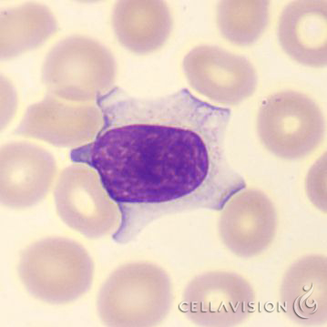
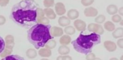
Variant Lymphocyte 15-30 microns Round, oval, indented, notched, folded, cleaved or lobulated nucleus None to multiple nucleoles Plentiful grey to blue cytoplasm Cytoplasm may be darker in the periphery and lighter near the nucleus |
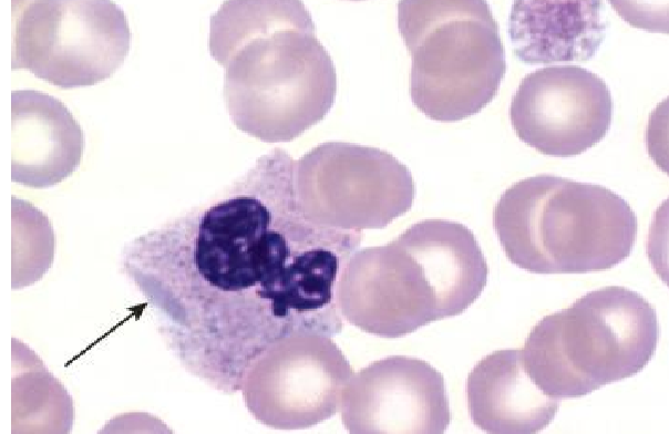
| Toxic Granulation Found in neutrophils Large irregular violet-purple granules Increase in staining density and numbers of granules Granules not evenly distributed | |
| Platelet Satellitism Found at neutrophils and monocytes Cell surface is surrounded with 4 or more platelets Morphology and function of the leukocyte and the platelet is normal In vitro phenomenon in EDTA anticoagulated blood | |
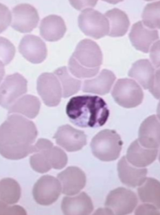
| Nucleated Red Blood Cell 8-10 microns Small, round or oval nucleus with coarser chromatin strands, pyknotic Pink cytoplasm Nucleos decreases in size as the cell ages No nucleoles | |
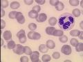
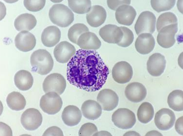
| Hypersegmented Neutrophil Increased size and lobulation 6 or more lobes The condition is often indicative of reduced DNA synthesis Associated with vitamin B12 deficiency, folate deficiency, anti-metabolite therapy or alcoholism | |
|
Image:
Image (binary/octet-stream)
|
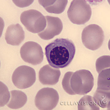
Nucleated Red Blood Cell 8-10 microns Small, round or oval nucleus with coarser chromatin strands, pyknotic Pink cytoplasm Nucleos decreases in size as the cell ages No nucleoles |
| Döhle Bodies 1-2 microns Light blue or blue-gray oval structure Usually found at the periphery of the neutrophil Consist of ribosomes and endoplasmic reticulum Found in bacterial infections and in a benign inherited condition known as May-Heggling Anomaly | |
| Döhle Bodies 1-2 microns Light blue or blue-gray oval structure Usually found at the periphery of the neutrophil Consist of ribosomes and endoplasmic reticulum Found in bacterial infections and in a benign inherited condition known as May-Heggling Anomaly | |
| Auer Rods 0.2-0.5 microns Azurphilic round or rod shaped cytoplasmic inclusions Mostly seen in immature granulocytes Found in blast cells associated with acute myeloid leukemia | |
|
Image:
Image (binary/octet-stream)
|
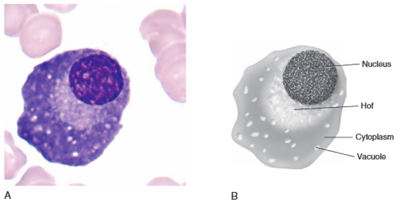
plasma cell |
|
Image:
Image (binary/octet-stream)
|
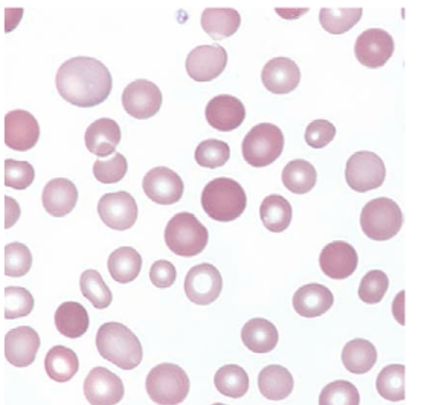
ancistocytosis |
|
Image:
Image (binary/octet-stream)
|
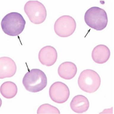
polychromatic erythrocyte |
|
Image:
Image (binary/octet-stream)
|
acanthrocyte (spur cell) |
|
Image:
Image (binary/octet-stream)
|
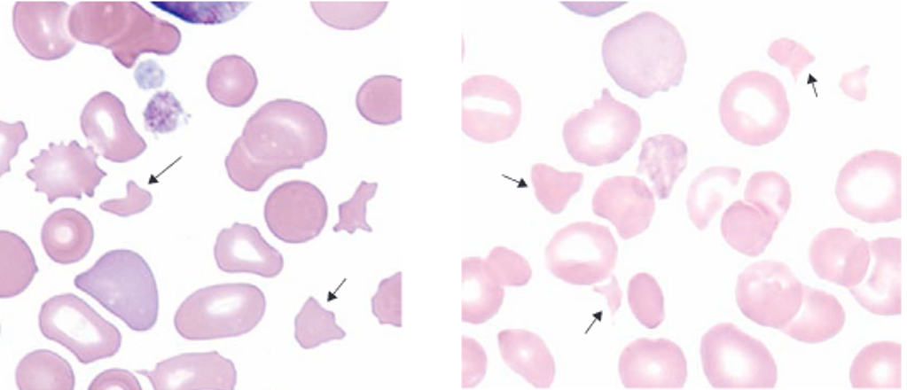
schistocyte |
|
Image:
Image (binary/octet-stream)
|
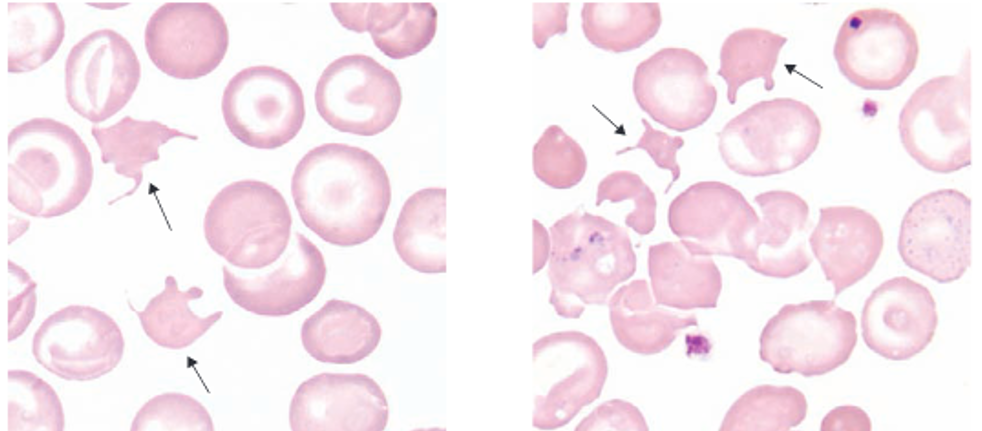
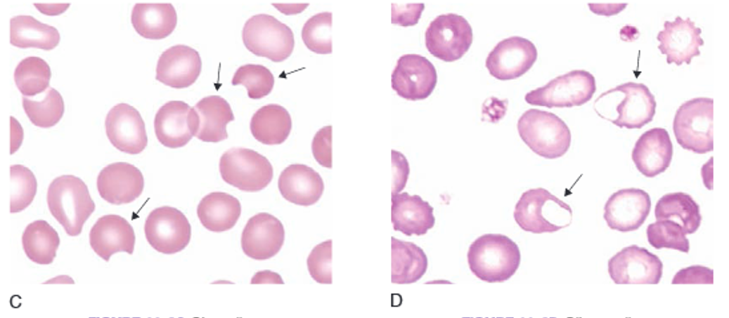
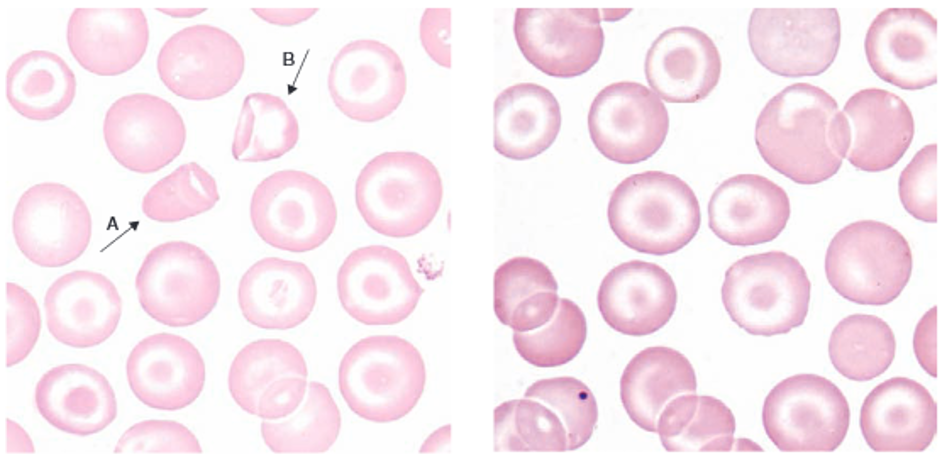
bite cell (helmet cell) blister cell |
|
Image:
Image (binary/octet-stream)
|
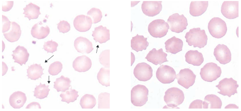
burr cell (echinocyte) |
|
Image:
Image (binary/octet-stream)
|
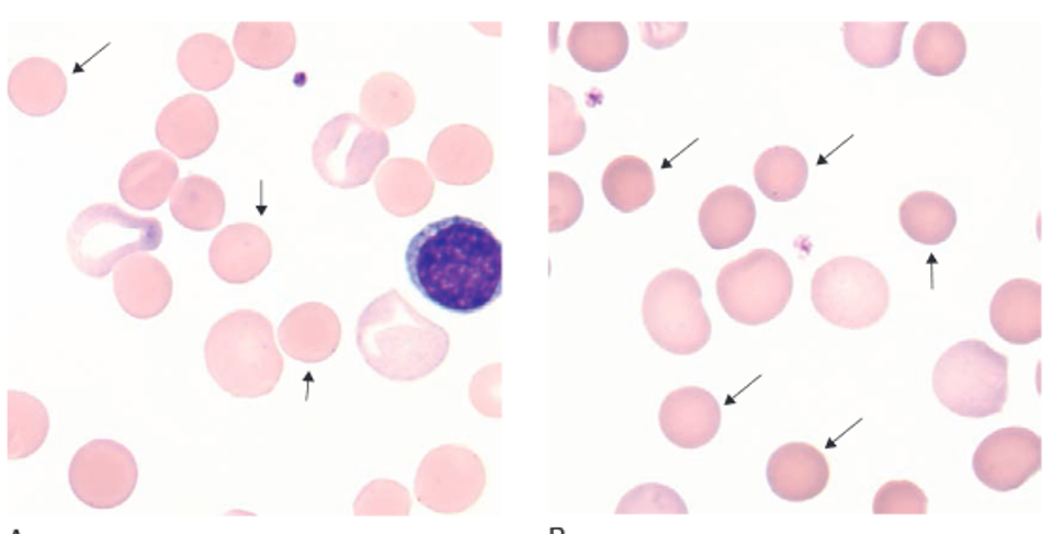
spherocyte |
|
Image:
Image (binary/octet-stream)
|
target cell |
|
Image:
Image (binary/octet-stream)
|
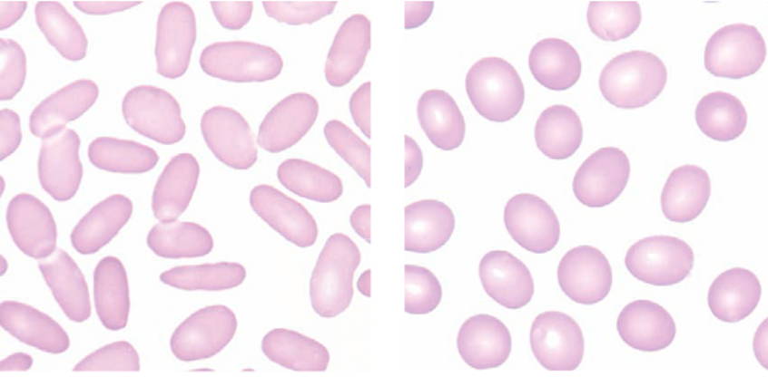
elliptocyte and ovalocyte |
|
Image:
Image (binary/octet-stream)
|
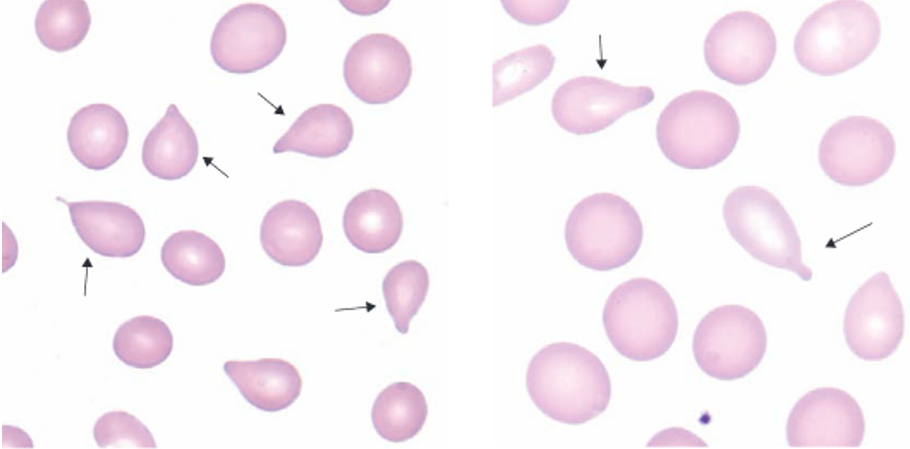
teardrops |
|
Image:
Image (binary/octet-stream)
|
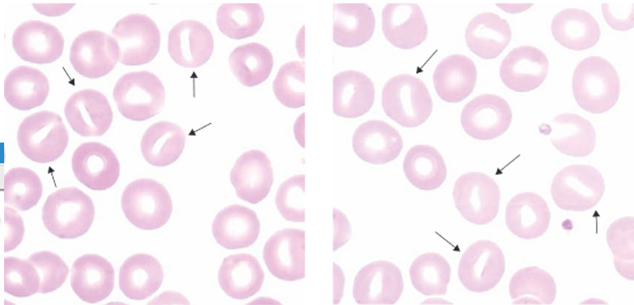
stromatocyte |
|
Image:
Image (binary/octet-stream)
|
sickle cell |
|
Image:
Image (binary/octet-stream)
|
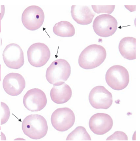
HJB |
|
Image:
Image (binary/octet-stream)
|
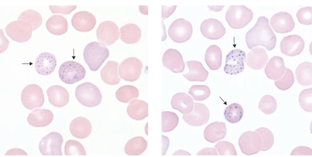
basophilic stippling |
|
Image:
Image (binary/octet-stream)
|
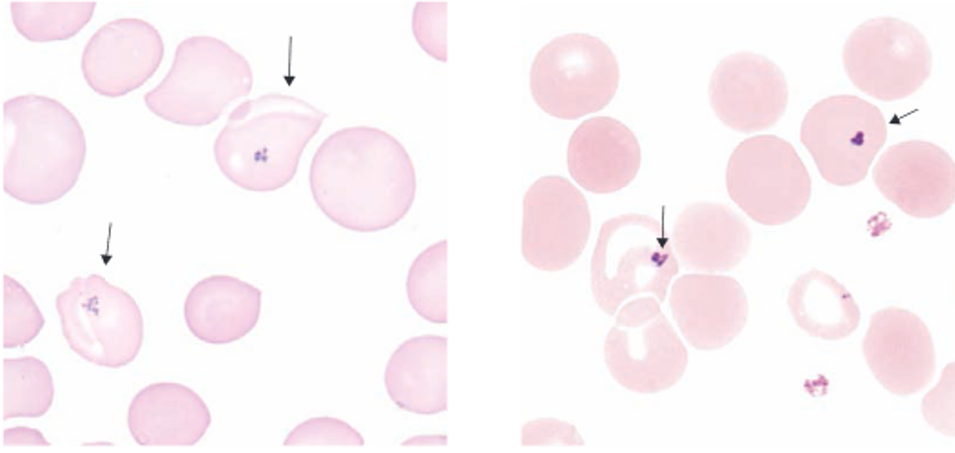
pappenheimer |
|
Image:
Image (binary/octet-stream)
|
Alder-rielly body |
|
Image:
Image (binary/octet-stream)
|
Chideki higashi body |
|
Image:
Image (binary/octet-stream)
|
May Hegglin body |
|
Image:
Image (binary/octet-stream)
|
apoptotic lymphocyte |
|
Image:
Image (binary/octet-stream)
|
apoptotic neutrophil |
|
Image:
Image (binary/octet-stream)
|
g |
![Prolymphocyte Img[1] (binary/octet-stream)](https://examtimeassets.s3.amazonaws.com/uploads/media/image/27730637/desktop_d4ec7f4d-ec93-4bb7-afb8-4a79ae4fca1b.jpg)
![Large Lymphocyte Img[1] (binary/octet-stream)](https://examtimeassets.s3.amazonaws.com/uploads/media/image/27730728/desktop_9177e280-0fb7-404a-9d03-c6eda87bc0d5.jpg)
![Plasma Img[1] (binary/octet-stream)](https://examtimeassets.s3.amazonaws.com/uploads/media/image/27730754/desktop_6911f1fc-0090-4ef0-a9f8-2df1bcc53cf3.jpg)
![Megakaryoblast1[1] (binary/octet-stream)](https://examtimeassets.s3.amazonaws.com/uploads/media/image/27730778/desktop_972aa4ea-5ac7-4ecd-b679-ec0c91fcd693.jpg)
![Megakaryocyte1[1] (binary/octet-stream)](https://examtimeassets.s3.amazonaws.com/uploads/media/image/27730820/desktop_b3bd6b62-7985-4009-80eb-d0d0236e3364.jpg)
![Myeloblast1[1] (binary/octet-stream)](https://examtimeassets.s3.amazonaws.com/uploads/media/image/27730976/desktop_e27a7294-d2ec-46c7-b69f-d179026d9346.jpg)
![Promyelocyte1[1] (binary/octet-stream)](https://examtimeassets.s3.amazonaws.com/uploads/media/image/27730993/desktop_4fbed303-0859-403b-9d6b-8ecfc52d1729.jpg)
![Metamyelocyte1[1] (binary/octet-stream)](https://examtimeassets.s3.amazonaws.com/uploads/media/image/27731052/desktop_ac4a2ddd-b850-43d4-bed5-540f902a3206.jpg)
![Band1[1] (binary/octet-stream)](https://examtimeassets.s3.amazonaws.com/uploads/media/image/27731079/desktop_824fd5eb-f0de-4a91-8c7c-d64a3e8bff10.jpg)
![Eos1[1] (binary/octet-stream)](https://examtimeassets.s3.amazonaws.com/uploads/media/image/27731126/desktop_4a91eca4-f6e5-49b8-ab16-bb3a6a6d7a40.jpg)
![Baso1[1] (binary/octet-stream)](https://examtimeassets.s3.amazonaws.com/uploads/media/image/27731149/desktop_d8e6709b-6392-464c-a420-06d506b731cf.jpg)
![Eos2[1] (binary/octet-stream)](https://examtimeassets.s3.amazonaws.com/uploads/media/image/27731183/desktop_794a312b-0f93-446a-a3af-57def69d4f55.jpg)
![Baso2[1] (binary/octet-stream)](https://examtimeassets.s3.amazonaws.com/uploads/media/image/27731220/desktop_f565c326-cea4-428e-a186-53d43b5ec237.jpg)
![Proerythroblast1[1] (binary/octet-stream)](https://examtimeassets.s3.amazonaws.com/uploads/media/image/27731249/desktop_dd127f0d-5387-4bf5-aa26-6146f29fae94.jpg)
![Basophilicerythroblast1[1] (binary/octet-stream)](https://examtimeassets.s3.amazonaws.com/uploads/media/image/27731275/desktop_ed85fc6a-3021-4e0f-8cf3-f9c4ac06fb2a.jpg)
![Polychromaticerythroblast1[1] (binary/octet-stream)](https://examtimeassets.s3.amazonaws.com/uploads/media/image/27731288/desktop_fa5bd5df-6cbb-44a6-bf67-4d3a0dd73986.jpg)
![Orthrochromaticerythroblast1[1] (binary/octet-stream)](https://examtimeassets.s3.amazonaws.com/uploads/media/image/27731310/desktop_a22898dc-ce1c-4673-8866-b1dec659c0d0.jpg)
![Polychromaticerythrocyte1[1] (binary/octet-stream)](https://examtimeassets.s3.amazonaws.com/uploads/media/image/27731333/desktop_d65f6df8-d84d-4202-8cf4-cae3ab39751a.jpg)
![Erythrocyte2[1] (binary/octet-stream)](https://examtimeassets.s3.amazonaws.com/uploads/media/image/27731355/desktop_182fb773-aadd-40d5-9ffd-6e102cc9ea17.jpg)
![Smal Llymph1[1] (binary/octet-stream)](https://examtimeassets.s3.amazonaws.com/uploads/media/image/27731397/desktop_039f8b3b-c3f8-42eb-8133-8e8a8773691c.jpg)
![Larg Elymph1[1] (binary/octet-stream)](https://examtimeassets.s3.amazonaws.com/uploads/media/image/27731419/desktop_7f640239-3583-47a8-b4a7-2de9ee805344.jpg)
![Baso3[1] (binary/octet-stream)](https://examtimeassets.s3.amazonaws.com/uploads/media/image/27731453/desktop_9abbba88-b6ae-48a5-a4ae-098b92bcbdd3.jpg)
![Toxisk Granulation1[1] (binary/octet-stream)](https://examtimeassets.s3.amazonaws.com/uploads/media/image/27731492/desktop_8d538664-67aa-4006-80d6-57e3563038ae.jpg)
![Platelet Satellitism1[1] (binary/octet-stream)](https://examtimeassets.s3.amazonaws.com/uploads/media/image/27731794/desktop_6b982643-17a7-4227-bc78-3d5d034542ff.jpg)
![Nucleated Red Blood Cell1[1] (binary/octet-stream)](https://examtimeassets.s3.amazonaws.com/uploads/media/image/27731817/desktop_31c86870-d56f-48ea-a2fc-d4a1aeb773b4.jpg)
![Hypersegmented Neutrophil1[1] (binary/octet-stream)](https://examtimeassets.s3.amazonaws.com/uploads/media/image/27731869/desktop_dc43d695-1b5a-409d-b95b-6df082c16577.jpg)
![Dhle Bodies1[1] (binary/octet-stream)](https://examtimeassets.s3.amazonaws.com/uploads/media/image/27731923/desktop_f6b7c62a-3abc-4fab-9949-8f68ed62ccc9.jpg)
![Dhle Bodies2[1] (binary/octet-stream)](https://examtimeassets.s3.amazonaws.com/uploads/media/image/27731948/desktop_39f62794-b637-4b64-af25-6cb9bc800358.jpg)
![Aur Rods2[1] (binary/octet-stream)](https://examtimeassets.s3.amazonaws.com/uploads/media/image/27731968/desktop_2547531d-cf5f-4f7e-b1fd-4c8616e73743.jpg)
Want to create your own Flashcards for free with GoConqr? Learn more.