3977468
Description
Flashcards by Chigozie Osammor, updated more than 1 year ago
|
|
Created by Chigozie Osammor
over 8 years ago
|
|
| Question | Answer |
| Which layer of the eye is the sclera found in? | The outer fibrous layer. |
| What fraction of the eye does the sclera cover? | 5/6. |
| The sclera is anteriorly covered with x and posteriorly connected to y | X- conjunctiva Y- the facial sheath |
| What is the thickness of the sclera at a) corneosclera b) behind the rectus muscles c) equator d) posterior | a) 0.8mm b) 0.3mm c) 0.6mm d)1mm |
| Why is the sclera thickest at the back? | Embryologic development. |
| Where is the sclera weakest and why? | At the optic nerve due to all the apertures for the optic nerves and blood supply. |
| Where is the lamina cribrosa? | This is where the optic nerve pierces the sclera a swell as other nerves. |
| Why are there apertures on the anterior of the sclera? | For the arteries leading to the ciliary body and for the insertions of the rectus muscles. |
| Why are there apertures on the middle of the sclera? | For the exit of the vortex veins (about 4mm posterior to the equator). |
| Why are there apertures on the posterior of the sclera? | For the long and short ciliary nerves and for the many other nerves (and for the main optic nerve), and also the long and short ciliary arteries. |
| Where is the canal of Schlemm? | Posterior to the conjunctival epithelium and the Tennons capsule (not directly). |
| *What is the function of the canal of Schlemm? | The canal of Schlemm collects aqueous humour from the anterior chamber and delivers it to the bloodstream by the ciliary veins. |
| Where is the scleral spur? | Posterior to the canal of Schlemm. |
| What is the funaction of the sclera? | • protection of the interocular contents from trauma and mechanical displacement • preserves the shape of the globe and maintains the position of the different parts of the optic system • the strength and firmness of the sclera provides a rigid insertion for extra ocular muscles • Maintains pressure towards the centre of the eye to counteract the outward pressure from the humours in the eye |
| How many layers does the sclera have? | 3. |
| What are the three layers of the sclera? (From anterior to posterior) | • episclera • stromal sclera • lamina fusca |
| Is the episclera loose or dense connective tissue? | Loose. |
| Where does the episclera get its blood supply? | Rich blood supply from the ciliary arteries. |
| Does the episclera a) finish abruptly and then the stroma starts, b) form the rest of the sclera or c) merge into the stroma? | C. |
| What is the innermost scleral layer? | Lamina fusca. |
| What is the purpose of the grooves in the lamina fusca? | To allow ciliary blood vessels and nerves to pass through. |
| What separates the lamina fusca from the choroid? | Perichoroidal space. |
| Is the choroid connected to the lamina fusca? If so how? | Yes, connected by fine collagen fibres. They are weak connections. |
| What makes the sclera so opaque? | There are a low number of GAGs, and therefore the sclera is very dehydrated. Also the variation in fibril size and the irregular distribution induce light scattering. |
| *What is the function of the sclera? | • Protection of the intraocular contents from trauma and mechanical displacement • Preserve the shape of the eyeball and maintain the exact position of the different parts of the optic system • The strength and firmness of the sclera provide a rigid insertion for the extraocular muscles |
| In which layer of the eye is the cornea found? | The fibrous outer layer. |
| What fraction of the eye is civer d by the cornea? | 1/6. |
| What is the horizontal diameter of the cornea? | 12mm. |
| What is the vertical diameter of the cornea? | 11mm. |
| What is the diameter of the posterior surface of the cornea? | 11.7mm. |
| What is the radius of curvature of the cornea? | 7.8mm. |
| What is the thickness of the cornea at its a) centre b) periphery | a) 0.53mm b) 0.71mm |
| *What are the functions of the cornea? | • to allow light into the eye • refract light onto the retina |
| Why is the cornea transparent? | • there is a high percentage of GAGs in the cornea, therefore the cornea is more hydrated • the collagen fibres are regular in both distribution and in size, therefore the cornea is smooth, therefore there is minimal light scattering • the fact that the cornea is avascular also aids the above fact |
| What is a) anterior to the cornea b) posterior to the cornea? | a) tear film b) aqueous humour |
| Which structure(s) is the cornea continuous with? | Conjunctiva and the sclera. |
| How many layers are in the cornea? | 5. |
| Name the layers of the cornea (anterior to posterior) | Corneal epithelium Bowmans layer Corneal stroma Descements membrane Corneal endothelium |
| How thick is the corneal epithelium a) in cells b) in micrometers | a) 5-7 cells thick (7 more so ant the periphery) b) 50μm |
| True or false- the corneal epithelium is continuos with the conjunctival epithelium | True. |
| How many layers are there in the corneal epithelium? | 3. |
| Name the layers of the corneal epithelium (anterior to posterior) | • surface epithelium • middle layer • basal epithelium |
| How thick is the corneal surface epithelium? (In cells) | 2 cells thick. |
| What type of cells are in the corneal surface epithelium layer? | Non keratinised squamous cells with flattered nuclei. |
| How is the corneal surface epithelium joined to the tear film? | By a glycolalix excreted by the plasma membrane. |
| What type of junctions join the surface corneal epithelium cells? | Zonula occludens. |
| Why is/isn't the surface epithelium of the cornea semi-permiable? | It is semi-permeable so that small molecules (eg oxygen) and other fluids can pass through the cells. |
| How many cells thick is the middle epithelium layer of the cornea? | 2-3. |
| What type of cells are in the middle cell layer of the cornea? | Polyhedral cells (aka wing cells), with convex anterior surfaces and concave posterior surfaces (•(. |
| What joins wing cells in the corneal epithelium together? | Desmosomes and gap junctions. |
| What joins wing cells to the surface epithelium and to the basement epithelium of the cornea? | Desmosomes and interdignations (regarding the basement membrane). |
| What is a desmosome? | A strong attachment between cells made of protein. |
| What is a gap junction? | A tunnel like connection that forms between two neighboring cells, allowing rapid communication. |
| What is a zonula occludens? | A belt that wraps around a cell, sticking it to the next (like double sided sellotape). |
| How many layers does the besement epithelium of the cornea have? | 1. |
| What type of cells are in the basement layer of the cornea? | Columnar cells. |
| What attaches the basal columnar cells of the cornea together? | Desmosomes and gap junctions. |
| How are the corneal epithelium cells replaced? | The old/dead cells at the top are sloughed off and replaced from the layers below. (Like a cell fountain) |
| Why is the Bowman's layer not always considerd to be a layer in the cornea? | It is very thin, it is not clearly defined and it is acellular. It is more of a transition between the corneal epithelium and the corneal stroma. |
| How thick is the Bowman's layer? | 8-12μm. |
| Can the Bowman's layer regenerate? Why doesn't this matter? | No. It doesn't matter because the Bowman's layer is very tough due to the slightly irregular collagen fibrils. |
| How thick is the corneal stroma? | 500μm. |
| What percentage of the cornea is the stroma? | 90%. |
| What type of cells are in the corneal stroma? | • keratocytes (corneal fibroblasts) • leukocytes |
| What are keratocytes? | Active cells that produce collagen and extra cellular matrix components. They can be found between lamellae, and they are more frequent in the anterior stroma. |
| What is the range of diameters for a collagen fibril (in the corneal stroma)? | 25-35nm. |
| True or false- fibrils in the lamellae of the corneal stroma run parallel to each other | True- they run parallel with regular spacing. |
| True or false- lamellae in the corneal stroma run parallel to each other | False- they run at angles to each other. Fibrils and the lamellae run parallel to the corneal surface. |
| Are the lamellae in the corneal stroma thicker anteriorly or posteriorly? | Posteriorly. |
| Are the lamellae in the coreneal stroma more regularly arranged posteriorly or anteriorly? | Posteriorly. |
| How is the corneal stroma bound to the Descements membrane? | The collagen fibres near the Descements membrane interlace more to form a collagen sheet which contributes to binding the two layers. |
| *What is the function of the corneal stroma ground substance? | • fills the space between fibres, lamellae and cells • contains marmolecules with a high GAG percentage, contributing to the transparency of the cornea |
| What makes the cornea transparent? | • high percentage of GAGs, which makes the cornea more hydrated • the regular size and distribution of the collagen fibrils, minimising the light scattering • small diameter of the collagen fibrils • spacing between the fibrils also minimises the light scattering |
| How thick is the Descements membrane? | No set thickness, as it grows over time. The range is 5-15μm. |
| How many lamellae layers make up the Descements membrane? | 2- the anterior and posterior lamellae. |
| What are the features of the corneal posterior lamellae? | • non banded • homogeneous • secreted by the epithelium throughout life • very tough • elastic properties • can be regenerated |
| What are the features of the corneal anterior lamellae? | • 3μm thick • banded in appearance • lattice work of collagen • secreted during embryonic development |
| How thick is the corneal endothelium? | 5μm thick. |
| What is posterior to the corneal endothelium? | Aqueous humor. |
| Which surface of the corneal endothelium lies on the Descements membrane? | The basal surface. |
| What shape are the majority of the corneal endothelial cells? | Hexagonal. |
| Are corneal endothelial cells arranged regularly to irregularly? | Regularly. |
| Do the corneal endothelial cells divide? | No they do not divide or replicate. |
| How are the corneal endothelial cells joined together? | By gap junctions and interdignations. |
| Is the corneal endothelium permeable? | Yes, large molecules such as glucose and proteins can enter. |
| How is the corneal endothelium hydrated? | By a metabolic pump. |
| How does the cornea receive nourishment? | Through the endothelium from the aqueous humor, and also from the conjunctival capillary network and episclera like capillary network . |
| Where are the nerves in the cornea? | • mid stroma • sub epithelial • epithelial |
| Where is the limbus? | At the corneoscleral junction. |
| Which structures terminate at the limbus? | • Bowman's layer • Decscement's membrane |
| The limbus is the transitional zone between which two pairs of structures? | • Cornea and sclera • Cornea and conjuntiva |
| What does the a) corneal epithelium become at the limbus, b) regular corneal stroma, c) corneal endothelium ? | a) Conjuctival epithelium b) irregular scleral stroma c) corneal endothelium becomes discontinuous and wraps around the trabecular meshwork |
| Which three structures begin at the limus? | • Conjunctival stroma • Episclera • Tenon's capsule • COnjunctival submucosa |
| How thick is the limbal epithelium? | 10-15 cells thick. |
| Why are some peoples limbi pigmented? | Melanocytes may be present in the basal layer. |
| Where is the scleral spur? | In the scleral stroma. |
| What are the Palisades of Vogt? | Radial projections of limbal epithelium and stroma . |
| *What is the function of the Palisades of Vogt? | They contain the limbal stem cells and corneal epithelial basal cell replacement (replacement epithelial basal cells move centripetally from the limbus to the cornea). |
| What makes up the limbal blood vessel network? | Ciliary artery loops that surround the cornea (from conjuctival and episcleral vessels). • this vessel network provides nourishment to the avascular corneal tissue |
| Where do the limbal veins drain? | Into the radial episcleral veins - which empty into the anterior ciliary veins. |
| *What are the two main functions of the limbus? | • Provide nourishment and metabolites for surrounding avascular tissue (e.g. cornea (posterior potion)) • Provides a drainage pathway for the aqueous humor (the internal scleral sulcus, the trabecular meshwork and the canal of Schlemm). |
| Which two structures is the iris suspended between? | • The cornea • The lens |
| What is the iris? | A pigmented, contractile, thin diaphragm with a centre aperture (pupil). |
| *What is the function of the pupil? | Allows the aqueous humor to flow from the posterior chamber to the anterior chamber WITH NO RESISTANCE, and to regulate the amount of light entering the eye. |
| What is the name of the termination of the iris? | The iris root (which joins the iris to the anterior aspect of the ciliary body. |
| Where is the pupillary margin? | On the lens, surrounding the pupil. |
| Where is the iris a) thickest, b) thinnest? | a) collarete (1.5mm) b) root (0.5mm) |
| What is the diameter of the iris? | 12mm. |
| What produces the iris colour? | The number of melanin granules within melanocytes and the area that the melanocytes occupy. |
| Which colour of iris has the a) most melanin granules, b) least melanin granules? | a) brown b) blue |
| What is the diameter of the pupil? | 1-9mm. |
| When is the iris a) miotic, b) mydriatic? | a) bright light (miotic - small pupil) b) dim light (mydriatic - large pupil) |
| What are the two main surfaces of the iris? | Anterior surface and posterior surface. |
| Where is the colarette? Describe its features. | The colarette is the ring of the iris closest to the pupil (not including the ruff). It forms a wavy line. Looking at the colarette you can see the crypts of Fuch where the pigmented strands of the iris are, as well as the traberculae. The colarette divides the iris into the central and peripheral pupillary zone. |
| What causes the appearance of the concnetric and contraction furrows? Where are they located? | Dilation of the pupil causes the conentric furrows, whilst constriction of the pupil causes the contraction furrows. They are on the iris. |
| Does the iris have an epithelium? | No. |
| What makes the ruff? | An extension of the pigmented posterior epithelium of the iris which folds around the edge of the iris on the pupil side. Excessive extension of the pigmented posterior epithelium is called ectropion uvae. This can indicate a tumor. |
| Describe the appearance of the posterior surface of the iris. | Black surface with radial contraction folds (like spokes on a bike wheel). These are the radial contraction furrows of Schwalbe. They run through the pars plicata. |
| Where are the radial contraction furrows of Schwalbe located? | On the posterior surface of the iris. |
| What are the histological layers of the iris? | Anterior border layer Iris stroma and sphincter muscle Anterior epithelium and dilator muscle Posterior epithelium |
| What cells and fibrils make uo the anterior border layer of the iris? | • fibroblasts • pigmented melanocytes • collagen fibrils - arranged in vertical columns that appear white in lightly coloured irises (fibroblasts are on the surface and melanocytes are beneath) • clumps of melanocytes cause freckles |
| Does the anterior border layer cover the iris? | No, it is absent at the crypts of Fuch. |
| Where does the anterior border layer of the iris terminate? | The anterior border layer terminates at the iris root. Some of the anterior border form finger shaped processes which extend into the trabecular meshwork. |
| What is the iris stroma? | Connective tissue composed of collagen fibrils, pigmented and non-pigmented cells and extensive ground substance. |
| What cells are pigmented in the iris stroma? | • melanocytes • clump cells (more concentrated near to the pupil) |
| What are the non-pigmented cells in the iris stroma? | • fibroblasts • lymphocytes • macrophages • mast cells |
| Where is the sphincter iris muscle? | In the iris stroma, posterior to the colarette (the pupillary zone of the stroma). |
| What type of muscle makes up the sphincter iris muscle? | Smooth muscle cells. |
| *What is the function of the sphincter iris muscle? | To constrict the pupil to reduce the amount of light entering the eye. |
| Is the sphincter muscle of the iris innervated by the sympathetic or parasympathetic nervous system? | Parasympathetic nervous system. |
| Name the layers of the anterior iris epithelium. | • Anterior iris epithelium • Posterior pigmented iris epithelium (the anterior iris epithelium is located posterior to the iris stroma) |
| What type of cells make up the apical portion of the anterior epithelium? | Cuboidal myoepithelial cells joined by tight junctions and desmosomes. |
| What makes up the basal portion of the anterior epithelium? | Elongated contractile smooth muscle processes, which extend into the stroma and form the dilator muscle of the iris also joined by tight junctions and desmosomes . |
| Where are the boundaries of the dilator muscle of the iris? | The iris root to the middle of the iris sphincter muscle. |
| How is the iris dilator muscle of the iris arranged in comparison to the iris sphincter muscle? | • Sphincter muscle is circular ring of fibres • Dilator muscle in concentric fibres of muscle |
| What type of muscle makes up the dilator muscle of the iris? | Smooth muscle cells. |
| *What is the function of the dilator muscle of the iris? | To dilate the pupil and allow more light to enter the eye. |
| Is the dilator muscle of the iris innervated by the sympathetic or parasympathetic nervous system? | Sympathetic nervous system. |
| *What is the function of the iris? | To control the amount of light that enters the eye. |
| What makes up the posterior (second posterior) epithelium of the iris? | A heavily pigmented single layer of columnar cells joined be tight junctions and desmosomes. (less pigmented at the periphery) |
| Why are the anterior and posterior iris epithelium layered apex to apex? | Due to the folding of the neural ectoderm in the embryologic development. |
| What are heterochromia and pigmentary dispersion syndrome? | Heterochromia - two different coloured irises, or multicoloured irises. Pigmentary dispersion syndrome - pigment granules are shed from the posterior iris and dispersed into the anterior chamber. |
| Where does the arterial supply of the iris come from? | The arterial supply comes from the radial vessels that lie in the iris stroma (contributing to the radial ridges seen in the iris), which come from the major arterial circle (consists of two long posterior ciliary arteries and seven anterior ciliary arteries). |
| Where does the venous supply of the iris come from? | The veins follow the arteries. The veins come from the major venous circle of the iris, however the radial veins only drain into the vorticose veins. |
| Why are the veins and arteries associated with the iris spiraled? | This makes it easier for the blood vessels to carry out their function whilst the iris is contracting and dilating. |
| Which nerves supply the iris, and what specifically do they innervate? | • Long ciliary nerves- innervate the dilator muscle and the blood vessels • Short ciliary nerves- innervates the sphincter muscle |
| In which layer of the eye is the iris found? | The uvea. |
| In which layer of the eye is the choroid found? | The uvea. |
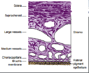
| Outline the dimensions of the choroid and its boundaries. | • The choroid is 0.22mm at the posterior pole (thickest part), and 0.1mm at the anterior pole (thinnest part) • The choroid extends from the optic nerve posteriorly and extends to the ora serata/ ciliary body anteriorly |
| Describe the inner and outer surfaces of the choroid (hint: take into account the layers that surround the choroid). | • The outer layer is rough abd firmly attached to the sclera, especially at the optic nerve, the ciliary arteries and nerves and the vortex veins • The inner layer is smooth and firmly attached to the retinal pigmented epithelium |
| Where is the perichoroidal space? | Between the sclera and the choroid. |
| Which two sheaths does the choroid become continuous with at the optic nerve? | The pia matter and the arachnoid. |
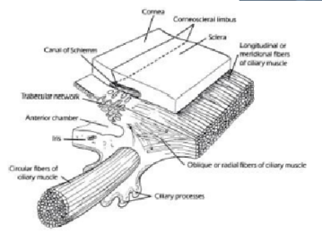
| Lable the diagram | |
| Name the layers of the choroid. | Suprachoroid Blood vessel layer Capillary layer Bruch's membrane |
| Name the cells and the types of blood vessels in the blood vessel layer of the choroid. | • Cells: melanocytes, fibroblasts, macrophages, lymphocytes and mast cells • Blood vessels: large and medium sized blood vessels |
| Where do the arteries and the veins in the vessle layer of the choroid come from/go to? | • Arteries- short posterior ciliary arteries • Veins- converge and go to the four vortex veins |
| What type of blood vessles are in the choriocapillaris? Where are they most concentrated and why? | Capillaries. They are most concentrated at the macula and therefore the macula will have the greatest blood supply and so more oxygen and other nutrients will be supplied to the macula. |
| Where do the capillaries in the choroid a) get their blood supply from and b) drain into? | a) arteries in the blood vessel layer b) veins in the blood vessel layer |
| How many layers are in the Bruch's membrane? What are they called? | 5:• Basement membrane of the endothelium of the capillaries of the capillary layer • Outer layer of collagen fibres • Meshwork of elastic fibres • Inner layer of collagen fibres • The basement membrane of the pigment epithelium of the retina |
| *What is the function of the Bruch's membrane? | To facilitate the passage of tissue fluid from the choroidal capillaries to the retina. |
| What is the main blood vessel that supplies the choroid with blood? | The posterior ciliary arteries (and a small amount form the anterior ciliary arteries). |
| Which blood vessels drain the choroid? | The 4 vortex veins. |
| How is the choroid innervated, and where do the nerves enter the eye? | Long and short ciliary nerves innervate the choroid. These nerves enter the eye at the optic nerve, piercing the sclera. |
| *What is the function of the choroid? | The choroid nourishes the outer layers of the retina with blood. Its other functions include: • a pathway for blood vessels to run through • heat production from blood exchange in the retina • regulation of IOP (using blood flow) • choroidal melanocytes absorb excess light that penetrates the retina to prevent reflection |
| In which layer of the eye is the ciliary body found? | The uvea. |
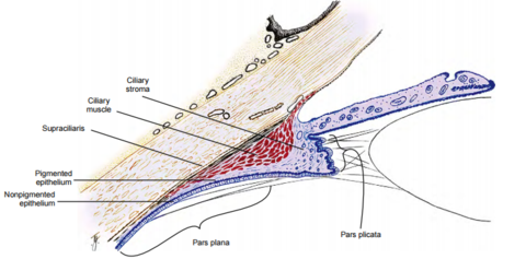
| Outline the dimensions of the ciliary body and its dimensions. | • 5.9mm- nasal side • 6.7mm- temporal side • The ciliary body terminates at the ora serrata, whilst the anterior extends into the posterior chamber |
| Lable the diagram | |
| What shape is often associated with the ciliary body? Which ocular structures do the parts of this shape lay nearest to? | A triangle. The ora serrata is at the apex, the iris root is in the middle of the base, the long flat side is next to the sclera, and the curved side is in contact with the posterior chamber and vitreous chamber. |
| Which two parts make up the ciliary body? | The pars plana (flat curved side) and the pars plicata (invaginated side (the base)) |
| What are the other names for the pars plana and the pars plicata? | Pars plana - corona ciliaris Pars plicata - orbicularis ciliaris |
| What are the dips inbetween the pars plicata's processes called? | Valleys of Khunt. |
| What are the boundaries of the pars plicata? | The iris root to the pars plana. |
| What are the boundaries of the pars plana? | The pars plicata to the ora serrata. |
| What are the portions inbetween the pars plana called? | The oral bays. |
| What are the zonula fibres? | They are the fibres that connect the lens to the ciliary body and they keep it suspended. |
| How many layers are in the ciliary bidy? What are they called? | There are 4: the suprciliaris, ciliary muscle, ciliary stroma and ciliary epithelium. |
| Describe the position of the supracilliaris. | In the outermost layer of the cilliary body, adjacent to the sclera. |
| Describe the features of the surpacilliaris (what it contains and its arrangement). | • Made of loose connective tissue which is arranged in ribbon like layers which allow the ciliary body to slide against the sclera without stretching or detaching • the cells in this layer are oigmented mealnocytes, fibroblasts and collagen bands |
| What type of muscle makes up the ciliary muscle? How is this muscle orientated? | Smooth muscle fibres, which are orientated longitudinally, radially and circularly. |
| Lable the diagram | |
| Where is the insertion of the longitudinal ciliary muscle of Brücke? | Adjacent to the supraciliaris and parallel to the sclera. The actual insertion is at the anterior 1/3 of the choroid. |
| Where are the radial fibers of the ciliary muscles? Where do they originate from? | Below the longitudinal fibres of Brücke. The radial fibres originate from the scleral spur. |
| At the radial fibres the type of muscle fibre transitions from a) to b) | a) Longitudinal fibres b) Circular fibres |
| What is Müllers annular muscle? Where is it located? | Müllers annular muscle is a bundle of circular muscle fibres that carry out a sphincter type action. It is located near the major circle of the iris |
| Which branches of the nervous system innervate the ciliary muscle? | The autonomic nervous system innervates the ciliary muscle. The parasympathetic nervous system innervates contraction, whilst the sympathetic nervous system innervates relaxation. The short ciliary nerves innervate the ciliary muscle. |
| Describe the ciliary stroma in relation to blood vessels, the type of connective tissue, its location *its function and the layers that it is continuous with. | The ciliary stroma is highlt avascular (because of the ciliary muscles). It also contains loose connective tissue. The cilioary stroma is located in between the ciliary muscle and the epithelial layers. The function of the ciliary stroma is to form the core of the ciliary processes. The ciliary stroma is continuous with both the iris and choroidal stroma, as well as the connective tissue that separates the bundles of the ciliary muscle. |
| Where is the major arterial circle of the iris? | Found in the ciliary stroma, near the iris root. |
| How many layers of ciliary epithelium are found in each eye? How are they postioned? | 2. They are positioned apex to apex, covering the ciliary body. |
| Describe the outer layer of the ciliary epithelium. | The outer layer of the ciliary epithelium is pigmented, cuboidal and joined by desmosomes and gap junctions. Anteriorly the outer epithelium is continuous with the anterior iris epithelium and posteriorly continuous with the RPE. |
| Describe the inner layer of the ciliary epithelium. | The inner layer is non-pigmented, and composed of columnar cellsin the pars plana and cuboidal cells in the pars plicata. Gasp junctions, desmosomes and zonula ocludens form a blood-aqueous barrier. Anteriorly the inner layer is continuous with the posterior iris epithelium and posteriorly continuous with the ora serrata and the basement of ther Brüch's membrane. |
| *What is the function of the ciliary body? | Production and secretion of the aqueous humor. |
| *What is the function of the ciliary muscles? | To control accomodation and to control aqueous flow |
| How many layers are in the retina? What are there names (in order)? | 1. RPE 2. Photoreceptor layer 3. The external limiting membrane 4. The outer nuclear layer 5. The outer plexiform layer 6. The inner nuclear layer 7. The inner plexiform layer 8. Ganglion cell layer 9. Nerve fibre layer 10. Internal limiting membrane |
| What is the function of the retina? | The retina is the first part of the visual system to detect light, motion and colour. It also provides sharp vision (at the fovea). |
| What are the three main regions of the retina? | The peripheral retina, the central retina (fovea) and the optic cup. |
| What is the retinal pigmented epithelium? | It is the first and outermost layer of the retina. It is only a single cell thick and it is made of hexagonal cells. tbc |
Want to create your own Flashcards for free with GoConqr? Learn more.