4275309
Description
Flashcards by CaitlinJoy, updated more than 1 year ago
|
|
Created by CaitlinJoy
over 8 years ago
|
|
| Question | Answer |
| What are the name of the two major Valves? | Atrioventricular ( Left) and Semilunar ( Right) |
| What is the important job of valves? | They stop the back flow of blood. |
| Why is the heart called a cardiac muscle? | As it consists of fibres and branches that help spread stimulus across the heart. This ensures a squeezing action. q1 |
| What is the cardiac cycle? | The cardiac cycle is the sequence of events in one heart beat. |
| How does the cardiac cycle work? | At low pressure blood flows into the atria when it is at the stage of diastole. As the blood feels up the atria they go into atria systole. Walls contract and blood is pushed through the atrioventricular valves into the ventricles. After a short delay the ventricles contract from the apex. Pressure in the ventricles increases due to ventricular systole. Due to the high pressure in the ventricles it forces the atrioventricular valves shut. Semilunar valves open due to high pressure and the blood leaves through the semilunar valve. When the ventricles relax they go into ventricular diastole which means the semilunar valves shut. |
| How does pressure change affect the valves? | When the pressure is high in the atria this allows the atrioventricular valves to be forced open and the semi lunar valves are forced shut. However when the pressure is high in the ventricles then the atrioventricular valves are forced shut due to the pressure of blood and the semilunar valves are forced open until the ventricles go into diastole. |
| Why must the heart need coordination? | The heart can intiate it's own contraction and is known as myogenic. The heart needs coordination as if it does not have sufficient pumping it could cause a condition called fibrillation. |
| What starts off initation and control of the heart beat? | The SAN ( Sino-atrial node) starts off the electrical impulses. It is a small patch of tissue which initates a wave of excitation 55-85 times a minute. This is at regular intervals. It is also known as the pacemaker. |
| How does the atria contract? | The wave of excitation from the SAN spreads over the walls of both atria. It travels along the membranes of the tissues which allows the cardiac to contract and therefore the heart to go into atrial systole. |
| How does the ventricles contract? | After a short delay ( This allows time for blood to flow down into the ventricles) the wave of excitation flows down the purkyne tissue. This run down the interventricular septum. At the apex the wave of excitation moves upwards to cause muscles to contract. This pushes blood out of the aorta. |
| What is an electrocardiogram? | An electrocardiogram is a trace that records the electrical activity of the heart. |
| What type of electrocardiogram are the next three cards? | .... |
| Bradycardia ( Brad Pitt progressively got hot over the years) | |
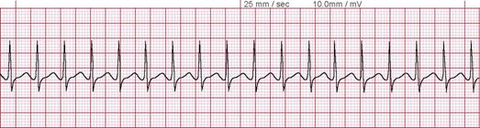
| Tachycardia ( Taches ( Moustaches) grow quickly on old men) | |
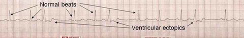
| Ectopic ( It's exciting) | |
| Sinus Rhythm ( We all have sinus and therefore it is a normal heart rate) | |
| What is haemoglobin? | A red pigment used to transport o2 in the blood. |
| How many subunits has Haemoglobin got and what are they made from? | Haemoglobin has four subunits it is made of 2 chains of alpha polypeptide and 2 chains of beta polypeptide. |
| What is a haem group? | A haem group is a non protein group which in the case of haemoglobin is a single iron ion. |
| How many oxygen molecules can each haem subunit carry? | Each subunit can carry one oxygen molecule ( 2 ATOMS) per subunit making that each haemoglobin molecule can carry 4 molecules of oxygen. |
| How is oxygen transported? | . Oxygen can be absorbed in the blood as it passes through the alveoli in the lungs. Oxygen molecules which diffuse into the blood plasma diffuse into the red blood cells. Due to the high conc of oxygen and high partial pressure the oxygen becomes associated ( binds) to the haemoglobin. This helps maintain a steep con. gradient. |
| How does Oxygen dissociate? | Oxygen is needed for aerobic respiration. Here the oxyhaemoglobin releases the oxygen for the cells to respire. |
| Explain what happens in an oxygen association curve? | A graph of oxygen saturation is plotted against a graph of oxygen tension. As haemoglobin associates with oxygen the graph often will be in form of a s shape. At the beginning of the curve the oxygen saturation is low as only one molecule has binded to the haemoglobin. This allows the haemoglobin to change shape and become biconcave. Then at the next point of the graph ( 90%) saturation with oxygen, more molecules combine with the haemoglobin meaning that the con. of oxygen increases. Where the graph levels off is where the haemoglobin molecule has reached full saturation and therefore it becomes harder to join the last molecule on. The haemoglobin has fully associated with the oxygen and is known as oxyhaemoglobin. |
| What is the difference between fetal and adult haemoglobin? | Fetal haemoglobin has a higher affinity for oxygen than adult haemoglobin meaning that the dissociation curve is to the left. Fetus absorb oxygen from the placenta as the oxygen tension level is low. This reduces the oxygen tension in the mother's blood and makes the mother release more oxygen ( Dissociation.) |
| How is carbon dioxide found in the body? | 5% is dissolved in the blood. 10% Is combined with plasma proteins and haemoglobin to form carbaminohaemglobin. 85% is transported as hydrogencarbonate ions. |
| What side of the heart carries deoxygenated and what side carries oxygenated blood? | Right= Deoxygenated Left= Oxygenated |
| What is the role of the septum? | This separates the ventricles from each other ensuring that the oxygenated and deoxygenated blood does not mix. |
| What are the features of the atria? | The muscle of the walls are very thin as they do not need to create as much pressure. There function is to receive blood and push it into the ventricles. |
| what are the features of the right and left ventricle? | The right ventricle has thicker walls than the atria as it needs to pump deoxygenated blood to the lungs. Whereas the left ventricle has to have thicker walls as it pumps oxygenated blood to the body via the aorta and needs a larger amount of pressure to overcome the resistance of systemic circulation. |
| What is plasma made up of? | Plasma contains may dissolved substances such as oxygen, carbon dioxide,minerals, glucose, amino acids, hormones and plasma proteins. |
| How is tissue fluid formed? | Tissue fluid is formed by the leaking of the capilliries. This bathes the cells and transports them with nutrients and oxygen they require. |
| What are the processes of the formation of tissue fluid? | An artery reaches the tissues it branches into smaller arterioles and then capillaries. This link up with the venules carry blood back into the veins. At the arterial end of the capillary the blood is high at hydrostatic pressure. This pushes blood fluid out of the capillaries through the capillary wall in tiny gaps. The fluid that leaves consists of plasma with dissolved oxygen and nutrients. WBC, RBC, platelets remain in the blood as they are too large to pass through the capillary wall. This tissue fluid surrounds the body cells for the exchange of gas and nutrients. The exchange occurs by diffusion. CO2 leaves cells. |
| How does it return to the blood? | The pressure at the venous end is lower this allows some of the tissue fluid to return to the capillary carrying waste products. Some tissue fluid is directed to the lymph system. This drains excess tissue fluid out of tissues and returns it to the blood system in the subclavian vein in the chest. |
| What is the hydrostatic and oncotic pressure like in the blood plasma, tissue fluid and lymph? | Blood plasma- Hydrostatic= High- Oncotic= more negative. Tissue fluid- H- Low-O-Less negative Lymph-H-Low-O-less negative. |
| What cells, proteins and fats has blood plasma have in it? | Cells- RBC, Neutrophils and lymphocytes. Proteins- Plasma proteins. Fats- Transported in lipoproteins. |
| What cells, proteins and fats has tissue fluid have in it? | Cells- some neutrophils Proteins and fats- Few |
| What cells, proteins and fats has Lymph have in it? | Cells- Lymphocytes Proteins- Few Fats- More fats especially near the digestive system. ydr |
| What is the difference between hydrostatic and oncotic pressure? | Hydrostatic- Pushes fluid into tissues and capillaries due to high pressure. Oncotic- Pulls water back into the blood and also pulls water into the tissue fluid as it is negative. |
Want to create your own Flashcards for free with GoConqr? Learn more.