5141369
| Question | Answer |
| What stages can a newborn cell enter into? | The newborn cell may drop out of cycle (so called G0 phase) or enter G1 |
| Describe the G1 stage | In early G1 the cell becomes committed to divide and proteins, organelles and macromolecules are produced. This can take weeks and results in the cell itself becoming bigger in size. |
| Describe the S phase of the cell cycle. | The cell commences genome replication in S phase, the only time when DNA is synthesised, and a copy of each chromosome is produced. This stage takes 8-10 hours and involves the uptake of deoxynucleotides. |
| Describe the G2 phase of the cell cycle. | This is a short stage (3-4 hours). It is the second growth stage of the cell and the same processes occur as the G1 phase. |
| How long is the mitotic phase of the cell cycle? | minutes- 1 hour |
| Give the order of the stages of cell cycle. | G0 <- newborn cell -> G1 -> S -> G2 -> M |
| What are the key decision points in the regulation of the cell cycle? | - the entry into S phase - the entry into M phase (known as the G1/S and G2/M checkpoints) |
| What is the overall goal of mitosis? | To generate 2 daughter cells and ensure that each inherits one copy (chromatid) of each chromosome |
| Name the phases of mitosis | (interphase) prophase, metaphase, anaphase, telophase, cytokinesis |
| Describe what occurs during prophase. | -2 pairs of chromosomes condense. -cohesin (a protein) forms rings which hold the sister chromatids together -The mitotic spindle (mircrotubule structure) forms. |
| What is the name of the transition phase from prophase to metaphase? | prometaphase |
| What occurs during prometaphase? | -the nuclear envelope breaks down -microtubules connect to each centromere via a protein complex called the kinetochore -microtubules begin to pull chromosomes towards spindle poles |
| Describe what happens during metaphase. | - chromosomes become aligned along cell equator by microtubule tension |
| State the different types of microtubles present in metaphase | -astral microtubles -Kinetochore microtubules -interpolar microtubules. |
| What do astral microtubules connect? | connect the spindle pole (tubules) to the cell membrane |
| What is the purpose of interpolar microtubules? | They connect the 2 spindle poles together. |
| What is the role of the kinetochore microtubules? | connect the chromosome to the spindle pole (attach to chromosome at centromere via kinetochore complex. |
| Describe the microtubule movements occuring in prometaphase | kinetochore microtubules shorten interpolar microtubules lengthen pulls chromosome pairs into the midline between spindle poles. |
| what happens in anaphase | -cohesin ring holding chromosome pairs together cut by enzyme separase -movement of microtubules pulls chromosomes apart. |
| describe the microtubule movements in anaphase | -further shortening of kinetochore microtubules -lengthening of interpolar tubules |
| Describe the regulation of the cutting of the cohesin ring | catalysed by the enzyme separase anaphase promoting complex promotes process Securin inhibits process. |
| Briefly describe Tubulin | -made of Alpha and Beta subunits -can polymerise and depolymerise |
| How are the spindle poles pulled apart during anaphase | -astral microtubules shorten -helped by 'pushing' from the lengthening interpolar tubules/ |
| Describe the process of telophase | -chromosomes arrive at cell poles -the spindle is broken down -nuclear membrane reforms around chromosomes |
| Describe the process of cytokinesis in animal cells | an actin/myosin ring forms around the cell equator, just under the cell membrane. The actin and myosin filaments slide over each other, drawing the ring shut and creating the ‘cleavage furrow’. Cells separated as 'purse string' draws shut |
| Describe the process of cytokinesis in plants | Vesicles form across the mitotic spindle, which then fuse to form a cell plate then eventually a new cell wall. phragmoplast -> cell plate -> cell wall |
| How is cell shape and movement facilitated in a dividing cell? | motor proteins such as kinesin and dyenin move along microtubules and the actin cytoskeleton (ie in cytokinesis) |
| In which direction does dyenin move? | + -> - |
| In which direction does kinesin move? | - -> + |
| state the 3 lines of research which gave clues to the regulation of progression through the cell cycle. | 1)cell fusion experiments 2)Sea Urchin Embryos 3)Yeast |
| Describe how cell fusion experiments were used to give clues to the nature of cell cycle control. | Fusing a cell in S phase with one in G1 results in G1 nucleus enterinb the S phase early. Fusing a cell from any other phase of the cycle with one in M phase drives the non mitotic nucleus into mitosis. |
| Therefore what can be concluded from the outcome of the cell fusion experiments? | That positive factors drive the entry into S and M phases |
| Describe how the sea urchin embryo experiments were conducted. | 1) isolate cells at same point in cycle (ie embryos dividing in synchrony) 2) radiolabel proteins and observe intensity differences by running out. |
| What were the results of the sea urchin embryo experiments? | revealed that 2 proteins accumulated during interphase and were lost at mitosis. these proteins were named 'cyclins'. cyclin levels fluctuate throughout experiment |
| What type of yeast was used in the yeast experiments? | -mutants arrested at specific points in the cell cycle (both in budding yeast -Saccharomyces cerevisiae and fission yeast- Saccharomyces pombe) |
| Describe the process carried out in the yeast experiments. | "Restoration of Function" 1) compare normal + mutant CdC mutant phenotypes 2) introduce unknown genes 3)gene which restores cell cycle is involved in G2 stage 4) protein CDK1 identified in correspondence with gene cdc2 |
| What does cdk 1 stand for? | cyclin dependent kinase 1 |
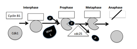
| what is the critical factor for entry into mitosis? | a complex of cyclin b and cdk1 |
| what is its mode of action? | phosphorylates a range of proteins regulating nuclear envelope breakdown, chromosome condensation, and spindle reorganisation, enabling mitosis to proceed. |
| describe the levels of regulation of cdk 1 | i) requirement for cyclinb which only accumulates prior to mitosis ii) cdk1 is inactive unless the tyrosine at position 15 is dephosphorylated iii) enzyme controls |
| why does tyrosine 15 phosphorylation inactivate cdk1? | tyrosine 15 phosphorylation blocks the ATP binding site of Cdk1, preventing kinase activity. |
| describe how enzymes are able to regulate cdk1 action. | 2 enzymes identified from genetic studies in yeast. Wee1- tyrosine kinase which phosphorylates Cdk1 inactivating it. Cdc25- a phosphatase which removes the phosphosphate group activating cdk1. |
| in which stage does cyclinb1 destruction take plate? | anaphase |
| in which stage is the mitotic entry stage switch turned 'on' | metaphase |
| Draw a diagram showing the progression through the mitotic cycle for the complex and enzymes. | |
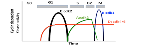
| Describe the regulation of entry into S phase. | regulated in similar way- but using different unrelated cyclins. (cyclinsA,D+E). and different cyclin dependent kinases. (eg Cdk2 which drives S phase). cyclin degradation and regulation cdk phosphorylation are critical for control. |
| Draw a diagram showing which stages each cdk:cyclin complex regulates. | |
| on top of cyclin binding and phosphorylation, what additional regulators are in position? | - cdk inhibitors such as the INK4 family and Cip/kip family, which bin to cdks and inhibit their activity. |
| What are Cdk inhibitors (Cdkis) used for in the cell? | used to halt the cell cycle in response to external signals or by internal quality control mechanisms (such as those which detect DNA damage). this is a critical defense against mutations that lead to cancer. |
| Describe the process that occurs preventing cells from entering the S phase if DNA damage has been detected. | 1) ATM and ATR kinases activated 2) both can activate p53 via phosphorylation. (either directly or indirectly) 3) p53 induces expression of genes including p21 (cdk2 inhibitor). 4)prevents cells from entering S phase by blocking cdk2 |
| Describe the process that occurs to halt cells which have already entered the S phase, when DNA damage has been detected. | 1) ATR and ATM activated 2) this activates Chk1 (checkpoint kinase) 3) Chk1 phosphorylates the phosphatase Cdc25 4) Cdc25 unable to dephosphorylate Cdk1 5) cell unable to enter into mitosis. |
| How long is cell cycle arrest maintained for? | Cell cycle arrest in G1, S or G2 phase is maintained until DNA is repaired, or if lesions are irreparable, apoptosis results. |
Want to create your own Flashcards for free with GoConqr? Learn more.