7445635
Description
Flashcards by Becca Schmeidler, updated more than 1 year ago
|
|
Created by Becca Schmeidler
over 7 years ago
|
|
| Question | Answer |
| Artery | Blood vessels that conduct blood away from the heart and into the circulation |
| Capillary | The smallest of the blood vessels and the sites of exchange between the blood and tissue cells |
| Vein | Blood vessels that return blood toward the heart from the circulation |
| Erythrocyte | Red blood cells |
| Thrombocytes | Platelet; cell fragment that participates in blood coagulation |
| Platelet | Cell fragment found in blood; involved in clotting |
| Leukocyte | PWhite blood cells; formed elements involved in body protection that take part in inflammatory and immune responses |
| Plasma | The nonliving fluid component within which formed elements and various solutes are suspended and circulated |
| Hematocrit | The percentage of total blood volume occupied by erythrocytes |
| Polycythemia | An abnormally high number of erythrocytes |
| Polycythemia Vera | Cancer of bone marrow where too many red blood cells are produced |
| Albumin | The most abundant plasma protein |
| Osmotic Pressure | The force drawing water down its concentration gradient; A measure of the tendency of water to move into a more concentrated solution |
| Interstitial Space | Related to or situated in the small, narrow spaces between tissues or parts of an organ |
| Edema | Abnormal increase in the amount of interstitial fluid due to low levels of albumin making water stay in interstitial spaces; causes swelling (primarily in the feet) |
| Plasmapheresis | A method of removing blood plasma from the body by withdrawing blood, separating it into plasma and cells, and transfusing the cells back into the bloodstream. It is performed especially to remove antibodies in treating autoimmune conditions |
| Alpha and Beta Globulins | Produced by the liver or immune system; Glycoprotein; Carrier proteins |
| Antibody (Gamma/ immunoglobulin) | Any of a class of proteins present in the serum and cells of the immune system, that function as antibodies |
| Fibrinogen | A soluble blood protein that is converted into insoluble fibrin during blood clotting |
| Spectrin | Proteins that help erythrocytes hold their shape |
| Bilirubin | Yellow pigment of bile |
| Urobilin | Chemical that gives urine its yellow color; Bilirubin is broken down into this |
| Jaundice | A medical condition with yellowing of the skin or whites of the eyes, arising from excess of the pigment bilirubin and typically caused by the obstruction of the bile duct, by liver disease, or by excessive breakdown of red blood cells |
| Hyperbilirubinemia | Excess bilirubin that occurs because the baby's liver isn't mature enough to breakdown and remove the bilirubin from the bloodstream |
| Phototherapy | Uses light (not UV light) to increase the breakdown of bilirubin |
| Erythropoiesis | Process of producing erythrocytes from stem cells in the red bone marrow |
| Hemoglobin | Oxygen-transporting protein of erythrocytes; Can carry 4 Oxygen molecules |
| Deoxyhemoglobin | Hemoglobin found in the veins which does not have oxygen molecules; dark red blood |
| Oxyhemoglobin | Hemoglobin found in the arteries which are carrying oxygen molecules; bright red blood |
| Sickle Cell Anemia | Under low oxygen conditions, hemoglobin chains link together, forming stiff rods that deform the erythrocyte; Blood transfusion is the standard treatment |
| Diapedisis | The first major process of leukocytes where leukocytes pass through the capillary wall into interstitial space |
| Chemotaxis | The second major process of leukocytes where leukocytes move toward a chemical stimulus (i.e. bacteria, toxin) |
| Phagocytosis | The third major process of leukocytes where the leukocyte engulfs exotic or extraneous material, and use an intracellular enzyme to digest them |
| Phagocytes | A type of cell within the body capable of engulfing and absorbing bacteria and other small cells and particles; "eater cell"; eat until they die |
| Granulocyte | Have granules in their cytoplasm and lobed nuclei; phagocytes; there are three types, named by their appearance when stained |
| Neutrophil | Neutral stained cytoplasm; Function is to attack bacteria; Granules are very fine (hard to see); Granules contain defensins |
| Eosinophil | Granules stain auburn; Granules contain digestive enzymes, but these enzymes do not digest bacteria; Most important function is to attack parasitic worms |
| Basophil | Granules stain blue (purplish black); Respond to allergens (like pollen or pet dander); Granules contain histamine |
| Defensins | Antimicrobial proteins; poke holes in the pathogen membrane |
| Allergens | Innocuous substances that the body thinks are dangerous |
| Histamines | Inflammatory chemical that causes the blood vessels to get bigger; A chemical messenger which causes vasodilation and increased capillary permeability |
| Vasodilator | Relaxation of the smooth muscles of the blood vessels, producing dilation |
| Mast Cell | Also release histamine, but are found in connective tissue instead of blood |
| Monocyte | Agranulocytes; Single kidney shaped nucleus; Largest of the leukocytes; Activate lymphocytes; convert into macrophages when they leave the bloodstream |
| Agranulocytes | No granules in the cytoplasm |
| Differentiate | to convert; Monocytes convert into macrophages |
| Macrophage | Big Eater; Protective cell type common in connective tissue, lymphoid tissue, and many body organs; phagocytizes tissue cells, bacteria, and other foreign debris; presents antigens to T cells in the immune response |
| Lymphocyte | Agranulocytes; large, spherical nucleus; only a few in the blood, most are found in the lymphoid tissues; Arises from bone marrow and becomes functionally mature in the lymphoid organs of the body; Two types are T and B cells |
| B Cell | Also called B lymphocytes; oversee humoral immunity; their descendants differentiate into antibody-producing plasma cells |
| T Cells | Lymphocytes that mediate cellular immunity; include helper, cytotoxic, regulatory, and memory cells; Also called T lymphocytes |
| Plasma Cell | Members of a B cell clone; effector B cells specialized to produce and release antibodies |
| Antigen | A substance or part of a substance (living or nonliving) that is recognized as foreign by the immune system, activates the immune system, and reacts with immune cells or their products. This is the site that the antibody binds to |
| Immunity | When the body makes an antibody that matches the invader; Ability of the body to resist many agents (both living and nonliving) that can cause disease; resistance to disease. |
| Hematopoiesis | Production of blood cells from a common precursor (stem cell); occurs in the red bone marrow |
| Myeloid Leukemia | Abnormal leukocytes are myeloblast descendants (granulocytes) |
| Lymphatic Leukemia | Overproduction of abnormal, nonfunctioning leukocytes which are lymphoid descendants (lymphocytes). |
| Acute | Quickly advancing |
| Chronic | Slowly advancing |
| Megakaryoblast | Fragments of giant cells |
| Hemostasis | Blood halting; Important in preventing "bleeding out" from cuts |
| Clotting Factors | Series of protein activations that lead to fibrin formation |
| Vitamin K | An important vitamin that is vital to blood clotting; It is sometimes injected into babies at birth to help prevent life-threatening bleeding |
| Hemophilia | Includes several bleeding disorders; Genetic; Loss of one of the clotting factors, so clotting does not occur; Treatment can include transfusion of plasma or of the appropriate clotting factor |
| Thrombocytopenia | Platelet deficiency; Can cause increased bleeding and bruising |
| Thrombosis | Formation of a blood clot inside of a blood vessel |
| Thrombus | A blood clot that develops and persists in an unbroken blood vessel |
| Anti-coagulants | Inhibit vitamin K production |
| Clot Retraction | Occurs after hemostasis; Further stabilizes the clot; Contractile proteins in platelets contract and pull the ruptured edges of the blood vessel together, and reduces clot size |
| PDGF (Platelet-derived Growth Factor) | produced by the platelets, stimulates smooth muscle cells and fibroblasts to divide and rebuild the vessel wall. |
| Fibrinolysis | Removal of clot after healing has occurred (begins ~2 days after clot is formed) |
| Plasminogen | protein incorporated into a blood clot; inactive precursor for the enzyme plasmin, present in the blood |
| tPA | Tissue plasminogen activator; an enzyme that helps dissolve clots |
| Plasmin | An enzyme that digests fibrin and breaks down a clot |
| Fibrin | Fibrous insoluble protein formed during blood clotting |
| Agglutination | Clumping of foreign cells; induced by cross-linking of antigen-antibody complexes |
| Karl Landsteiner | Found that when blood from different people is mixed, it can lead to blood clumping; Identified 3 blood types based on clumping |
| Type A Blood | Has A antigen present on its blood cells; has an antibody to B type blood cells; Can't receive B or AB Blood |
| Type B Blood | Has B antigen present on its blood cells; has an antibody to A type blood cells; Can't take A or AB Blood |
| Type AB Blood | Has both A and B antigens present on its cells; Has no antibodies and is the universal recipient |
| Type O Blood | Has no antigen present on its cells; Has both A and B antibodies present and is the universal donor; Can only receive Type O Blood |
| Agglutinogen | RBC antibodies that have the ability to cause clumping in cells |
| Agglutinin | The antibody present on a red blood cell |
| Transfusion Reaction | A reaction to wrong type of donor blood cells that causes the clumping of the recipient's red blood cells; This is due to the antibody present in the recipient blood cells |
| Rh Factor | Rh positive blood has the antigen D present on its cells; Rh negative blood can only receive negative blood, because if it is exposed to Rh positive blood cells, it will create antibodies to the cells |
| Rh Incompatibility | If an Rh negative woman is impregnated by an Rh positive man and the baby inherits the Rh positive antigen, a small amount of the fetal blood will enter the mother's circulation at birth. The woman can develop antibodies to the Rh antigen and when she becomes pregnant with her second Rh positive fetus, her cells will attack the fetus's blood cells. The treatment for this is an injection of RhoGAM right after the birth of the first child to prevent the mom from creating antibodies. |
| Erythroblastosis Fetalis | The same thing as Rh Incompatibility |
| RhoGAM | An immune globulin that is injected into an Rh Negative mother after child birth to create her from creating antibodies to an Rh Positive antigen |
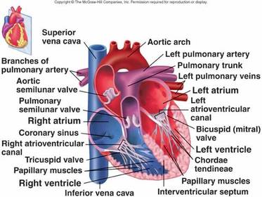
| Septum | Separates the heart into the left and right halves |
| Pericardium | Double walled sac that encloses the heart. Has three layers: 1) Fibrous pericardium-protects the heart, anchors the heart, prevents overfilling of the heart with blood 2) Parietal layer of serous pericardium 3) Visceral layer of serous pericardium |
| Myocardium | muscular tissue of the heart |
| Endocardium | Inner endothelial lining of the heart |
| Trabeculae | "Little beams"; if the heart walls were flat, suctioning would occur during pumping |
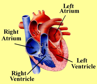
| Atrium | The areas of the heart where blood enters |
| Ventricle | The areas of the heart where blood exits |
| Sulcus | -Interventricular sulcus: marks the separation of the left and right ventricles -Coronary sulcus: marks the separation of the atria and ventricles; encircles the heart like a crown (corona) |
| Pulmonary Circuit | The right side of the heart which takes blood to the lungs |
| Systemic Circuit | Left side of the heart which takes blood to the rest of the body |
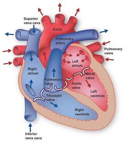
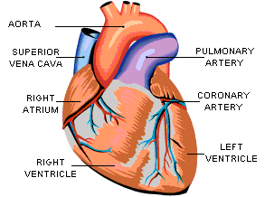
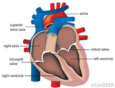
| Right atrium (Right Auricle) | Where deoxygenated blood enters the heart from the superior vena cava and the inferior vena cava |
| Right Ventricle | Where deoxygenated blood goes after going through the right atrium. Later pushes through the pulmonary artery toward the lungs where the blood is oxygenated |
| Left Atrium | Where oxygenated blood enters the heart from the lungs through the pulmonary vein. |
| Left Ventricle | Where oxygenated blood flows after going through the left atrium. Blood then leaves the heart through the aorta to be distributed to the rest of the body |
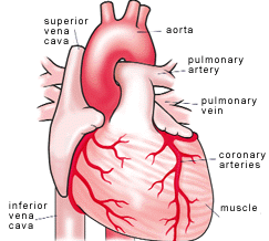
| Superior Vena Cava | Large vein carrying deoxygenated blood to the right side of the heart (the top one) |
| Inferior Vena Cava | Large vein carrying deoxygenated blood to the right side of the heart (the bottom one) |
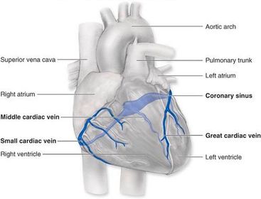
| Coronary Sinus | Empties blood directly into the right atrium |
| Pulmonary Artery | Carries blood from the right ventricle to the lungs to be oxygenated |
| Pulmonary Veins | Carry oxygenated blood from the lungs to the left atrium of the heart |
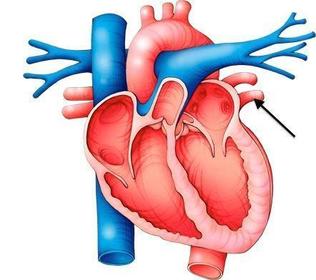
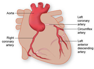
| Aorta | Artery where oxygenated blood is forced out of the left ventricle to travel toward the tissues of the body |
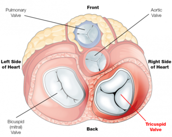
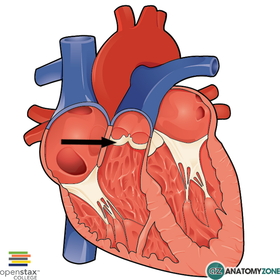
| Atrioventricular Valves (AV) | Tricuspid valve and the mitral valve. They separate the atria and the ventricles. |
| Semilunar Valves (SL) | Pulmonary valve and aortic valve which are shaped like three crescent moons (semilunar) |
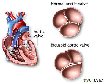
| Aortic Valve | Valve between the left ventricle and the aorta |
| Tricuspid Valve | The valve between the opening of the right atrium of the heart into the right ventricle and that resembles the mitral valve in structure but contains three membranous flaps |
| Pulmonary Valve | Between the right ventricle and the pulmonary artery |
| Bicuspid (Mitral) Valve | The valve between the left atrium and the left ventricle of the heart, consisting of two tapered cusps |
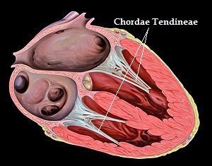
| Chordae Tendineae | "tendinous chords"; "heart strings"-anchor AV valves to the ventricle wall |
| Auscultation | Listening to the heart sounds, typically with a stethoscope |
| S1 | LUB; Tricuspid and bicuspid valves closing, loudest at the apex of the heart |
| S2 | DUB; Pulmonary and aortic valves closing, loudest at top of the heart (base) |
| Diastole | Period of the cardiac cycle when either the ventricles or the atria are relaxing |
| Systole | Period of the cardiac cycle when either the ventricles or the atria are contracting |
| Infarct | Dead tissue resulting from a loss of blood supply |
| Myocardial Infarction | Heart Attack; Condition characterized by dead tissue areas in the myocardium; caused by interruption of blood supply to the area. |
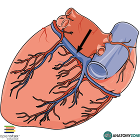
| Coronary Artery | For most people, brings oxygenated blood to the right/left atrium and ventricle by the right/left artery. For 15% of people the left artery brings blood to both ventricles (while the right still brings blood to the right atrium). 4% of people only have one of these arteries which brings blood to the entire heart |
| Coronary Vein | Empties blood from the walls into the coronary sinus of the heart |
| Striated | When muscle fibers are arranged in a latticework, with the fibers dividing, recombining, and then spreading again (like skeletal muscle!) |
| Sarcomere | The reason that cardiac muscle fibers are striated. Composed of myosin and actin. (The smallest contractile unit of muscle) |
| Endomysium | Thin connective tissue surrounding each muscle cell. (Loose connective tissue matrix between cells) |
| Fibrous Cardiac Skeleton | Endomysium connects to this dense connective tissue, which acts as the origin and insertion sites, giving the cardiac cells something to pull against |
| Intercalated Discs | Specialized connections between myocardial cells containing gap junctions and desmosomes |
| Gap junctions | Allow communication between two cells; they transmit the current (ions) across the entire heart |
| Desmosomes | Act like interlocking velcro and hold two cells together; They prevent cells from separating during contraction |
| Functional Syncytium | A single coordinated unit; It's actually two syncytium, the atria and the ventricles. This allows the atria to contract slightly before the ventricles. |
| Pacemaker | Generate the impulse (tell the heart to contract); Also is a device which electrically stimulates the heart muscle if the SA or AV nodes aren't working. |
| Intrinsic | The conduction system inside the body, stimulated by the heart's pacemaker |
| Sinus Rhythm | The rhythm of the heart (contractions of the ventricles) |
| Heart Rate | Determined by the sinus rhythm; The number of heartbeats per unit of time, usually per minute; It is based on the number of contractions of the ventricles. |
| Electrocardiogram | Detects the electrical currents the heart generates and transmits throughout the body; 12 leads, each looking at the heart from a separate angle |
| Arrhythmia | Heart's rhythm is abnormal. They can be caused by heart attack, drug overdose, genetic disease, or old age. |
| Rapid Ventricular Filling | 80% of ventricle filling occurs, as blood flows passively into the atria through open AV valves into the ventricles. |
| End-Diastolic Volume (EDV) | Maximum volume of blood the ventricles can hold |
| Isovolumetric Contraction Phase | All valves are closed for a split second during ventricular systole. |
| End-Systolic Volume (ESV) | Blood left in the ventricles after ventricular systole. |
| Isovolumetric Relaxation Phase | All valves are closed for a split second during early diastole |
| Vagus Nerve | Decreases the heart beat |
| Cardiac Output | The amount of blood pumped out by each ventricle in one minute |
| Cardiac Reserve | The difference between a person's Cardiac Output and their maximal Cardiac Output. Non-athletes: usually 4-5 times resting (~20-25 L/min) Athletes: can be 7 times resting (~35 L/min) |
| Stroke Volume | The difference between end-diastolic volume (EDV) and end-systolic volume (ESV) |
| Norepinephrine | Increases the number of action potentials; binds to Beta-adrenergic receptor on cells of the SA node |
| Beta-adrenergic Receptor | Norepinephrine binds to this and causes the threshold to be reached more quickly, and an increase in calcium entry |
| Acetylcholine | Released by the vagus nerve and hyperpolarizes the membranes of the cells in the SA and AV nodes. This slows down the number of signals the nodes send, which slows the heart rate. |
| Vagal Tone | The vagus nerve is continuously sending signals, so the heart beat is slower than the rate of the SA node on its own (75 vs 100). |
| Epinephrine (adrenaline) | -Secreted during times of high stress -Acts like norepinephrine and increases heart rate |
| Thyroxine | -Secreted from the thyroid gland -Increases the rate at which the body uses energy (and thus increases body heat) -Increases heart rate |
| Tachycardia | "Heart Hurry" -Abnormally fast heart rate (>100 bpm) -Causes include fever, stress, certain drugs, or heart disease |
| Bradycardia | Heart rate slow (<60 bpm) -Occurs in athletes as heart gets bigger (hypertrophies), stroke volume increases, allowing a lower resting heart rate while still having a high cardiac output -When it occurs in non-athletes, for example because of problems with the SA node, it can cause inadequate blood circulation to the tissues |
| Congestive Heart Failure | Myocardium weakens, so heart doesn't pump blood efficiently, and tissues don't get the blood that they need |
| Coronary Atherosclerosis | Blockage in the coronary arteries so heart doesn't get the blood (nutrients/oxygen) it needs, causing it to pump ineffectively (Can lead to myocardial infarction) |
| Dilated Cardiomyopathy | -Ventricles stretch and thin, and the myocardium deteriorates -Causes unknown, may be genetics, or drug abuse or toxin exposure |
| P | If the left side of the heart fails; The right side sends blood to the lungs, but the left side can't send oxygenated blood to the tissues. |
| Pulmonary Edema | Edema caused by blood getting "stuck" in the lungs - if it's left untreated it can cause suffocation |
| Peripheral Congestion | If the right side of the heart fails and blood backs up into the systemic system, causing edema of the extremities. |
| Angioplasty | Surgical repair or unblocking of a blood vessel, especially a coronary artery. |
| Stents | A tubular support placed temporarily inside a blood vessel, canal, or duct to aid healing or relieve an obstruction |
| Bypass Surgery | A surgical procedure to restore normal blood supply to the heart by creating new routes for the blood to travel into the heart when one or both of the coronary arteries have become clogged or obstructed. |
| Arteriole | A small branch of an artery leading into the capillaries |
| Venule | A very small vein, where the capillaries converge leading into the veins |
| Tunica Intima | Contains the simple squamous endothelium that lines the artery lumen |
| Tunica Media | -Circular smooth muscle (elastic fibers only in arteries) -Important for blood pressure regulation because it has the ability for vasoconstriction and vasodilation (vasomotor nerve fibers) |
| Tunica Externa | -Collagen fibers-protect and anchor the arteries |
| Elastic Artery | -Large (1.0-2.5cm), thick-walled arteries close to the heart (includes aorta and pulmonary artery) -Contain a high amount of elastic fibers -Allows artery to stretch when the ventricle ejects blood |
| Muscular Artery | -Deliver blood to specific body organs -Internal diameter ranges from that of a little finger to that of pencil lead (0.1-10mm) -Have a thick tunica media (muscle layer) -Active in the process of vasoconstriction and vasodilation |
| Pre-Capillary Sphincter | A band of smooth muscle that adjusts blood flow into capillaries |
| Arterial Pulse | -When the left ventricle contracts, it causes a pulse wave of blood through the arteries, which expands the arteries -After this expansion, the arteries will contract to push the blood into the capillaries -These pulse waves can be palpated at specific body sites on the body ("pulse points") |
| Pericytes | Smooth muscle-like cells that stabilize the capillary wall and help control what can enter and exit the capillary |
| Continuous Capillary | -Most common; found in skin and muscles -Is the least permeable type of capillary because the endothelial cells that line the capillary are joined by tight junctions -some areas are missing the tight junctions-the intercellular clefts -important for passage of fluids and small solutes |
| Intercellular Cleft | NOT present in the continuous capillaries of the brain-thus forming the blood-brain barrier |
| Pinocytotic Vesicle | (pino = "to drink") shuttle the fluids across the capillary wall |
| Fenestrated | Has pores |
| Fenestrated Capillary | -Are more permeable than continuous capillaries because of the presence of fenestrations (pores) -Found at sites of active absorption/filtration -intestines (nutrients) -endocrine glands (hormones) -kidneys (removal of waste) |
| Sinusoid Capillary | -Most permeable of the capillaries ("leaky") -Have lots of fenestrations and few tight junctions (large intercellular clefts) -Found in the liver, bone marrow, and spleen -Large gaps allow blood cells and bacteria to pass through -Macrophages and other phagocytes sit just outside the capillaries and remove any foreign invaders |
| Systolic Blood Pressure | Maximum pressure in the veins-occurs when the ventricles are contracted |
| Diastolic Blood Pressure | Minimum pressure in the arteries-occurs when ventricles are relaxed |
| Peripheral Resistance | The amount of friction the blood encounters as it passes through the vessels (most resistance is found away from the heart-periphery) |
| Baroreceptor | Stretch receptors located in the carotid sinuses (dilations in the carotid arteries), in the aorta, and the large arteries of the neck and chest. They respond to increased blood pressure (60-100 mmHg) -Respond more to a change in blood pressure than a constant increase |
| Chemoreceptor | In the carotid artery and aorta; respond to changes in carbon dioxide levels, oxygen levels, and pH levels. They respond to low blood pressure (40-80 mmHg) |
| Epinephrine/Norepinephrine | Increase cardiac output and promote vasoconstriction |
| Angiotensin II | In response to low blood pressure, the kidney releases renin, which is converted into this. This stimulates vasoconstriction. |
| Hypertension | -Chronically elevated blood pressure (over 140/90) -Heart must work harder to pump against the increased resistance -90% of people, there is no one specific underlying cause, but lots of risk factors: heredity, diet, obesity, age, diabetes, stress, smoking |
| Nitric Oxide | Dilates the blood vessel |
Want to create your own Flashcards for free with GoConqr? Learn more.