7906289
Description
Flashcards by Joanne North, updated more than 1 year ago
|
|
Created by Joanne North
about 7 years ago
|
|
| Question | Answer |
| What is a transport system required to do? | To take materials from cells to exchange surfaces and from exchange surfaces to cells. |
| What are the two factors that a transport system is based on? | a. The surface area to volume ratio. b. Activity of the organism. |
| Which type of animals require a transport system? | Animals with a lower surface area to volume ration and those that are more active. |
| What are the main features of transport systems? | - A suitable medium to carry materials e.g. blood. - A form of mass transport in which the transport medium is moved around the body. - A closed system of tubular vessels that contains the transport medium and branches to deliver to all parts of the organism. - A mechanism for moving the transport medium within vessels which requires a pressure difference. - A mechanism to maintain the mass flow movement in one direction e.g. valves. - A means of controlling the flow of the transport medium to suit the changing needs of different parts of the organism. - A mechanism for the mass flow of water or gases e.g. intercostal muscles and diaphragm in mammals. |
| What are the two ways in which mass transport can be achieved? | a. Animals use muscular contraction either of the body muscles or a specialised pumping organ e.g. the heart. b. Plants rely on natural, passive processes e.g. evaporation of water |
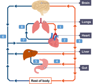
| What type of system do mammals have? | A closed, double circulatory system in which the blood is confined to vessels and passes twice through the heart for each complete circuit of the body. |
| Why is blood passed through the heart twice? | To maintain the pressure to prevent circulation being to slow which is necessary to keep up with the metabolism of a mammal. |
| Label the diagram. | 1. Pulmonary Vein 2. Aorta 3. Arteries 4. Renal Artery 5. Renal Vein 6. Veins 7. Vena Cava 8. Pulmonary Artery |
| Which side of the heart carries deoxygenated blood? | Right |
| Which side of the heart carries oxygenated blood? | Left |
| The heart has two pumps which each have two chambers. What are these chambers and describe them? | 1. The atrium - thin walled, elastic and stretches as it collects blood. 2. The ventricle - thicker muscular wall to be able to contract. |
| Where does the right ventricle pump blood to? | The lungs. |
| Where does the left ventricle pump blood to? | The rest of the body. |
| Why does the left ventricle have a thicker muscular wall? | It has to contract to create enough pressure to pump blood to the rest of the body |
| What are the two valves between the atria and the ventricles called? | - Right atrioventricular valve - Left atrioventricular valve |
| The ventricles pump blood away from the heart into..? | The arteries. |
| The atria receive blood from the..? | Veins. |
| What are the vessels connecting to the heart called? | Pulmonary vessels. |
| What are the 4 vessels connecting to the heart? | 1. Aorta 2. Vena Cava 3. Pulmonary Artery 4. Pulmonary Veins |
| What is the aorta connected to and what does it carry? | Connected to the left ventricle and carries oxygenated blood to all parts of the body accept for the lungs. |
| What is the vena cava connected to and what does it carry? | Connected to the right atrium and brings deoxygenated blood back from the tissues of the body (except for the lungs). |
| What is the pulmonary artery connected to and what does it carry? | Connected to the right ventricle and carries deoxygenated blood to the lungs. |
| What is the pulmonary vein connected to and what does it carry? | Connected to the left atrium and bring oxygenated blood back from the lungs. |
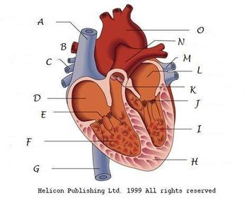
| A - Anterior vena cava B - Right pulmonary artery C - Right pulmonary vein D - Right Atrium E - Right atrioventricular valve F - Right ventricle G - Posterior vena cava H - Thick muscular wall I - Left ventricle J - Left atrioventricular valves K - Semi-lunar valves L - Left atrium M - Pulmonary veins N - Left pulmonary artery O - Aorta | |
| What blood vessels supply the heart? | Coronary arteries |
| What is the resting heart rate of humans? | 70 bmp |
| What are the three stages of the cardiac cycle? | 1. Diastole (relaxation of the heart). 2. Atrial Systole (contraction of the atria). 3. Ventricular Systole (contraction of the ventricles). |
| What happens in diastole? | - Blood returns to the atria via the pulmonary veins and vena cava. - As the atria fill the pressure rises causing the atrioventricular valves to open and blood to go into the ventricles. |
| What happens in atrial systole? | - The contraction of the atrial walls forces the remaining blood into the ventricles from the atria. - Throughout this stage the muscle of the ventricle walls are relaxed. |
| What happens in ventricular systole. | - After a short delay the ventricular walls contract increasing the pressure which shuts the atrioventricular valves and prevents backflow of blood into the atria. - Pressure in the ventricles further increases and exceeds the pressure of the aorta and pulmonary arteries. - Therefore the semi-lunar valves open and blood is forced through.. |
| What is the function of a valve? | To prevent blood backflow. |
| What are the three main types of valves? | 1. Atrioventricular valves 2. Semi lunar valves 3. Pocket valves |
| Where do atrioventricular valves occur and what is there function? | Occur between the left atrium and ventricle and the right atrium and ventricle. These valves prevent the backflow of blood when the contraction of the ventricles means that ventricular pressure exceeds atrial pressure. Closure of these valves ensures that blood moves to the aorta and pulmonary artery rather than back to the atria. |
| Where do semi lunar valves occur and what is there function? | Occur in the aorta and pulmonary artery. These prevent the backflow of blood into the ventricles when the pressure in these vessels exceeds that in the ventricles. |
| Where do pocket valves occur and what is there function? | Occur in the veins throughout the venous system. These ensure that when the veins are squeezed, blood flows back towards the heart rather than away from it. |
| What are valves made up of? | A number of flaps of tough, but flexible, fibrous tissues which are cusp-shaped. |
| What is the equation for cardiac output? | Heart rate x Stroke volume |
| What is cardiac output measured in? | dm3 min-1 |
| Describe the pressure changes in the atria. | Always has a relatively low pressure due to the thin walls not being able to create high force. Pressure is at its highest when they are contracting but drops when the atrioventricular valves close and the walls relax. There is also a gradual build up of pressure when blood is entering the atria. |
| Describe the pressure changes in the ventricle. | Is low at first but gradually increases as the ventricles fill with blood. When the left atrioventricular valves close the pressure increases dramatically as the walls begin to contract and blood is forced into the aorta or pulmonary artery. Pressure falls as the ventricles relax. |
| Describe the pressure changes in the aorta. | Rises when the ventricles contract as blood is forced into the aorta. Gradually begins to fall. Slightly increases when the elastic walls recoil to move the blood. |
| Describe the ventricular volume in the cardiac cycle. | Rises as the atria contract and the ventricles fill up with blood. Drops as the blood is forced out of the ventricles. |
| What are the four different types of blood vessels? | 1. Arteries 2. Arterioles 3. Capillaries 4. Veins |
| What is the function of the arteries? | To carry blood away from the heart and into arterioles then to the tissues under high pressure. |
| What is the function of the arterioles? | Are smaller arteries that control blood flow from arteries to capillaries under low pressure. |
| What is the function of the capillaries? | Tiny vessels that link arterioles to veins. To exchange metabolic materials such as oxygen between the blood and the cells. |
| What is the function of veins? | Carry blood from capillaries back to the heart under low pressure. |
| What is the basic structure of arteries, arterioles and veins? | a. Tough fibrous outer layer - to resist pressure changes. b. Muscle layer - can contract and control the flow of blood. c. Elastic layer - helps to maintain blood pressure by stretching and springing back. d. Thin inner lining (endothelium) - smooth to reduce friction and allow for diffusion. e. Lumen - central cavity. |
| How is the arterys structure related to its function? | a. The muscle layer is thick compared to veins - constriction and dilation. b. The elastic layer is thick compared to veins - high blood pressure. c. The overall thickness of the wall is great - resist bursting under pressure. d. There are no valves - due to high pressure. |
| How is the arterioles structure related to its function? | a. The muscle layer is relatively thicker than arteries - allows for the constriction of the lumen. b. The elastic layer is relatively thinner than in arteries - lower pressure. |
| How is the veins structure related to its function? | a. The muscle layer is relatively thin. b. The elastic layer is relatively thin - low pressure. c. The overall thickness of the wall is small - no risk of bursting. d. There are valves at intervals - low pressure. |
| How is the capillaries structure related to its function? | a. Their walls consist mostly of the lining layer - diffusion pathway is short. b. They are numerous and highly branched - large surface area. c. They have a narrow diameter - no cell is far away. d. Their lumen is so narrow - red blood cells are squeezed flat. e. There are spaces between the lining (endothelial) cells - allows white blood cells to escape. |
| What is tissue fluid? | A watery liquid that contains glucose, amino acids, fatty acids, ions in solution and oxygen. Means by which materials are exchanged between the blood and cells. |
| Where is tissue fluid formed? | Blood plasma |
| What is the composition of blood plasma controlled by? | Homeostatic systems. |
| How is tissue fluid formed? | - Pumping of the heart creates hydrostatic pressure at the arterial end of the capillaries. - This hydrostatic pressure causes tissue fluid to move out of the blood plasma. - This pressure is only enough to force small molecules out of the capillaries laving cells and proteins in the blood as they are too large to cross. - This process is called ultrafiltration. |
| How does tissue fluid return to the circulatory system? | - The loss of the tissue fluid from the capillaries reduces that hydrostatic pressure inside them. - Therefore at the venous end there is a lower hydrostatic pressure. - Tissue fluid is forced back into the capillaries by the higher hydrostatic pressure outside. - The blood plasma has lost water and still contain proteins so has a lower water potential. - Water leaves the tissues by osmosis down a water gradient. - The returning tissue fluid has lost oxygen and nutrients but has gained carbon dioxide and wastes. |
| What happens to the tissue fluid that doesn't return to the capillaries? | It is carried back by the lymphatic system which later drain their contents back into the blood stream via two ducts that join veins close to the heart. |
| How are the contents of the lymphatic system moved? | - Hydrostatic pressure - by tissue fluid that leaves the capillaries. - Contraction of bodily muscles - squeeze lymph vessels. |
Want to create your own Flashcards for free with GoConqr? Learn more.