8666633
| Question | Answer |
| Exchange Surfaces | There are 3 factors that affect the the need of an exchange system: Size Surface area to volume ratio Level of activity |
| Size: | In small organisms such as single celled organisms, the cytoplasm is very close to the environment. Diffusion will supply enough oxygen and nutrients to keep the cells alive and active. Multicellular organisms have several layers of cells. Any oxygen or nutrients diffusing in from the outside have a longer diffusion pathway. Diffusion is too slow to enable a sufficient supply to the innermost cells. |
| Surface area to volume ratio: | • Small organisms have small surface area and volume. Surface area is large compared to volume. Large enough to supply all their cells with sufficient oxygen, • Large organisms have large surface area and volume. As size increases the volume raises more quickly than the surface area. Surface area is small compared to volume ratio (SA: V). |
| Surface area to volume ratio: | Some organisms increase their surface area by adopting a different shape. Animals such as a flatworm (thin body). This gives it a larger surface area to volume ratio which limits the body size an animal can reach. Large organisms need a range of tissues to give the body strength and support. Volume increases as body gets thicker, but surface area does not increase as much. Large organism’s surface area to volume ratio is small. |
| Level of activity: | Metabolic activities use energy from food and require oxygen to realise the energy in aerobic respiration. The cells of an active organism need good supplies of nutrients and oxygen to supply the energy for movement. This needs for energy is increased in those animals such as mammals, that keep themselves warm. |
| Features of a good exchange system: |
Large surface area provides more space for molecules to pass through. This is often achieved by folding the walls and membranes involved (root hair in plants).
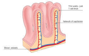
A thin barrier to reduce the diffusion distance and that barrier must be permeable to the substances being exchanged (alveoli of the lungs)
Good blood supply. This can bring fresh supplies of molecules to one side, keeping concentration high. Important to maintain a steep concentration gradient so diffusion occurs quickly (gills in fish).
Damp surface areas
Image:
Images (image/jpeg)
|
| Mammalian Gas Exchange System: | The gaseous exchange system in mammals consists of the lungs and associated airways that carry air into and out of the lungs. Air can pass into the lungs through the lungs along the trachea (windpipe), Bronchi and bronchioles. Finally it reaches tiny air-filled sacs called alveoli. These are the surfaces where gas exchange takes place. The lungs are protected by the ribcage. These ribcage are held together by the intercostal muscles. The action of these muscles and the diaphragm helps to produce ventilation. |
| Gaseous exchange in the lungs: | Gases pass by diffusion through the thin walls of the alveoli. Oxygen passes from the air in the alveoli to the blood in the capillaries. Carbon dioxide passes from the blood to the air in the alveoli. The lungs must maintain a steep concentration gradient in each direction in order to ensure that diffusion can continue. |
| Large surface area to provide more space for molecules to pass through: | The individual alveoli are 100- 300 micrometres. However they are numerous and the total surface area of the lungs is much larger than the skin. The total surface area of the exchange surface in humans is 70 m2. The alveoli are lined by a thin layer of moisture, which evaporates when we breathe out. The lungs must produce a surfactant that coats the internal surface of the alveoli to reduce the cohesive forces between the water molecules, as these forces tend to make the alveoli collapse. |
| The barrier to exchange is permeable to oxygen and carbon dioxide: | The barrier to exchange is comprised of the wall of the alveolus and the wall of the blood capillary. The cells and their plasma membrane readily allow the diffusion of oxygen and carbon dioxide, as molecules are small and non-polar. |
| Thin barrier to reduce the diffusion distance: | There are a number of adaptations that reduce the distance that gasses have to diffuse; The alveolus wall is one cell thick The capillary wall is one cell thick Both walls consist of squamous cells (flattened or thin) The capillaries are in close contact with the alveolus walls. The capillaries are narrow and the red blood cells are squeezed against the capillary wall- making them closer to the air in the alveoli and reducing the rate of flow (Total barrier to diffusion is only two flattened cells and is less than 1 micrometre thick). |
| A good blood supply: | Blood supply helps to maintain a steep concentration gradient so that the gasses continue to diffuse. • The blood system transports carbon dioxide from the tissues to the lungs. This ensures that the concentration of carbon dioxide in the blood is higher than that in that in the air of the alveoli. Therefore carbon dioxide diffuses into the alveoli. • The blood transports oxygen away from the lungs. This ensures that the concentration of oxygen in the blood is lower than that is alveoli- so that oxygen diffuses into the blood. |
| Ventilation: | The breathing movements ventilate the lungs. This replaces the used air with fresh air, bringing in more oxygen and removing carbon dioxide. Ventilation ensures that the concentration of oxygen in the air of the alveolus remains higher than that in the blood and the concentration of carbon dioxide in the alveoli remains lower than that in the blood. Therefore, concentration gradient necessary for diffusion is maintained. |
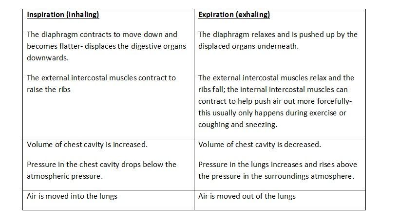
| Inhaling and Exhaling: |
Image:
Inhalation (image/jpeg)
|
| Lung tissue: | The alveolus walls contain elastic fibres that stretch during inspiration but then recoil to help push air out during expiration; the alveolus walls are so thin that it may not be possible to distinguish separate cells under a light microscope. |
| The airways: | Airways must be; Large enough to allow sufficient air to follow without obstruction Supported to prevent collapse when the air pressure inside is lower during inspiration Flexible in order to allow movement |
| Airways are: | The airways are lined by ciliated epithelium, which contributes to keeping the lungs healthy. Goblet cells in the epithelium releases mucus which traps pathogens. The cilia then move the mucus up the top of the airway, which it is swallowed. The glandular tissue in the loose tissue also produces mucus. |
| The trachea and bronchi: | The trachea and bronchi walls have similar structures. However bronchi are narrower than the trachea. These airways are supported by cartilage which prevents collapse during inspiration. The rings of cartilage in the trachea are C-shaped rather than a complete ring which allows flexibility and space for food to pass down the oesophagus. |
| The bronchioles: | The bronchioles are much narrower than the bronchi. The larger bronchioles may have some cartilage but smaller ones have none. The wall is comprised mostly of the smooth muscle and elastic fibres. The smallest bronchioles end in clusters of alveoli. |
| Smooth muscle and elastic tissue: | The smooth muscle can contract; this action will constrict the airway. This makes the lumen of the airway narrower. Constriction of the lumen can restrict the flow of air to and from the alveoli. Controlling the flow of the air of the alveoli might be important if there are harmful substances in the air. The contraction of the smooth muscle and control of airflow is not a voluntary act and may occur as a result of an allergic reaction. Once the smooth muscle has contracted, it cannot reverse this effect on its own. The smooth muscle is elongated again by the elastic fibres. When the muscle contracts, it deforms the elastic fibres recoil to their original size and shape. This acts to dilate the airway. |
| Using a spirometer: | This is a device that measures the movement of air in and out of the lungs as a person breathes. A float-chamber spirometer consists of a chamber of air or medical-grade oxygen floating on a tank of water. During inspiration air is drawn from the chamber so that the lid moves down. During expiration, the air returns to the chamber, raising the lid. These movements can be recorded on a data blogger. The carbon dioxide- rich air exhaled is passes through a chamber of soda lime, which absorbs the carbon dioxide. This allows the measurement of oxygen consumption but not the rate. |
| Precautious that must be taken when using a spirometer: | The subject should be healthy and, in particular, free from asthma The soda lime should be fresh and functioning There should be no air leaks in the apparatus, as this would give invalid or inaccurate results. The mouthpiece should be sterilised. The water chamber must not be overfilled. |
| Lung volumes: | This is measured by taking a deep breath and expiring all the air possible from the lungs. Vital capacity depends on a number of factors such as the size of a person, their age and gender and their level of exercise. Usually between 2.5- 5.0 dm. The residual volume- is the volume of air that remains in the lungs even after forced expiration. This air remains in the airways and alveoli. This is approximately 1.5 dm. Tidal volume- is the volume of air moved in and out with each breath. It is normally measured at rest (0.5dm). This is sufficient to supply all the oxygen required in the body at rest. |
| Lung Volumes | ital capacity- can be measured Tidal volume- cannot be measured Vital capacity- is the maximum volume of air that can be moved by the lungs in one breath. |
| Oxygen uptake: | Breathing supplies oxygen for respiration and removes carbon dioxide produced in respiration. As a person breathes from the spirometer, oxygen is absorbed by the blood and replaced by carbon dioxide. This carbon dioxide is absorbed by the soda lime in the spirometer, so that the volume of air in the chamber decreases. This decrease can be observed and measured on this spirometer trace. We can assume that the volume of carbon dioxide released and absorbed by the soda lime equals the volume of oxygen absorbed by the blood. Therefore measuring the gradient of the decrease in volume enables us to calculate the rate of oxygen uptake. |
| Breathing rate and oxygen uptake: | The breathing rate can also be measured from a spirometer trace. Count the number of peaks each minute. Breathing rate at rest is usually about 12-14 breaths per minute. |
| Oxygen Uptake: | Oxygen uptake will depend upon a number of factors. A higher oxygen uptake will result from increased demand, such as during exercise when the muscles are respiring more, increased oxygen uptake will result from increased breathing rate and deeper breaths. |
| Calculating oxygen uptake from a spirometer trace: | On trace a, draw a line from point A down a horizontal axis and another line by from point B to the horizontal axis. Measure the length of time between these points (55 seconds). Measure the difference in volume between A and B (0.3dm) Divide by the time taken for this decrease (55s) The unit will be in dm s . Reate of oxygen uptake on trace a = 0.3/55 = 0.0055 dm s . |
| Bony fish: | They use gills in order to absorb oxygen dissolved in the water and release carbon dioxide into the water. The oxygen concentration gradient will be typically much lower than is found in the air. Most bony fish have 5 pairs of gills which are covered by a bony plate called the operculum. Each gill consists of two rows of gill filaments (primary lamellae attached to a bony arch. The filaments are very thin, and their surface is folded into many secondary lamellae (gill plates). This provides a very large surface area. Blood capillaries carry deoxygenated blood close to the surface of the secondary lamellae where exchange takes place. |
| Countercurrent flow: | Blood flows along the gill arch and out along the filaments to the secondary lamellae. The blood then flows through capillaries in the opposite direction to the flow of water over the lamellae. This argument creates a Countercurrent flow that absorbs the maximum amount of oxygen from the water. |
| Ventilation in bony fish: | Bony fish can keep water flowing over the gills by using a buccal-opercular pump. The bucucal cavity can change volume. The floor of the mouth moves downwards, drawing water into the buccal cavity. The mouth closes and the floor is raised again pushing water through the gills. Movements of the operculum are coordinated with the movements of the buccal cavity, the operculum moves outwards. This movement reduces the pressure in the opercula cavity, helping water to flow through the gills. |
| Insects: | Insects do not transport oxygen in blood. Insects have an open circulatory system in which the body fluid acts as blood and tissue fluid. Circulation is slow and can be affected by body movements. Insects possess an air-filled tracheal system which supplies air directly to all the respiring tissues. Air enters the system via a pore in each segment, called a spiracle. The air is transported into the body through a series of tubes called tracheae. These divide into smaller and smaller tubes called tracheoles. The ends of tranchioles are open and filled with a fluid called tracheal fluid. Gaseous exchange occurs between the air in the tracheoles and the tracheal fluid. Some exchange can also occur across the thin walls of the tracheoles. Many insects are active and need a good supply of oxygen. When tissues are active, the tracheal fluid can be withdrawn into the body fluid in order to increase the surface area of the tracheoles wall exposed to air. This means that more oxygen can be absorbed when the insect is active. |
| Ventilation in insects: | Larger insects can also ventilate their tracheal system by movements of the body. This can be achieved in a number of ways. In many insects, sections of the tracheal system are expanded and have flexible walls. These act as air sacs which can be squeezed by the action of flight muscles. Repetitive expansion and contraction of these sacs ventilate the tracheal system. In some insects, movements of the wings alter the volume of the thorax. As the thorax volume decreases, air in the tracheal system is put under pressure and is pushed out of the tracheal system. When the thorax increases in volume, the pressure inside drops and air is pushed into the tracheal system from outside. Some insects have developed this ventilation even further. Locusts can alter the volume of their abdomen by specialised breathing movements. These are coordinated with opening and closing valves in the spiracles. As the abdomen expands, spiracles at the front end of the body open and air enters the tracheal system. As the abdomen reduces in volume, the spiracles at the rear end of t |
Want to create your own Flashcards for free with GoConqr? Learn more.