9758496
Description
Flashcards by Candice Young, updated more than 1 year ago
|
|
Created by Candice Young
over 6 years ago
|
|
| Question | Answer |
| 3 meninges (in Humans) | protective tissue that protect CNS; 3 layers include Dura Mater (outer), Arachnoid Mater (middle), and Pia Mater (inner) |
| Cerebrospinal Fluid | fluid-filled container that the brain floats in; NOT directly connected to blood |
| (in mammals) CNS includes: | brain and spinal cord |
| (in mammals) PNS includes: | neurons that communicate between CNS and periphery |
| afferent | incoming to the brain |
| efferent | outgoing from the brain |
| Cerebral Cortex features | sulci (folds) & gyri (smooth regions inbetween), both in a left and right hemisphere |
| Spinal Nerves | a part of the PERIPHERAL nervous system, carry signals between spinal cord and body |
| Dorsal Roots | where sensory information comes into the spinal nerves |
| Dorsal Root Ganglia | cell bodies of sensory neurons located here (in PNS) |
| Ventral Roots | motor information leaves from here (in CNS) |
| Congregations of neural cell bodies | Ganglia |
| Nuclei | Many ganglia within the brain |
| Nissl Stain | stains neurons, shows white vs grey matter in visual cortex |
| White Matter | long bits of axon communication tracts between neurons |
| Grey Matter | contains the cell bodies, dendrites, and axon terminals of neurons |
| "The Neuron Doctrine" | The idea that the brain is composed of individual cells |
| Glial Cells | cells that surround neurons in the CNS |
| Glial Cell Functions | provide support and protection to neurons, remove waste, supply nutrients, lay down axon tracts & provide charge |
| Soma | cell body of neuron |
| Dendrite | branched part of neuron |
| Myelin | type of glial outgrowth that wraps around axons; helps speed up conduction and avoid degradation of signal |
| Direction of information flow along neuron bodies | FROM the dendrites, along the axon, and TO the axon terminals |
| Conductance | Measure of an ion's ability to cross the membrane, can change by opening or closing ion channels |
| How mV is measured: | difference in charge "between the inside and the outside" |
| Concentration Differences | Influences ions to diffuse down concentration gradient through open channels |
| Electrical potential difference | Negative charged ions along inside and positive ions on outside --> attracts and repels ions across a membrane |
| Ion Pumps | Na+/K+ ATPase pumps Na+/K+ against their concentration/electrical gradient through this |
| Concentration of Na+ | higher concentrations OUTSIDE the cell |
| Concentration of K+ | higher concentrations INSIDE the cell |
| At "rest" the membrane is permeable to this ion | K+ |
| Resting Membrane Potential | a -70 mV charge produced by K+ leaving neurons through leak channels at rest & involvement of Na+/K+ ATPase |
| Voltage Gated Channels | Receptors that change conformation in response to cell depolarization/change in membrane potential |
| Graded potentials | a small change in voltage, will decay along cell fibre if only passively conducted |
| Action Potential | conformational change of voltage gated channel proteins releases Na+ into cell; activation gate opens after depolarization (-55mV) and then inactivation gate closes (at about +30mV); is the signal traveling across synapses! |
| Threshold Potential | the critical level to which a membrane potential must be depolarized in order to initiate an action potential |
| Absolute Refractory period | period where it is impossible to start a second action potential, since a "reset" must first occur (activation gates close and inactivation gates open) |
| Relative Refractory period | after absolute refractory period, possible to generate AP but large stimulus needed |
| hyper-polarization | brief period before neuron reaches resting membrane potential once again, more polarized than at rest |
| Direction of travel of Action Potential | From axon hillock to terminal boutons |
| Unmyelinated Axons | Signal will degrade across these unless the axon diameter is large enough |
| Types of glial cells that provide myelination for axons | Oligodendrocytes and Schwann cells |
| Nodes of Ranvier | gaps in myelin where voltage gated channels are located |
| Myelin is composed of this | bilayers of phospholipids in glial cells |
| Saltatory Conduction | propagation of action potentials along myelinated axons from one node to the next; can be considered "jumping" |
| Synapses | points of contact between nerve/nerve cells or nerve/muscle cells |
| Electrical Synapses | gap junctions between plasma membranes of adjacent cells, bidirectional, instantaneous and direct transmission |
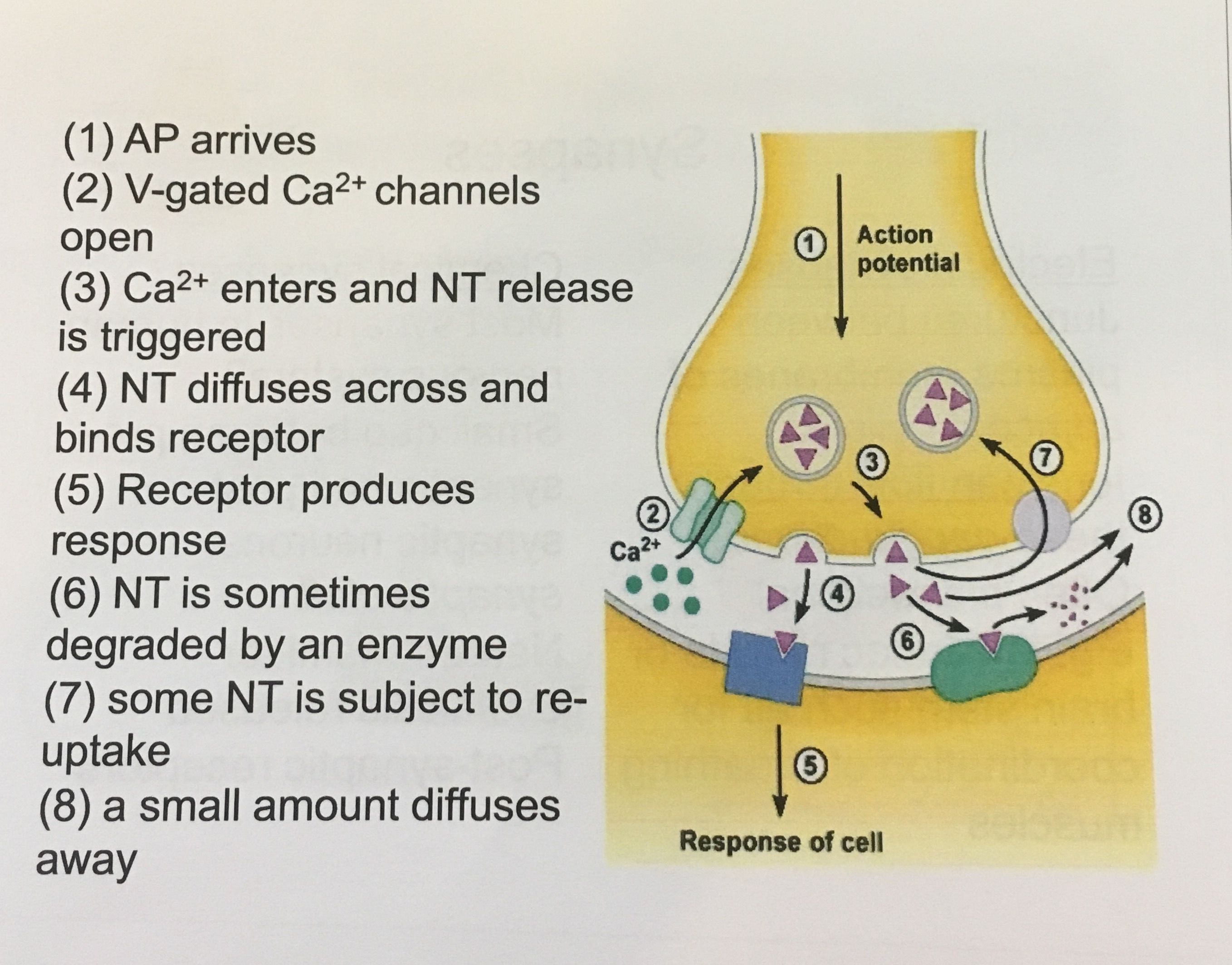
| Chemical synapses | synaptic clefts between pre- and post-synaptic neurons, neurotransmitter chemical released, unidirectional, delayed and excitatory/inhibitory transmission, humans mostly have this |
| Activation of Chemical Synapses (step by step) | |
| Neurotransmitters | chemicals that are released by one neuron and interact with a receptor on the synaptic partner |
| Acetylcholine (ACh) | main neurotransmitter at neuromuscular junctions, acts at nAChR and mAChR ion channel receptors |
| Nicotine | an agonist at nAChRs |
| Muscarine | an agonist at mAChRs |
| Atropine | an antagonist at mAChRs |
| GABA-A receptor | ligand gated ion channel, when ligand bonds it conducts Cl- through its pore which hyper-polarizes the neuron and inhibits an action potential *Picrotoxin blocks channel, can lead to convulsions* |
| D1 receptors | Metabotropic receptor that responds to bonding of dopamine --> G protein activates --> bonds to adenylyl cyclase (using ATP) --> produces cAMP--> downstream effects |
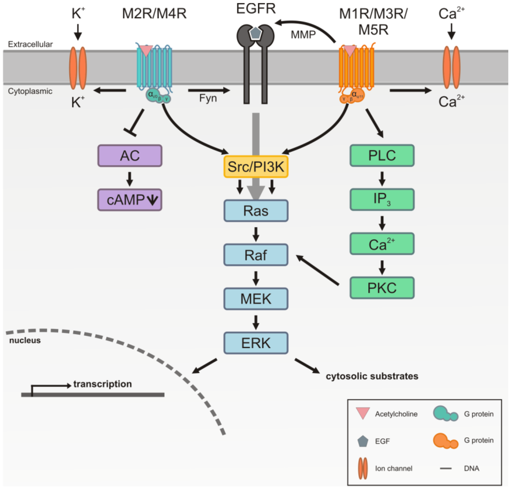
| mAChRs | 5 subtypes of metabotropic receptors, inhibit cAMP and activate GTFs |
| Photoreceptors | cells in the eye that respond to light by change in voltage |
| Rods | responsible for vision at low light levels |
| Cones | responsible for vision at higher light levels, capable of color vision and responsible for high spatial acuity |
| Rhodopsin | membrane proteins that respond to different wavelengths of light |
| Vitreous Humour | gel space in between the lens (front) and the retina (back) |
| Bipolar cells | transmit signals from photoreceptor cells to ganglion cells *amacrine and horizontal cells help integrate the information* |
| Retinal Ganglion Cells | neurons in the inner surface of the retina; receive visual information from photoreceptors and then fire action potentials |
| Optic Nerve | composed of retinal ganglion and glial cells; transmits all visual information from retina to the brain; FORM the optic nerve |
| fovea | small pit packed with cones, responsible for sharp central vision |
| Optic Chiasm | where the "crossing over" of information occurs when being transmitted to the visual cortex |
Want to create your own Flashcards for free with GoConqr? Learn more.