
Mind map
Leg Injuries
Map content
Leg Injuries
In the injuries of athletes the leg is the fourth most involved
- It involves the calf musculature and achilles tendon
- Two acute injuries involve the same unit musculotendinous
- A tension of the gastrocnemius has few medical consequences
- Achilles tendon rupture is painless but involves 6 months of rehabilitation
- It involves the calf musculature and achilles tendon
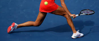
Gastrocnemius Strain (Tennis Leg)
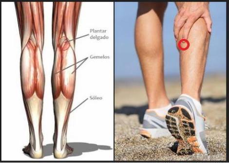
- is a tension that involves the musculotendinous union
- medial head of the gastrocnemius and achilles tendon
- higher frequency in athletes over 30 years and women
- Not only in tennis is this problem but in a variety of activities
- includes walks, basketball and various forms of dance
- if it is not recognized or handled inappropriately it can cause disability months
- is mistakenly diagnosed as plantar rupture
- Not only in tennis is this problem but in a variety of activities
- History
- Mechanism Of Injury
- is generated by a powerful propulsion movement ankle plantar flexion
- associated with muscle fatigue
- the injury follows a bout of achilles tendinitis or an ankle injury or sprain
- the injury occurs in two parts
- a minor strain that is ignored, followed minutes later by complete rupture
- associated with muscle fatigue
- is generated by a powerful propulsion movement ankle plantar flexion
- Symptoms
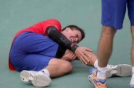
- The athlete feels or hears a pop in the medial appear of the calf
- feels like he has hit his calf with a tennis ball or racket
- a spasm of the calf musculature occurs rapidly
- swelling and ecchymosis not apparent the first hours of the injury
- ecchymosis may appear distally around the ankle and even on the foot
- 24 to 48 hours to induce the patient to seek medical attention
- Mechanism Of Injury
- Physical Examination
- tenderness towards the medial side of the muscle-tendon calf junction
- Swelling and ecchymosis are present distal to the site of injury
- Motion is limited, and the foot is held in plantar flexion
- it possible to palpate a defect in the muscle , but the defect fills rapidly with blood
- Motion is limited, and the foot is held in plantar flexion
- Swelling and ecchymosis are present distal to the site of injury
- tenderness towards the medial side of the muscle-tendon calf junction
- Radiographic Examination
- radiographic study is not required
- Treatment
- Inicial Treatment
- The ankle should be placed at a neutral angle and should be maintained with a splint
- stay with repeated ice and focal compression
- the efforts to recover the movement of the ankle
- stay with repeated ice and focal compression
- force the athlete to walk although gentle and active stretching is important
- Oral administration of anti-inflammatory medications should be started inmediately
- The ankle should be placed at a neutral angle and should be maintained with a splint
- Definitive
- the objectives is the recovery of normal strength and flexibility of the musculature
- Three to five daily physiotherapy sessions to improve movement return
- high intensity galvanic stimulation ice, active and passive stretching
- Crutches without weight support and compression first 24 hours to calm bleeding
- high intensity galvanic stimulation ice, active and passive stretching
- Three to five daily physiotherapy sessions to improve movement return
- strengthening with theraband 3 sets of 10 repetitions
- use a compression bandage
- It is employed after removal of the cast
- the objectives is the recovery of normal strength and flexibility of the musculature
- Inicial Treatment
- Other Diagnostic Test
- It is nor necessary
- is a tension that involves the musculotendinous union
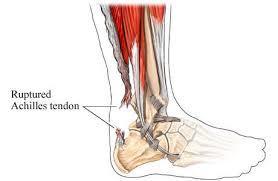
Achilles Tendon Strain (Rupture)
- Usually occurs in an abrupt, dramatic fashion
- orthopedic surgeon, and analyze the treatment of injury
- Hystory
- Mechanism of Injury
- Push-off with the forefoot (Forceful plantar flexion of the ankle )
- Basketball, racquetball, softball, and tennis are sports most often associated with the injury
- The injury often occurs at a time of relative fatigue
- usually occur in athletes older than 30 years
- Is less common in women
- usually occur in athletes older than 30 years
- Push-off with the forefoot (Forceful plantar flexion of the ankle )
- Symptoms
- sensation of being struck in the region of the tendon
- This usually accompanied by an audible pop or snap
- Mechanism of Injury
- Physical Examination
- lack of reflex plantar flexion when the calf is sharply squeezed
- A positive Thompson test
- Substantial loss of strength in plantar flexion
- Careful palpation usually reveals gap or loss of continuity in the tendon
- Bleending and swelling may be substantial in 24 to 48 hours
- Gait is antalgic and apropulsive
- lack of reflex plantar flexion when the calf is sharply squeezed
- Radiographic Examination
- is not indicated
- Other Diagnostic Test
- Magnetic resonance imaging may be useful to demonstrate the extent and exact location
- Treatment
- Definitive
- Treatment should be left to the treating orthopedist
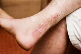
- the patient will be immobilized in a cast or orthopedic device for 2 to 3 months
- Initial
- should be placed in a compression wrap
- the ankle should be in a position of gravity- assisted plantar flexion
- a posterior plaster splint compression dressing helps maintain the plantarflexed
- Definitive
- Usually occurs in an abrupt, dramatic fashion
Acute Compartment Syndromes
- condition in which inside a osteofascial compartment they are compromised by the pressure
- 4 compartements in the leg, the anterior is by far the most commonly involved
- History
- Mechanism of Injury
- Any macrotrauma or microtrauma including
- fractures
- Tendon ruptures
- Muscle strains
- Contusions
- associated with bleeding or edema formation within
- Any macrotrauma or microtrauma including
- Symptoms
- Severe pain
- Paresthesias
- Mechanism of Injury
- Physical Examination
- Swelling and tendernessover the compartment are early signs
- Paresis, pallor and pulselessness are late and ominous signs
- Diagnostic Tests
- direct measurement of intracompartmental pressure
- Treatment
- Initial
- mainly of rest and the application of ice
- Compression is contraindicated
- Elevation is also probably contraindicated
- Compression is contraindicated
- mainly of rest and the application of ice
- Definitive
- Multiple compartment syndrome may be treated by proximal fibulectomy
- Initial
- condition in which inside a osteofascial compartment they are compromised by the pressure
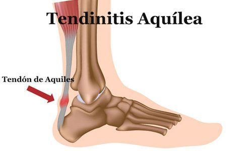
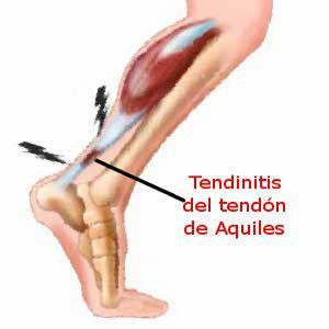
Achilles Tendinitis
- Inflammation of the Achilles tendon and even predispose to later rupture
- this condition is twofold
- The injury often seen only when it has become chronic
- The rehabilitation is a time-consuming and frustrating process
- The pathoanatomy includes of chronically inflamed partial rupture
- Stenosis and calcification within the tendon
- this condition is twofold
- History
- Mechanism of Injury
- Result of too much of an unaccustomed activity
- Hill running and changing of heel height
- Result of direct trauma such a pressure from the top of the heel counter of a shoe
- High-arched foot, a flat or pronated foot or a tight achilles tendon
- Result of too much of an unaccustomed activity
- Symptoms
- Pain and stiffness are the usual initial complaints
- The athlete notices the presence of crepitation often of the squeaking or snowball type
- Mechanism of Injury
- Physical Examination
- Palpation or squeezing the tendon produces tenderness
- passively dorsiflexing the ankle with the knee extended often produces pain
- A diffusely, grossly enlarged, dense, tender tendon usually indicated the presence of peritendinous fibrosis and scarring
- Radiographic Examination
- In chonic cases a lateral view of the leg
- May reveral the presence of calcification or ossification within the tendon
- In chonic cases a lateral view of the leg
- Other Diagnostic Tests
- A radionuclide bone scan reveals whether the process is still active
- MRI, and to a lesser degree sonography
- A radionuclide bone scan reveals whether the process is still active
- Treatment
- Initial
- Total cessation of weight load
- heel elevation, non-steroidal anti-inflammatory drugs and repeated application of ice
- sleep at right angles
- high intensity galvanic stimulation, ultrasonography, cryotherapy and even cyriax massage
- sleep at right angles
- heel elevation, non-steroidal anti-inflammatory drugs and repeated application of ice
- Total cessation of weight load
- Definitive
- Stretching exercises should be done repeatedly throughout the day and strengthening
- painless range of motion
- Stretching exercises should be done repeatedly throughout the day and strengthening
- Initial
- Inflammation of the Achilles tendon and even predispose to later rupture
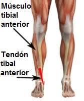
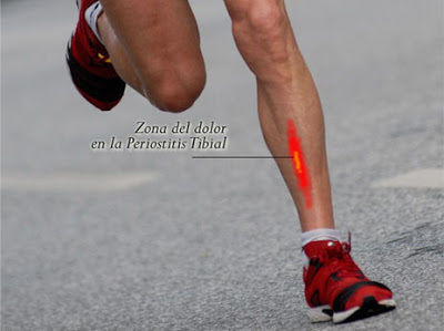
Chronic Anterior Leg Pain (Shin Splints)
- pain between knee and ankle
- not involving the calf/ achilles tendon
- History
- Mechanism of Injury
- Result of unaccustomed activity or overuse
- Biomechanical abnormalities, such as hyperpronation of the foot
- Result of unaccustomed activity or overuse
- Symptoms
- The primary symptom for all these conditions is pain
- Pain in the distal lateral aspect of the leg often indicates a fibular stress fracture
- Pain in the entire anterior lateral area of the leg may indicate presence of a chronic compartment syndrome
- Pain over the anterior surface of the tibia in the midleg indicated a tibial stress fracture
- Pain in the entire anterior lateral area of the leg may indicate presence of a chronic compartment syndrome
- Diffuse swelling, often described as a tense sensation
- Paresthesia or hypesthesia
- Pain in the distal lateral aspect of the leg often indicates a fibular stress fracture
- The primary symptom for all these conditions is pain
- Mechanism of Injury
- Physical Examination
- tenderness over either the tibia or the fibula
- Muscle testing is helpful in some conditions
- Careful palpation of the anterior compartment may reveal incresed tightness
- The presence of a high-arched foot, or signficant pronation
- Careful palpation of the anterior compartment may reveal incresed tightness
- Muscle testing is helpful in some conditions
- tenderness over either the tibia or the fibula
- Radiographic Examination
- Anteroposterior and lateral radiographs of the lef often reveal stress fractures
- Other diagnostics tests
- Radionuclide bone scanning
- Treatment
- Initial
- interruption of activities that produce pain
- wandering without crutches
- use modalities such as icing, ultrasonography and high intensity galvanic
- wandering without crutches
- interruption of activities that produce pain
- Definitive
- Stretching and strengthening exercises
- Arch Supports
- Band or counterforce bracing
- Arch Supports
- Stretching and strengthening exercises
- Initial
- pain between knee and ankle
Images and files in the map
Information
- Description
- Mind Map on Leg Injuries, created by Laura Grajales on 04/03/2018.
- Created by
- Laura Grajales
- Views
- 45
- Rating