
Mind map
Alimentary System (Nahiid)
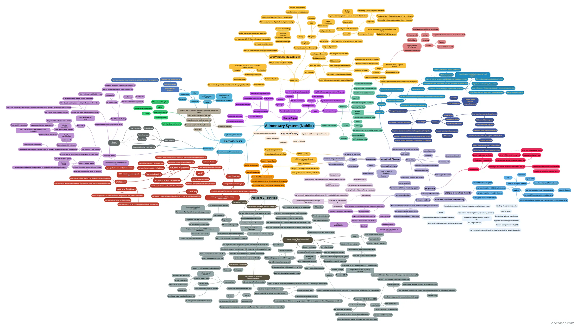
Mind map: Alimentary system
Map content
Alimentary System (Nahiid)
Routes of Entry
- Ingestion
- Direct Extension
- Expectorated from lungs and swallowed
- Systemic blood borne infections
- Parasitic migration
Defense Mechanisms
- Physical
- Normal motility
- Vomiting/Diarrhea
- Saliva/mucus/gastric juice/bile
- Gastric pH
- Intestinal Proteolytic enzymes
- High epithelial turnover/barrier
- Gut flora/fauna (biofilm)
- Innate
- Mast Cells, mØs, neutrophils, paneth cells
- Autophagy
- PRRs
- Complements, defensins, TLRs
- Adaptive
- Immunoglobulins
- DC, T, B, Plasma cells
- Physical
Clinical Signs
- Non-specific (region)
- Malaise/Lethargy/weakness
- Inappetance/anorexia/pain
- Dehydration
- Hemorrhage/melena
- Lower tract disease
- Abdominal pain/colic
- Polyphagia/hyperphagia
- Upper (includes forestomachs)
- Dysphagia
- Ptyalism
- Halitosis
- Bloat in ruminants
- And D+ secondary to lactic acidosis
- Upper Tract
- Halitosis + Ptyalism
- Inflammation
- Stomatitis/Gingivitis/Cheilitis/Glossitis/Pharyngitis/Tonsillitis
- Morphological changes
- Cattarrhal (mucous), fibrinonecrotic, granulomatous
- Vesicular
- Proliferative
- Erosive/ulcerative
- Inflammation
- Viral Vesicular Stomatitides
- Viruses, form vesicles, erode, granulate and heal
- All 4 lesions look the same
- can rupture and look like erosion/ulcer stomatitides
- BVDV, bluetongue, malignant catarrhal
- can rupture and look like erosion/ulcer stomatitides
- FMD, V. Exanthema, Swine VD, VS
- Endothelial damage
- Thrombosis, vasculitis
- ischemic necrosis
- oedema/hemorrhage
- ischemic necrosis
- Thrombosis, vasculitis
- Proliferative Lesions (Oral cavity)
- Neoplasias
- Benign
- fibromatous epilus of periodontal ligament origin
- common (can be multicentric), contain bone
- Acanthomatous ameloblastoma
- invasive, no metastasis
- fibromatous epilus of periodontal ligament origin
- Malignant
- SCC
- v. invasive
- Fibrosarcoma
- Malignant melanoma
- rapid metastasis
- SCC
- Benign
- Papillomas
- Spontaneous or viral (young dogs and cattle)
- Gingival Hyperplasia
- Viral Papular Stomatitis
- Scabby mouth/orf
- Bovine papular stomatitis
- Neoplasias
- Teeth and gums
- very common
- Malocclusions
- Teeth development anomalies
- Enamel/dentin defects (CDV/BVDV)
- Adontia/oligodontia/polyodontia
- Dental attrition and abnormal wear
- Feline Odontoclastic resorptive lesions (idipathic)
- Periodontal Disease
- Plaque
- bacterial mass + organic matrix
- Caries/infundibular impaction
- gingivitis/periodontitis/pulpitis/alveolar osteomyelitis
- Calculus
- mineralised plaque
- Plaque
- Rumenitis
- Basically means lactic acidosis
- Inflammation
- Degeneration/coagulative necrosis of ruminal epithelium
- Stellate scars
- Degeneration/coagulative necrosis of ruminal epithelium
- Inflammation
- Can be secondary to mechanical/chemical injury
- Primary viral diseases
- BVDV/MCF/FMD/bluetongue
- Sequelae
- Secondary bacterial/mycotic infection
- Fusobacterium -> Haematogenous to liver -> Abscess
- Aspergillus -> Haematogenous to liver -> Hepatitis
- Secondary bacterial/mycotic infection
- Basically means lactic acidosis
- Halitosis + Ptyalism
- Lower
- Vomiting/nausea
- Diarrhea
- Haematochezia
- Dyschezia/constipation/tenesmus
- Gas bloat/borborygmi/eructation/flatulence
- Faecal incontinence
- Lower Tract
- Monogastrics/abomasum
- Critical functions
- motility
- pyloric outlet patency
- mucosal integrity
- Clinical Signs
- Anemia/Haematemesis/melena/inappetance/weight loss
- Melena = significant blood loss in short period of time
- Stays in stomach/small int. long enough for digestion
- Anemia with hypoalbuminemia
- GIT blood loss
- Melena = significant blood loss in short period of time
- Vomiting
- Hypovolemia
- metabolic acidosis
- HypoK/HypoChloremia
- abdominal pain (colic/bruxism)
- Decreased appetite and weight loss
- Abdominal Ostertagiosis
- L3,4,5 in abomasal glands
- Glandular hyperplasia/metaplasia
- Increased luminal pH, less pepsin, fermentation
- osmotic diarrhea
- Adults damage mucosa -> inflammation
- Weight loss, diarrhea, hypoproteinemia
- Adults damage mucosa -> inflammation
- osmotic diarrhea
- Increased luminal pH, less pepsin, fermentation
- Glandular hyperplasia/metaplasia
- L3,4,5 in abomasal glands
- Abdominal Ostertagiosis
- Anemia/Haematemesis/melena/inappetance/weight loss
- Critical functions
- DAMNITV
- Inflammation
- Gastritis/Enteritis/Colitis/Proctitis
- Morphological Changes
- Catarrhal/fibrinonecrotic/granulomatous/necrotic/haemorrhagic
- Ulcerative/erosive
- Poorly understood: imbalance b/w acid secretion and protection
- mucosal barrier breached, acid/pepsin damage deeper tissue
- inflammation/pain
- SEVERE: perforation (septic peritonitis)
- Secondary to neoplasia (lymphoma) in all species
- inflammation/pain
- mucosal barrier breached, acid/pepsin damage deeper tissue
- Common in foals and pigs (dogs often subclinical)
- Pigs: Fatal hemorrhage, deep ulcers
- Mast Cell tumors (histamine release) -> haematemesis/melena
- Foals: cannot vomit, idiopathic, ill thrift and pain
- Adults: often subclinical
- Calves: dietary changes/stress/irritation
- Poorly understood: imbalance b/w acid secretion and protection
- Proliferative (Hyperplasia/Neoplasia)
- Inflammation
- Intestinal Disease
- Clinical Signs
- Varied, wide
- Acute/explosive to chronic/intractable
- Altered appetite
- Vomiting
- Diarrhea
- Pathological Mechanisms
- Malassimilation
- Results in weight loss: despite big appetite
- Also pyrexia/pain
- Maldigestion
- Incomplete breakdown of large molecules
- Not absorbed, accumulate in lumen
- Osmotic diarrhea
- Rapid fermentation
- More osmotic pressure, decreased pH worsens absorption
- Worse diarrhea
- More osmotic pressure, decreased pH worsens absorption
- Not absorbed, accumulate in lumen
- e.g. poor milk replacer, lactose intolerance, EPI, impaired bile salt mechanism
- Can lead to low Vitamin K
- Coagulopathy
- Produced by fermentation and gut microbes
- Incomplete breakdown of large molecules
- Malabsorption
- Structural abnormalities in mucosa impairing absorption
- ALWAYS due to intestinal disease
- villous atrophy
- Canine Parvovirus
- Targets crypt epithelium -> necrosis
- Canine Parvovirus
- increased intraluminal osmotic load
- Diarrhea
- enhanced fermentation
- villous atrophy
- Usually accompanies maldigestion
- Results in weight loss: despite big appetite
- Hypersecretion
- More ions: bacterial toxins/inflamm. mediators/endogenous laxatives
- e.g enterotoxigenic collibacillosis
- non invasive, blocks NaCl absorption
- e.g enterotoxigenic collibacillosis
- More ions: bacterial toxins/inflamm. mediators/endogenous laxatives
- Increased intestinal permeability
- Acute inflamm (bacteria), chronic, neoplasia, lymphatic obstruction
- Acute
- Enteroinvasive enteritis (salmonella/bacteria)
- Swine dysentery, Clostridium perfringens, coccidia
- Mechanisms increasing tissue pressure (e.g. chronic)
- Johne's Disease/Eosinophilic enteritis etc.
- IBD, fungal enteritis
- Starlings (Oedema) mechanics
- Fluid to lumen
- Severe loss = plasma protein loss
- Hypoalbuminemia/hypoproteinemia
- Protein-losing enteropathy (PLE)
- e.g. Intestinal lymphangiectasia in dogs (congenital), or lymph obstruction
- Protein-losing enteropathy (PLE)
- Hypoalbuminemia/hypoproteinemia
- Severe loss = plasma protein loss
- Fluid to lumen
- Acute
- Acute inflamm (bacteria), chronic, neoplasia, lymphatic obstruction
- Changes in intestinal motility
- Rarely primary cause of diarrhea
- Increased motility = possibly diarrhea
- Decreased motility = SIBO (Small intestine)
- Causes of change in gut flora
- hypomotility
- neonates before gut flora establish
- antibiotics
- EPI
- Leads to
- Bile Salt depletion -> Fat malassimilation
- osmotic diarrhea
- Decreased cobalamin (binding and inactivation of vitamin to bacteria)
- Increased folate due to increased bacteria
- Bile Salt depletion -> Fat malassimilation
- Causes of change in gut flora
- Can lead to intussusception
- Malassimilation
- Consequences
- Dehydration, hypovolemia
- Shock/inadequate perfusion
- Hypoglycemia, ketoacidosis, decreased pH
- loss of pH dependent enzymes
- Acidosis, fecal bicarb loss
- From loss of kidney perfusion
- Electrolyte imbalance, intracellular more H+, less K+
- lose control of myocardial contraction
- Vicious cycle --> Hypovolemic shock
- lose control of myocardial contraction
- Dehydration, hypovolemia
- Pathological Mechanisms
- Dehydration/lethargy/depression
- Weight loss/ill thrift
- Tenesmus (Cramping rectal pain)
- Abdominal distension
- Abdominal pain
- Borborygmi/flatulence
- Varied, wide
- Small vs Large
- Small
- Mild increase in frequency
- Increased fecal volume
- Melena
- No fecal mucus
- Large
- Alot more frequent defecation
- Decreased fecal volume
- Fecal mucus present
- Hematochezia
- Small
- NSAIDS and Ulcers
- Inhibits prostaglandin (pg) synthesis
- alters circulation
- Delays GIT healing (turnover and mucus)
- more gastrin, increased acid production
- Delays GIT healing (turnover and mucus)
- alters circulation
- Horses: Gastroduodenal/colitis
- Dogs: Colonic perforation
- Inhibits prostaglandin (pg) synthesis
- Proliferations
- Usually dogs and cats
- Benign or Malignant
- Hyperplastic polyps
- Epithelial: Adenoma/carcinoma
- Stromal: gastrointestinal stromal tumor, leiomyoma
- Round Cell tumor: Lymphoma, mast cell tumor
- Clinical Signs
- Omentum/Peritoneum
- Inflammation (Peritonitis
- Clinical Signs
- Abdominal Pain
- Ascites
- Usually due to multiple organ disease
- Aseptic abdominocentesis to characterise fluid
- Peritonitis
- Causes:
- Rupture
- Systemic Infection (FIP)
- Clinical Signs
- Inflammation (Peritonitis
- Monogastrics/abomasum
- Non-specific (region)
Gastric Ulcerations
- Erosion: loss of epithelium, BM intact
- heals fast
- Ulcer: loss of epithelium and BM
- Second intention healing + scarring
- Leads to perforations/strictures/stenosis in tubular GIT organs
- Erosion: loss of epithelium, BM intact
Diagnostic Tests
- Minimum Database
- Haematology/Biochem/Urinalysis
- Albumin + Globulin
- Panhypoproteinemia usually with PLE
- Compare with hepatic insufficiency/PLN (hypoalbuminemia alone typical)
- Insensitive indicators of protein loss
- Increased protein usually dehydration
- Hyperglobulinemia can be inflammation
- Panhypoproteinemia usually with PLE
- Serum Bile Acids
- SIBO increases unconjugated SBA
- Na/K
- Electrolyte abnormalities
- Vomiting, obstructions, secretory diarrhea
- Electrolyte abnormalities
- Liver Enzymes
- Leakage and Induced enzymes
- Primary GI disease with secondary hepatopathy
- Link with SBA to differentiate primary GI with primary hepatopathy
- Leakage and Induced enzymes
- Cholesterol
- Increased cholesterol with PLN (ddx for hypoalbuminemia)
- Decrease seen with diseases causing fat malabsorption (ddx hepatic insufficiency)
- Urea and Creatinine
- V+/D+/inappetance all associated with dehydration and pre renal azotemia
- increased urea/creat, hypersthenuria
- Increased urea can be gastric/upper intestine hemorrhage
- V+/D+/inappetance all associated with dehydration and pre renal azotemia
- Ca and Mg
- Decreased Ca/Mg seen with PLE (reduced Ca can be hypoalbuminemia)
- Cats with Chronic GIT Disease
- Serum T4 hormone, FeLV, FIV status are useful
- Albumin + Globulin
- Haematology/Biochem/Urinalysis
- Diagnostic Imaging
- US/XR
- Obstructions/Neoplasia
- Endoscopy/Biopsy
- Gold Standard, but invasive
- Scintigraphy/Fluoroscopy
- CT/MRI
- US/XR
- Serum markers of function/inflamm
- Fecal samples
- Infectious agents
- Faecal Culture and Sample
- Suspect a specific pathogen
- Isolate and ID
- Test Ab susceptibility
- Isolate and ID
- Use when Acute GI signs: haemorrhagic D+, pyrexia, inflamm leukogram, neutrophils
- Screening test for chronic
- Fresh feces, do not refrigerate/store with formalin fixed
- Suspect a specific pathogen
- Adjunct Isolate Testing
- Specific enteropathogens with GI disease
- Most are commensals, must be isolated
- Test for virulence genes
- Test for toxins
- Determines isolate's clinical importance, or typed for epidemiologic surveys
- Specific enteropathogens with GI disease
- Faecal PCR
- Detects DNA/RNA, not cells
- DNA extraction is hard, can have false negatives
- Looking for set pathogens
- Higher sensitivity
- DNA contamination is a problem
- False positives possible
- DNA contamination is a problem
- Only use a lab with expertise in this
- Detects DNA/RNA, not cells
- Canine Faecal Parvoviral Antigen (ELISA)
- ELISA: Fresh Feces > Swab
- For Young, unvaccinated, exposed
- Use if: D+, anorexia, haematemesis, melena/haematochezia, pyrexia, leukopenia, neutropenia
- False Negatives: Virus shed briefly in feces, small window
- If tested too early. Retest later if suspected
- False Positives: modified live vacc
- Fecal Parasite Testing
- Dependent on quantity/quality of feces
- Fecal Egg Count
- Quantitative
- Not for trematode eggs or some tapeworms
- Use with worm egg count guides (livestock)
- Fecal concentration (cysts/ova)
- Qualitative
- Incl. Giardia
- Immunofluorescence Antibody Test
- Faecal Culture and Sample
- Small Animals
- Wet microscopy
- Gram Stain
- Occult blood
- Concentrations for cysts and ova
- Large Animals
- Fecal Egg Count
- Infectious agents
- Assessing GIT Function
- Permeability Testing
- Dz causes epithelial damage
- Increased intestinal permeability
- Movement of antigens/pathogens through mucosa
- Use of Markers
- Exogenous non-metabolisable probes
- No degradation/metabolism
- If appear in serum/urine, THEN increased permeability
- Not useful to localise sites
- CANNOT tell decreased absorption
- If appear in serum/urine, THEN increased permeability
- No degradation/metabolism
- Sugar Probes
- Metabolised/degraded
- Mixtures of sugar probes can localise
- More in serum/urine = permeability increased
- Less = decreased absorptive capacity
- Mixtures of sugar probes can localise
- Metabolised/degraded
- Exogenous non-metabolisable probes
- Increased intestinal permeability
- Dz causes epithelial damage
- Evaluation of Intestinal Protein Loss (PLE)
- Serum albumin and total plasma protein
- Panhypoproteinemia usually
- sometimes only hypoalbuminemia
- Compare with A:G ratio/conc.
- sometimes only hypoalbuminemia
- Rule out: blood loss, PLN, hepatic failure, exudative dermatopathy
- Panhypoproteinemia usually
- Faecal a-1 proteinase inhibitor in fecal samples
- Validated for K9/F9 only in USA/Europe
- Lost with albumin in PLE, so increased fecal concentration = PLE
- Potential early indicator of PLE in dogs
- Lost with albumin in PLE, so increased fecal concentration = PLE
- Validated for K9/F9 only in USA/Europe
- 51Cr albumin recovery in fecal samples
- Gold standard
- IV radioactive substance
- hard to do (radioactive)
- Serum albumin and total plasma protein
- Biomarkers of Gastric/Intestinal Inflamm.
- Serum Gastrin
- For dogs/cats with unexplained gastric erosion/ulcers/hyperplasia
- Chemiluminescence immunoassay - LH serum serum immediately, freeze
- Persistent increase with CS = suggests gastrinoma
- Secretin stimulation test to confirm Dx
- Proton pump inhibitors can interfere
- Check about patient meds first
- Persistent increase with CS = suggests gastrinoma
- Faecal Occult Blood
- If GI bleeding suspected but NOT apparent
- e.g. NO melena/haematochezia
- Poor sensitivity/specificity
- Take 3 consecutive motions
- Detects pseudoperoxidase activity (Hb/Myb)
- But ALSO dietary haemoglobin (meat)
- If GI bleeding suspected but NOT apparent
- Plasma Pepsinogen
- Zymogen of gastric protease pepsin
- From chief cells
- Use in Ruminants
- Indicates abomasum damage
- Increases with ostertagiasis (esp. type II)
- Can also increase with azotemia
- Increases with ostertagiasis (esp. type II)
- Indicates abomasum damage
- Zymogen of gastric protease pepsin
- Ruminal Fluid pH
- Acidosis
- Normal = 6.5 - 7
- Lactate in Horses with colic
- Simultaneously measured blood lactate
- Indicates strangulation/ischemia
- Serial blood lactate measurements > 1 measurement
- prognostic indicator of postop recovery
- Simultaneously measured blood lactate
- Others
- Increased plasma L-lactate
- Plasma citrulline
- Inflamm. markers (CRP etc.)
- Serum Gastrin
- Evaluation of GIT motility
- Radiography
- Ultrasound
- Assessment of Intestinal function/pathology
- Lab detection of malassimilation
- Fecal Analyses
- microscopy of smears
- Cytology --> Infectious agents/neutrophils
- Fecal fat, starch, fibre
- Unassimilated materials
- Strictly Qualitative
- Cheap, but unreliable/rare
- Total fecal fat
- Steatorrhea
- Fecal proteolytic (trypsin) activity
- ID fecal proteases (mostly pancreatic)
- Unreliable, superseded by TLI to r/o EPI
- microscopy of smears
- Trypsin-like immunoreactivity (TLI)
- Canine
- Sensitive/specific to K9 EPI
- Feline
- Species specific
- Decreased renal excretion can also increase TLI, but those are mild even in severe renal failure
- Canine
- Intestinal absorption/tolerance tests
- Detect increased intestinal permeability relative to reduced SA/enterocyte dysfunction
- Feed and sample serum for absorbed substance
- Lipid assimilation tests
- Inaccuracies can be delayed gastric emptying, or poor micelle formation from low bile salts
- Carbohydrate assimilation tests
- Inaccuracies due to delayed emptying, reduced SI blood flow, abnormal motility, decreased GFR etc.
- Lipid assimilation tests
- Fecal Analyses
- Lab detection of malassimilation
- Assessment of SI dysbiosis (SIBO)
- Serum Cobalamin (B12)
- EPI decreases availability
- SIBO, bacteria binds to B12, prevents absorption
- Absorbed in ileum, severe SI disease decreases absorption
- Serum Folate
- Carriers necessary for absorption
- Only in proximal SI
- Increase with SIBO and EPI
- Artefact: Increases with haemolysis: red cell folate
- Breath Hydrogen
- Detects Carbohydrate malabsorption, or SIBO
- Bacteria in GI metabolises carbs to Hydrogen (not mammalian cells)
- Increased H with increased LI fermentation/SIBO
- BUT: variation in measured values, no standardised protocol, not readily available
- Limited utility
- Detects Carbohydrate malabsorption, or SIBO
- Serum Cobalamin (B12)
- Permeability Testing
- Cytology
- Neoplasia vs. Inflamm.
- ID aetiological agents
- Techniques
- FNA
- Impressions
- Scrapings
- Avoid ulcerated areas
- Exploratory Laporotomy
- Biopsy/Adjunct Methods
- Endoscopic biopsy
- Take multiple, quality matters
- Place on mount in mesh cassette in formalin jar, NOT on gauze/free
- Endoscopic biopsy
- Minimum Database
Information
- Description
- VET377 Pathology Mind Map on Alimentary System (Nahiid), created by Bertrand Ng on 22/10/2017.
- Created by
- Bertrand Ng
- Views
- 3
- Rating