
Mind map
Glaucoma
Map content
Glaucoma
TREATMENT (Ney, 2016; Turkoski, 2012
- Pharmacological (Ney, 2016; Turkoski, 2012)
- Medications that decrease outflow of aqueous humor
- Prostaglandin analogs
- Beta-adrenergic antagonists
- Alpha2-adrenergic agonists
- Carbonic anhydrase inhibitors
- Miotics
- GOAL
- Reduce adverse effects
- Avoid vision loss
- Medications that increase outflow of aqueous humor
- Alpha Antagonist
- Dipivefrin
- Brimonidine
- Cholinomimetics
- Combination Products
- Combigan
- Cosopt
- Combination Products
- Alpha Antagonist
- Medications that decrease outflow of aqueous humor
- Non-pharmacological
- Surgical Treatment (To reduce IOP) (Ney, 2016)
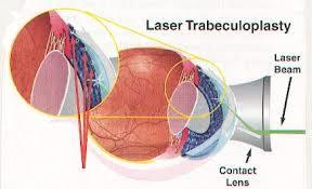
- Laser Trabeculoplasty
- Reduces IOP by creating better outflow of aqueous humor through the remodeling of the trabecular meshwork
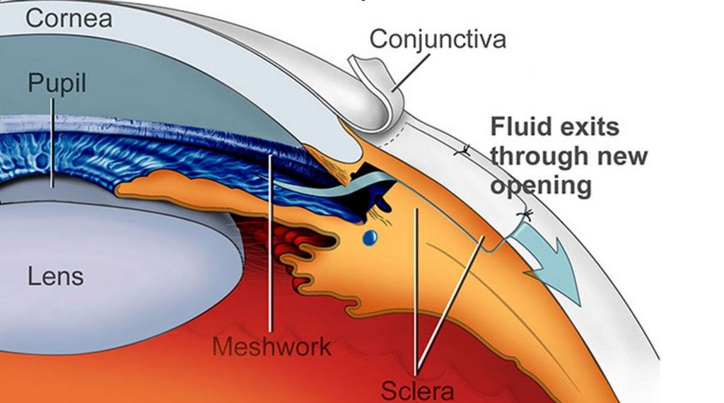
- Trabeculectomy
- Surgical fistula is created to drain aqeous humor into subconjunctival pocket
- Laser Trabeculoplasty
- Surgical Treatment (To reduce IOP) (Ney, 2016)
- Pharmacological (Ney, 2016; Turkoski, 2012)
PATHOGENSIS (Peate & Jones, 2014; Nowak et al., 2015)
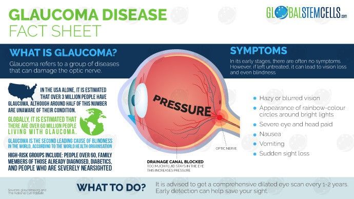
- What is Glaucoma?
- Optic Neuropathy that results in Visual Impairment
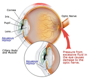
- Build up of fluid (aqueous humor)
- Ocular Hypertension
- Increased damage to blood vessels and optic nerve
- Results in axon loss, retinal ganglon cell death (RCG), excavated appearance of the optic nerve head
- Increased damage to blood vessels and optic nerve
- Ocular Hypertension
- What is Glaucoma?
DIAGNOSIS
- Medical Diagnosis (Peate & Jones, 2014; Ney, 2016; Turoski, 2012; Lewis et al., 2014)
- ophthalmologist examination
- Evidence of increased cup-to-disc ratio
- Use of Optical coherence tomography (OCT)
- Visual Field testing
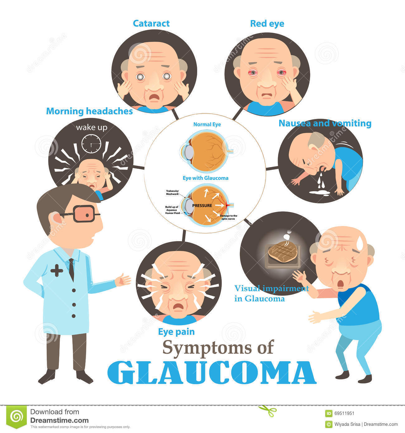
- Conjunctival injection (red eye)
- Corneal oedema
- Mid-dilated, non-reactive pupil.
- Glaucoma Test
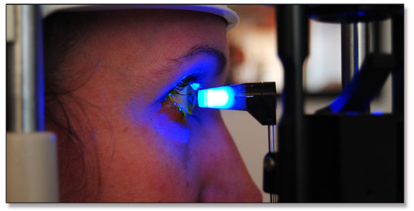
- 1. Tenometry
- 2. Perimetry
- 3. Direct Opthalmoscopy
- Measures the IOP and examines the head of optic nerve, aqueous humor, and cornea
- 4. Gonioscopy
- Uses a slit lamp or operating microscope to gain a view of the iridocorneal angle
- Measures and identifies defects in visual field
- 3. Direct Opthalmoscopy
- Measures IOP and determines how much pressure it needs to flatten central cornea. Often used with Direct Opthalmoscopy
- 2. Perimetry
- 1. Tenometry
- ophthalmologist examination
- Non Medical Diagnosis (Peate & Jones, 2014; Ney, 2016; Turoski, 2012; Lewis et al., 2014)
- Patient History
- History of Blurring vision with haloes
- Assessment of Ocular Pain
- Normal IOP: 12-22 mm Hg
- Ocular Hypertension: IOP greater than 22 mm Hg
- Normal IOP: 12-22 mm Hg
- Past eye surgery and eye injury
- Patient History
- Medical Diagnosis (Peate & Jones, 2014; Ney, 2016; Turoski, 2012; Lewis et al., 2014)
RISK FACTORS (Ney, 2016; Perry, 2014;Lewis et al., 2014)
- Family History
- Individuals of African Origin
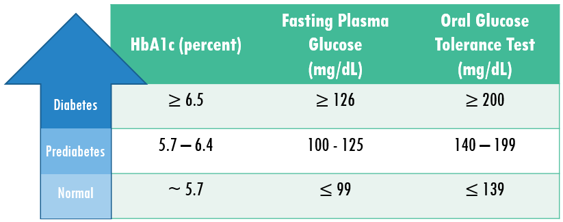
- Diabetes
- Ocular Hypertension (greater than 21 mmHg)
- Central Cornea Thickness
- Those over the age of 65 years old
- long-term use of steroid eye drops
TYPES OF GLAUCOMA (Peate & Jones, 2014; Turkoski, 2012)
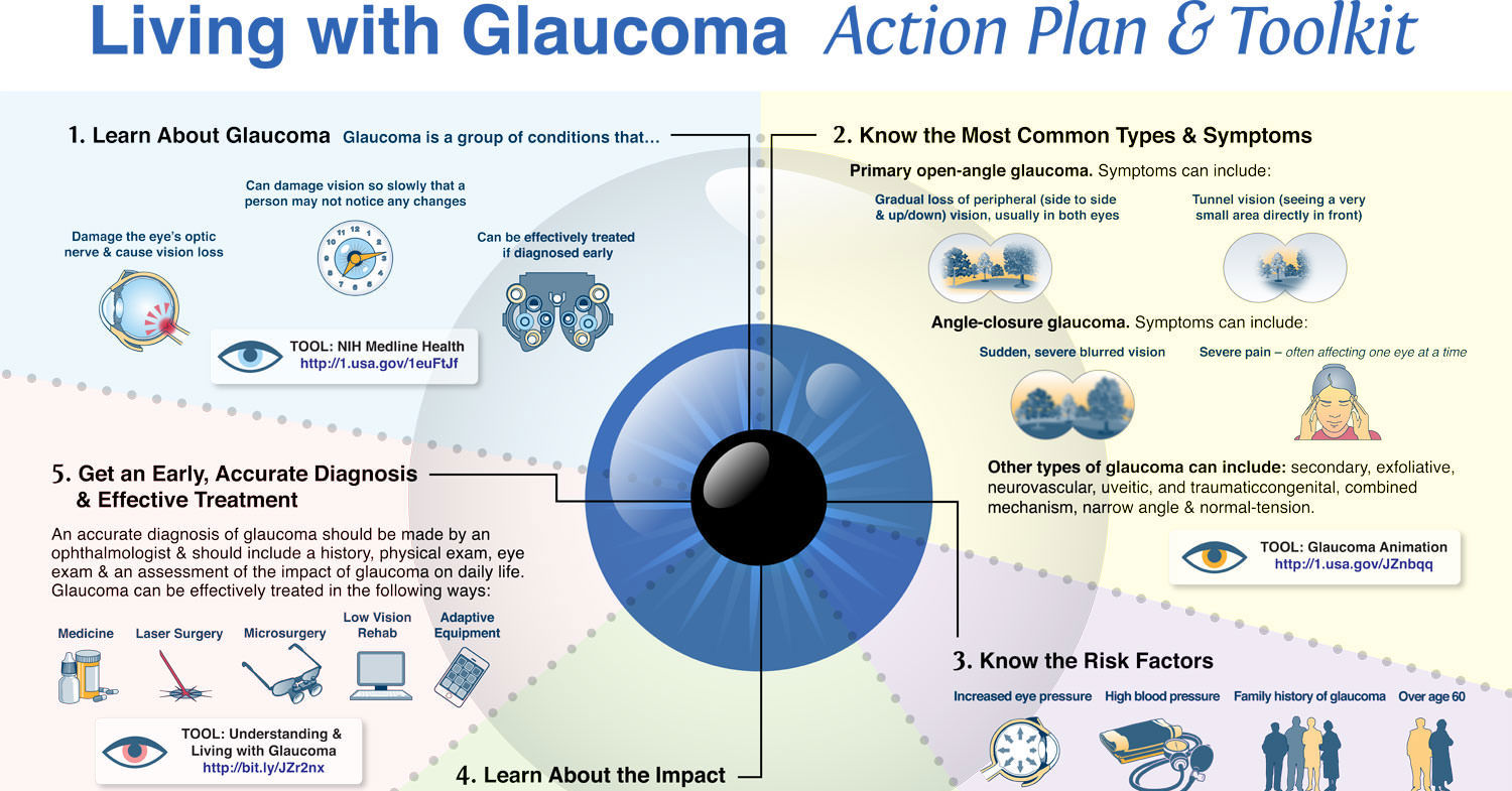
- Primary Open Angled Glaucoma
- Signs and Symptoms
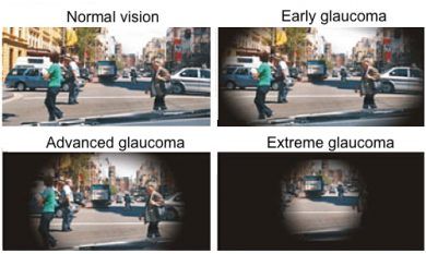
- No symptoms of pain occurs as result of the optic nerve damage
- Shadow in vision
- Decreased visual acuity
- Effects
- Extensive amount of vision loss occurs
- Elevated Intraocular Pressure
- People often lose their vision even before they are aware of their condition.
- Causes
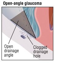
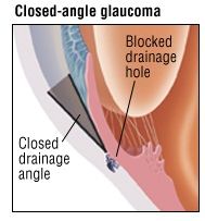
- Trabecular Meshwork (drainage angle) is partially blocked
- Slower fluid drainage out of the eye
- The angle between the cornea and iris is open (Turkoski, 2012).
- Insidious development
- It is the most common type of glaucoma
- Signs and Symptoms
- Primary Closed Angled Glaucoma
- Signs and Symptoms
- Pains
- Blurred Vision
- Colored Haloes
- Malaise
- Effects
- Elevated Intraocular Pressure
- Causes
- Iris Bulges
- Decreased fluid drainage
- Optic nerve damage
- The angle between the cornea and iris is very narrow
- Acute and gradual development
- It is a medical emergency
- Signs and Symptoms
- Primary Open Angled Glaucoma
Signs and Symptoms (NHS, 2016; Lewis et al., 2014)
- uveitis
- Cogenital Eye Problem
- vision loss
- blurred vision
- colored rings around lights
- redness in the eye
- Nausea
- Vomiting
- a "hazy" appearance to the eye
COLLABORATIVE CARE (CA0, 2014; Lewis et al., 2014)
- Age 65 and over must take eye exam annually
- Keep IOP low to prevent optic nerve damage
- Acute care for angle closed glaucoma
- Topical cholinergic agent
- Hyperosmotic agent
- Laser peripheral iridotomy
- Surgical iridotomy
- Client Centered Care (RNAO, 2002).
- Involve clients throughout the caring and service process.
- Maintain respect and value for clients through listening and practicing openness.
- The client’s goals are central to the coordination, continuity, and consistency of care
- Care plan must be aimed at achieving the clients’ desired goals and needs
- Collobrate with interprofessional team such as optometrist to create an action plan
NURSING INTERVENTION (Parikh & Parikh, 2011)
- Enhance adherence to medications
- Reduce the number of medication through combination therapy
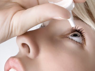
- Demsonstrate effective eye drop instillation
- Providing education on glaucoma
- Encourage physical activitiy
- Stress coping strategies
- Decrease alochol intake
- Decrease caffine intake
- Enhance adherence to medications
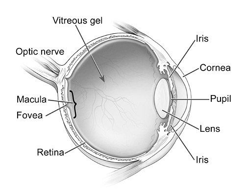
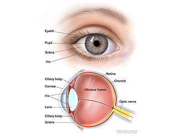
ANTAOMY OF THE EYE (Peate & Jones, 2014)
- Cornea, Iris and Pupil
- Aqueous humor, Trabecular Meshwork and lens
- Retina and Sclera
- Rods and Cones
By: Hamdi Mohamed & Asha Ahmed
Images and files in the map

Information
- Description
- Mind Map on Glaucoma, created by Hamdi Mohamed on 27/10/2017.
- Created by
- Hamdi Mohamed
- Views
- 124
- Rating