
Mind map
From Retina to Cortex
Map content
From Retina to Cortex
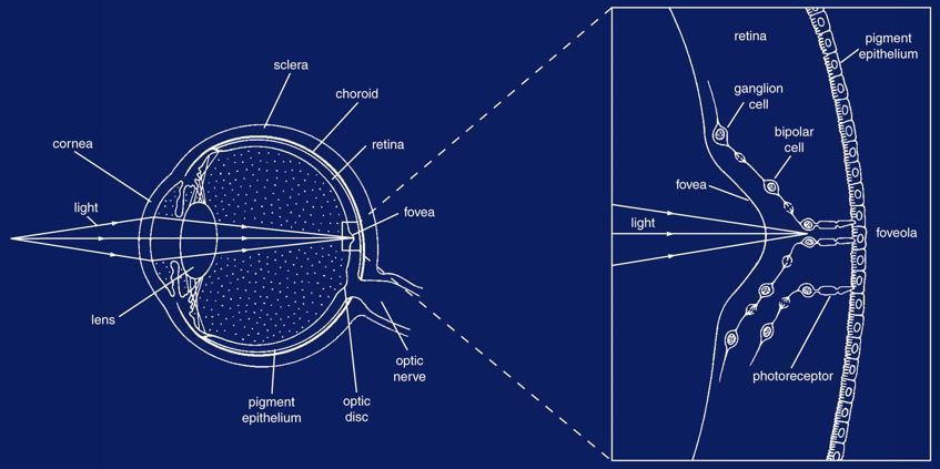
Gross anatomy of eye
- Stimuli
- Wavelength
- Hue (quality)
- Saturation (mix w/ b/w)
- Intensity
- (energy/time/area)
- Wavelength
- range 400 (v)-700 (r)
- Refraction
- cornea
- Focus
- lens
- fovea
- lose= functionally blind
- fovea
- lens
- Focus
- cornea
- Stimuli
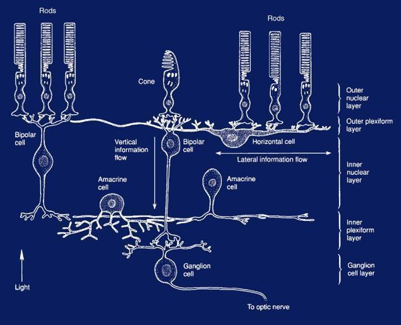
cellular organization
- Rod*
- Many rods 1 bipolar
- horizontal cell
- amacrine
- bipolar to ganglion
- amacrine
- low acuity
- horizontal cell
- more photopig
- can detect single photons
- dim
- saturate in daylight
- everywhere but foveola
- slower
- achromatic
- Many rods 1 bipolar
- Cone*
- 1:1 with bipolar
- no convergence
- sharp
- high acuity
- no convergence
- less photopig
- require hundreds photons
- Bright
- Saturate only intense
- [fovea]
- faster
- colors
- 1:1 with bipolar
- Rod*
Receptors
- Rhodopsin
- 7 TM
- Residue 296
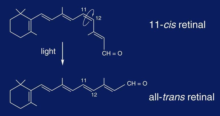
- attachment site for retinal
- Trans when activated
- no longer fits binding pocket
- Trans when activated
- attachment site for retinal
- floating disc
- Gi
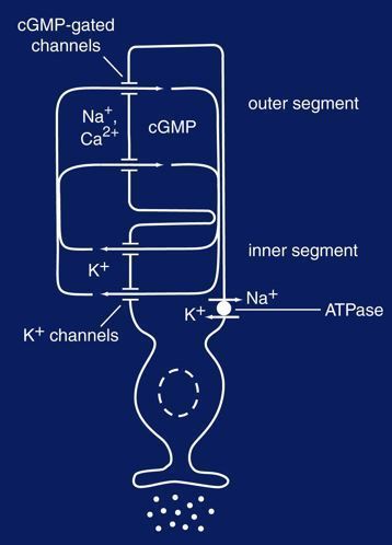
- inact cGMP phosphodiesterase
- releasing glutamate at resting*
- only shifts when illumination changes
- cGMP inact
- Close channel
- 0 pA
- reduced glutamate release
- 0 pA
- Close channel
- cGMP inact
- -40
- hyperpol
- -70*
- hyperpol
- cGMP act
- influx Na+
- -50 pA
- glutamate release
- -50 pA
- influx Na+
- only shifts when illumination changes
- response to light
- retinotopic map
- Rhodopsin
Receptive fields
- bipolar
- convergence
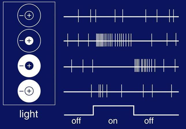
- antagonistic center-surround receptive field
- on=depol
- so TECHNICALLY every photoreceptor = "off" receptor
- even illumination +/- balance
- BUT depending on bipolar cell (mGluR), this signal may activate or inactive
- so TECHNICALLY every photoreceptor = "off" receptor
- sometimes color-specific
- red/green
- blue/yellow
- ganglion
- action potential!
- Magno+parvo
- baseline activation
- on
- increase
- off
- ZERO
- contrast needed*
- not all or nothing
- balances on/off signals, winner wins
- not all or nothing
- on
- more convergence!
- parvocellular
- magnocellular
- bipolar
cortical cells
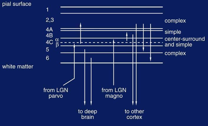
- LGN
- ventral 2
- Dorsal 4
- segregation by visual field
- centrifugal (descending) inputs
- ~50%=fovea
- V1*
- 4C-beta
- 4C-alpha
- neurons sensitive to lines, edges, movement
- neurons with binocular input
- caudal=center
- ~50%=fovea
- simple vs. complex
- Simple
- particular on/off strips
- complex
- specific ANGLE, not on/off region
- SOME movement sensitive
- hypercolumn
- bipolar! communication!
- Simple
- LGN
Images and files in the map
Information
- Description
- Mind Map on From Retina to Cortex, created by Emily Johnson on 05/02/2019.
- Created by
- Emily Johnson
- Views
- 0
- Rating