
Mind map
L5 - Optical approaches to studying circuits
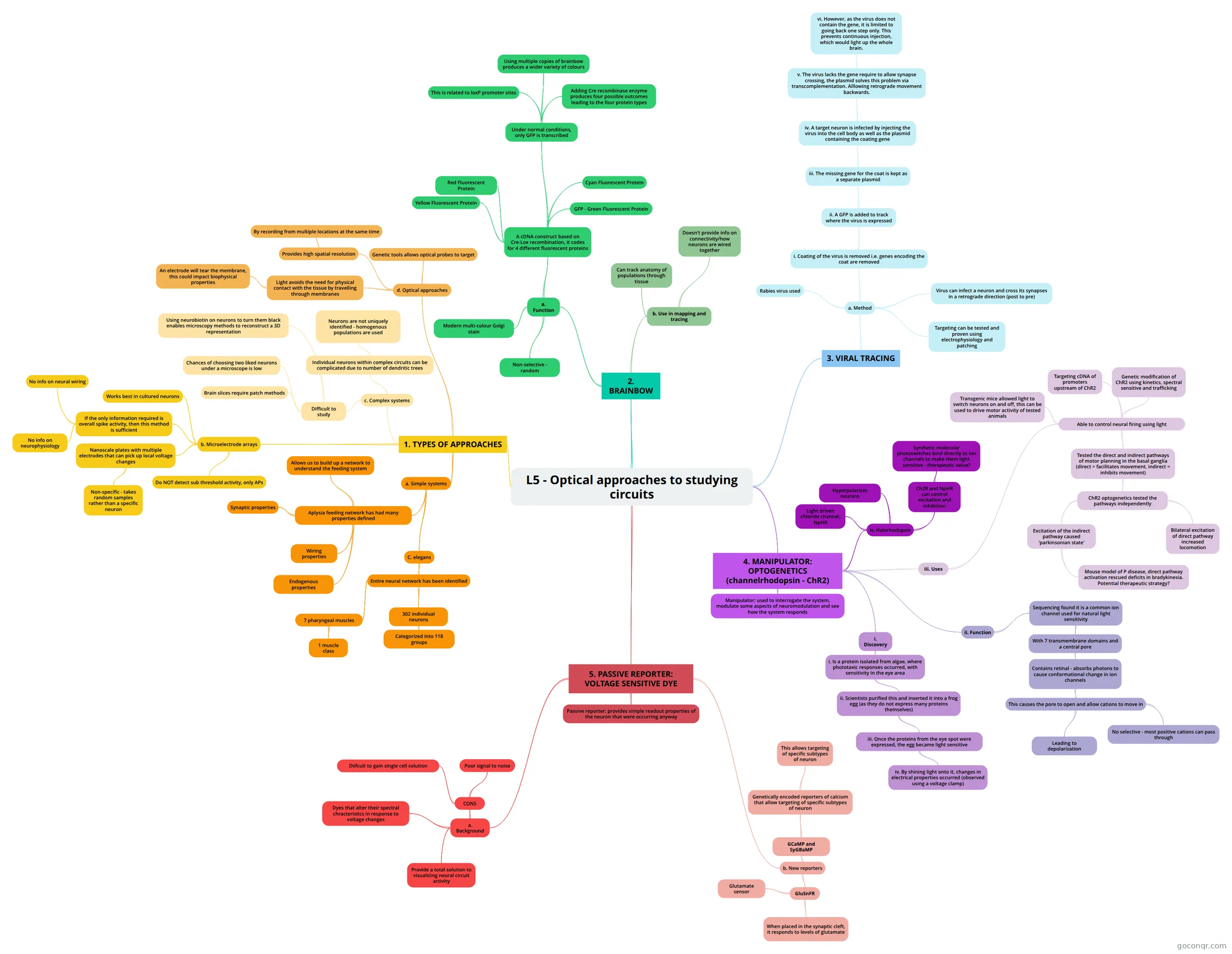
Mind map: optical approaches to studying circuits
Map content
L5 - Optical approaches to studying circuits
1. TYPES OF APPROACHES
- a. Simple systems
- Aplysia feeding network has had many properties defined
- Synaptic properties
- Wiring properties
- Endogenous properties
- Allows us to build up a network to understand the feeding system
- C. elegans
- Entire neural network has been identified
- 302 individual neurons
- Categorized into 118 groups
- 7 pharyngeal muscles
- 1 muscle class
- 302 individual neurons
- Entire neural network has been identified
- Aplysia feeding network has had many properties defined
- c. Complex systems
- Individual neurons within complex circuits can be complicated due to number of dendritic trees
- Neurons are not uniquely identified - homogenous populations are used
- Difficult to study
- Brain slices require patch methods
- Chances of choosing two liked neurons under a microscope is low
- Using neurobiotin on neurons to turn them black enables microscopy methods to reconstruct a 3D representation
- Individual neurons within complex circuits can be complicated due to number of dendritic trees
- b. Microelectrode arrays
- If the only information required is overall spike activity, then this method is sufficient
- No info on neural wiring
- No info on neurophysiology
- Nanoscale plates with multiple electrodes that can pick up local voltage changes
- Non-specific - takes random samples rather than a specific neuron
- Do NOT detect sub threshold activity, only APs
- Works best in cultured neurons
- If the only information required is overall spike activity, then this method is sufficient
- d. Optical approaches
- Light avoids the need for physical contact with the tissue by travelling through membranes
- An electrode will tear the membrane, this could impact biophysical properties
- Provides high spatial resolution
- By recording from multiple locations at the same time
- Genetic tools allows optical probes to target
- Light avoids the need for physical contact with the tissue by travelling through membranes
- a. Simple systems
2. BRAINBOW
- a. Function
- Modern multi-colour Golgi stain
- A cDNA construct based on Cre-Lox recombination, it codes for 4 different fluorescent proteins
- GFP - Green Fluorescent Protein
- Red Fluorescent Protein
- Yellow Fluorescent Protein
- Cyan Fluorescent Protein
- Under normal conditions, only GFP is transcribed
- This is related to loxP promoter sites
- Adding Cre recombinase enzyme produces four possible outcomes leading to the four protein types
- Using multiple copies of brainbow produces a wider variety of colours
- Non-selective - random
- b. Use in mapping and tracing
- Can track anatomy of populations through tissue
- Doesn't provide info on connectivity/how neurons are wired together
- a. Function
3. VIRAL TRACING
- a. Method
- Rabies virus used
- Virus can infect a neuron and cross its synapses in a retrograde direction (post to pre)
- i. Coating of the virus is removed i.e. genes encoding the coat are removed
- ii. A GFP is added to track where the virus is expressed
- iii. The missing gene for the coat is kept as a separate plasmid
- iv. A target neuron is infected by injecting the virus into the cell body as well as the plasmid containing the coating gene
- v. The virus lacks the gene require to allow synapse crossing, the plasmid solves this problem via transcomplementation. Alllowing retrograde movement backwards.
- vi. However, as the virus does not contain the gene, it is limited to going back one step only. This prevents continuous injection, which would light up the whole brain.
- v. The virus lacks the gene require to allow synapse crossing, the plasmid solves this problem via transcomplementation. Alllowing retrograde movement backwards.
- iv. A target neuron is infected by injecting the virus into the cell body as well as the plasmid containing the coating gene
- iii. The missing gene for the coat is kept as a separate plasmid
- ii. A GFP is added to track where the virus is expressed
- Targeting can be tested and proven using electrophysiology and patching
- a. Method
4. MANIPULATOR: OPTOGENETICS (channelrhodopsin - ChR2)
- Manipulator: used to interrogate the system, modulate some aspects of neuromodulation and see how the system responds
- i. Discovery
- i. Is a protein isolated from algae, where phototaxic responses occurred, with sensitivity in the eye area
- ii. Scientists purified this and inserted it into a frog egg (as they do not express many proteins themselves)
- iii. Once the proteins from the eye spot were expressed, the egg became light sensitive
- iv. By shining light onto it, changes in electrical properties occurred (observed using a voltage clamp)
- iii. Once the proteins from the eye spot were expressed, the egg became light sensitive
- ii. Scientists purified this and inserted it into a frog egg (as they do not express many proteins themselves)
- i. Is a protein isolated from algae, where phototaxic responses occurred, with sensitivity in the eye area
- ii. Function
- Sequencing found it is a common ion channel used for natural light sensitivity
- With 7 transmembrane domains and a central pore
- Contains retinal - absorbs photons to cause conformational change in ion channels
- This causes the pore to open and allow cations to move in
- No selective - most positive cations can pass through
- Leading to depolarization
- This causes the pore to open and allow cations to move in
- Contains retinal - absorbs photons to cause conformational change in ion channels
- With 7 transmembrane domains and a central pore
- Sequencing found it is a common ion channel used for natural light sensitivity
- iii. Uses
- Able to control neural firing using light
- Genetic modification of ChR2 using kinetics, spectral sensitive and trafficking
- Targeting cDNA of promoters upstream of ChR2
- Transgenic mice allowed light to switch neurons on and off, this can be used to drive motor activity of tested animals
- Tested the direct and indirect pathways of motor planning in the basal ganglia (direct = facilitates movement, indirect = inhibits movement)
- ChR2 optogenetics tested the pathways independently
- Excitation of the indirect pathway caused 'parkinsonian state'
- Mouse model of P disease, direct pathway activation rescued deficits in bradykinesia. Potential therapeutic strategy?
- Bilateral excitation of direct pathway increased locomotion
- Excitation of the indirect pathway caused 'parkinsonian state'
- ChR2 optogenetics tested the pathways independently
- Able to control neural firing using light
- iv. Halorhodopsin
- Light driven chloride channel, NpHR
- Ch2R and NpHR can control excitation and inhibition
- Synthetic molecular photoswitches bind directly to ion channels to make them light sensitive - therapeutic value?
- Hyperpolarizes neurons
5. PASSIVE REPORTER: VOLTAGE SENSITIVE DYE
- b. New reporters
- GluSnFR
- Glutamate sensor
- When placed in the synaptic cleft, it responds to levels of glutamate
- GCaMP and SyGBaMP
- Genetically encoded reporters of calcium that allow targeting of specific subtypes of neuron
- This allows targeting of specific subtypes of neuron
- Genetically encoded reporters of calcium that allow targeting of specific subtypes of neuron
- GluSnFR
- Passive reporter: provides simple readout properties of the neuron that were occurring anyway
- a. Background
- Dyes that alter their spectral chracteristics in response to voltage changes
- Provide a total solution to visualizing neural circuit activity
- CONS
- Poor signal to noise
- Diifcult to gain single cell solution
- b. New reporters
Information
- Description
- university Neural Circuits Mind Map on L5 - Optical approaches to studying circuits, created by Tara Cox on 22/05/2015.
- Created by
- Tara Cox
- Views
- 0
- Rating
More on Optogenetics and Cellular Neuroscience Methods