
Mind map
Parasitology 2:
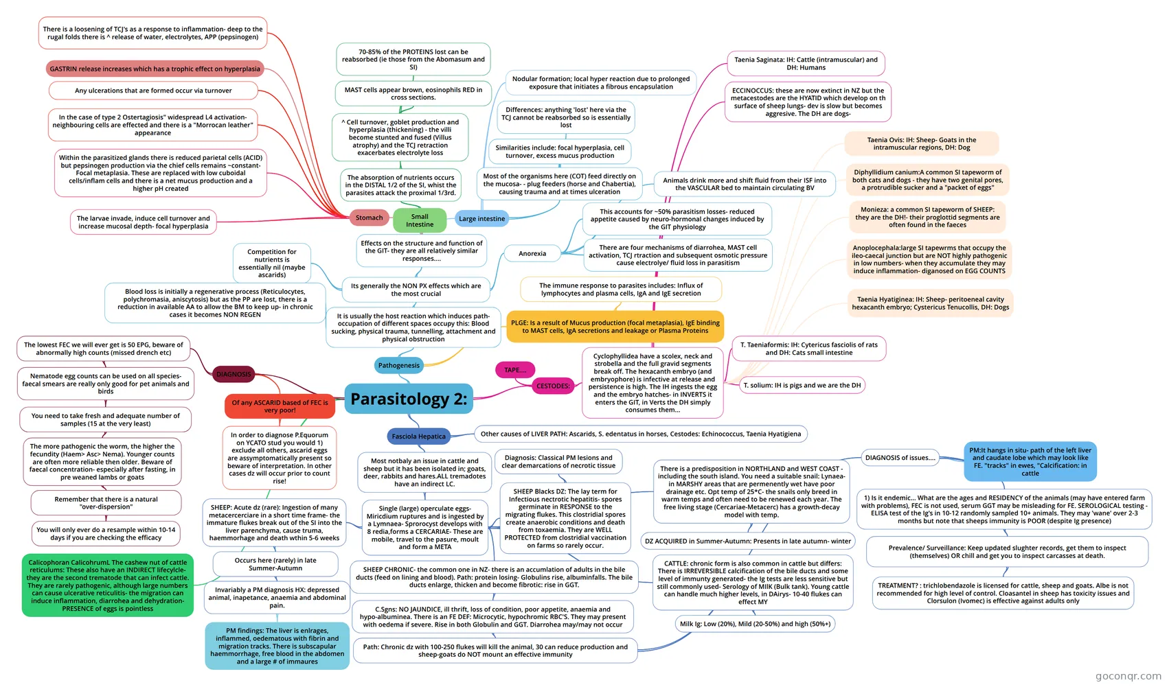
Mind map: Parasitology 2
Map content
Parasitology 2:
Pathogenesis
- It is usually the host reaction which induces path- occupation of different spaces occupy this: Blood sucking, physical trauma, tunnelling, attachment and physical obstruction
- Its generally the NON PX effects which are the most crucial
- Competition for nutrients is essentially nil (maybe ascarids)
- Effects on the structure and function of the GIT- they are all relatively similar responses....
- Stomach
- The larvae invade, induce cell turnover and increase mucosal depth- focal hyperplasia
- Within the parasitized glands there is reduced parietal cells (ACID) but pepsinogen production via the chief cells remains ~constant- Focal metaplasia. These are replaced with low cuboidal cells/inflam cells and there is a net mucus production and a higher pH created
- In the case of type 2 Ostertagiosis" widespread L4 activation- neighbouring cells are effected and there is a "Morrocan leather" appearance
- Any ulcerations that are formed occur via turnover
- GASTRIN release increases which has a trophic effect on hyperplasia
- There is a loosening of TCJ's as a response to inflammation- deep to the rugal folds there is ^ release of water, electrolytes, APP (pepsinogen)
- Small Intestine
- The absorption of nutrients occurs in the DISTAL 1/2 of the SI, whist the parasites attack the proximal 1/3rd.
- ^ Cell turnover, goblet production and hyperplasia (thickening) - the villi become stunted and fused (Villus atrophy) and the TCJ retraction exacerbates electrolyte loss
- MAST cells appear brown, eosinophils RED in cross sections.
- 70-85% of the PROTEINS lost can be reabsorbed (ie those from the Abomasum and SI)
- ^ Cell turnover, goblet production and hyperplasia (thickening) - the villi become stunted and fused (Villus atrophy) and the TCJ retraction exacerbates electrolyte loss
- The absorption of nutrients occurs in the DISTAL 1/2 of the SI, whist the parasites attack the proximal 1/3rd.
- Large intestine
- Most of the organisms here (COT) feed directly on the mucosa- - plug feeders (horse and Chabertia), causing trauma and at times ulceration
- Similarities include: focal hyperplasia, cell turnover, excess mucus production
- Differences: anything 'lost' here via the TCJ cannot be reabsorbed so is essentially lost
- Nodular formation; local hyper reaction due to prolonged exposure that initiates a fibrous encapsulation
- Most of the organisms here (COT) feed directly on the mucosa- - plug feeders (horse and Chabertia), causing trauma and at times ulceration
- Stomach
- Anorexia
- This accounts for ~50% parasitism losses- reduced appetite caused by neuro-hormonal changes induced by the GIT physiology
- There are four mechanisms of diarrohea, MAST cell activation, TCJ rtraction and subsequent osmotic pressure cause electrolye/ fluid loss in parasitism
- Animals drink more and shift fluid from their ISF into the VASCULAR bed to maintain circulating BV
- Blood loss is initially a regenerative process (Reticulocytes, polychromasia, aniscytosis) but as the PP are lost, there is a reduction in available AA to allow the BM to keep up- in chronic cases it becomes NON REGEN
- Its generally the NON PX effects which are the most crucial
- PLGE: Is a result of Mucus production (focal metaplasia), IgE binding to MAST cells, IgA secretions and leakage or Plasma Proteins
- The immune response to parasites includes: Influx of lymphocytes and plasma cells, IgA and IgE secretion
- It is usually the host reaction which induces path- occupation of different spaces occupy this: Blood sucking, physical trauma, tunnelling, attachment and physical obstruction
DIAGNOSIS
- The lowest FEC we will ever get is 50 EPG, beware of abnormally high counts (missed drench etc)
- Nematode egg counts can be used on all species- faecal smears are really only good for pet animals and birds
- You need to take fresh and adequate number of samples (15 at the very least)
- The more pathogenic the worm, the higher the fecundity (Haem> Asc> Nema). Younger counts are often more reliable then older. Beware of faecal concentration- especially after fasting, in pre weaned lambs or goats
- Remember that there is a natural "over-dispersion"
- You will only ever do a resample within 10-14 days if you are checking the efficacy
- Remember that there is a natural "over-dispersion"
- The more pathogenic the worm, the higher the fecundity (Haem> Asc> Nema). Younger counts are often more reliable then older. Beware of faecal concentration- especially after fasting, in pre weaned lambs or goats
- You need to take fresh and adequate number of samples (15 at the very least)
- Nematode egg counts can be used on all species- faecal smears are really only good for pet animals and birds
- Of any ASCARID based of FEC is very poor!
- In order to diagnose P.Equorum on YCATO stud you would 1) exclude all others, ascarid eggs are assymptomatically present so beware of interpretation. In other cases dz will occur prior to count rise!
- The lowest FEC we will ever get is 50 EPG, beware of abnormally high counts (missed drench etc)
Fasciola Hepatica
- Most notbaly an issue in cattle and sheep but it has been isolated in; goats, deer, rabbits and hares.ALL tremadotes have an indirect LC.
- Single (large) operculate eggs- Miricdium ruptures and is ingested by a Lymnaea- Sprorocyst develops with 8 redia,forms a CERCARIAE- These are mobile, travel to the pasure, moult and form a META
- SHEEP: Acute dz (rare): Ingestion of many metacercerciare in a short time frame- the immature flukes break out of the SI into the liver parenchyma, cause truma, haemmorhage and death wthin 5-6 weeks
- Occurs here (rarely) in late Summer-Autumn
- Invariably a PM diagnosis HX: depressed animal, inapetance, anaemia and abdominal pain.
- PM findings: The liver is enlrages, inflammed, oedematous with fibrin and migration tracks. There is subscapular haemmorrhage, free blood in the abdomen and a large # of immaures
- Invariably a PM diagnosis HX: depressed animal, inapetance, anaemia and abdominal pain.
- Occurs here (rarely) in late Summer-Autumn
- SHEEP Blacks DZ: The lay term for Infectious nectrotic hepatitis- spores germinate in RESPONSE to the migrating flukes. This clostridial spores create anaerobic conditions and death from toxaemia. They are WELL PROTECTED from clostridial vaccination on farms so rarely occur.
- Diagnosis: Classical PM lesions and clear demarcations of necrotic tissue
- SHEEP CHRONIC- the common one in NZ- there is an accumlation of adults in the bile ducts (feed on lining and blood). Path: protein losing- Globulins rise, albuminfalls. The bile ducts enlarge, thicken and become fibrotic: rise in GGT.
- C.Sgns: NO JAUNDICE, ill thrift, loss of condition, poor appetite, anaemia and hypo-albuminea. There is an FE DEF: Microcytic, hypochromic RBC'S. They may present with oedema if severe. Rise in both Globulin and GGT. Diarrohea may/may not occur
- Path: Chronic dz with 100-250 flukes will kill the animal, 30 can reduce production and sheep-goats do NOT mount an effective immunity
- DIAGNOSIS of issues....
- 1) Is it endemic... What are the ages and RESIDENCY of the animals (may have entered farm with problems), FEC is not used, serum GGT may be misleading for FE. SEROLOGICAL testing - ELISA test of the Ig's in 10-12 randomly sampled 10+ animals. They may 'wane' over 2-3 months but note that sheeps immunity is POOR (despite Ig presence)
- Prevalence/ Surveillance: Keep updated slughter records, get them to inspect (themselves) OR chill and get you to inspect carcasses at death.
- TREATMENT? : trichlobendazole is licensed for cattle, sheep and goats. Albe is not recommended for high level of control. Cloasantel in sheep has toxicity issues and Clorsulon (Ivomec) is effective against adults only
- Prevalence/ Surveillance: Keep updated slughter records, get them to inspect (themselves) OR chill and get you to inspect carcasses at death.
- PM:It hangs in situ- path of the left liver and caudate lobe which may look like FE. "tracks" in ewes, "Calcification: in cattle
- 1) Is it endemic... What are the ages and RESIDENCY of the animals (may have entered farm with problems), FEC is not used, serum GGT may be misleading for FE. SEROLOGICAL testing - ELISA test of the Ig's in 10-12 randomly sampled 10+ animals. They may 'wane' over 2-3 months but note that sheeps immunity is POOR (despite Ig presence)
- DIAGNOSIS of issues....
- Path: Chronic dz with 100-250 flukes will kill the animal, 30 can reduce production and sheep-goats do NOT mount an effective immunity
- C.Sgns: NO JAUNDICE, ill thrift, loss of condition, poor appetite, anaemia and hypo-albuminea. There is an FE DEF: Microcytic, hypochromic RBC'S. They may present with oedema if severe. Rise in both Globulin and GGT. Diarrohea may/may not occur
- There is a predisposition in NORTHLAND and WEST COAST - including the south island. You need a suitable snail: Lynaea- in MARSHY areas that are permenently wet have poor drainage etc. Opt temp of 25*C- the snails only breed in warm temps and often need to be renewed each year. The free living stage (Cercariae-Metacerc) has a growth-decay model with temp.
- DZ ACQUIRED in Summer-Autumn: Presents in late autumn- winter
- CATTLE: chronic form is also common in cattle but differs: There is IRREVERSIBLE calcification of the bile ducts and some level of immunty generated- the Ig tests are less sensitive but still commonly used- Serology of MIlK (Bulk tank). Young cattle can handle much higher levels, in DAirys- 10-40 flukes can effect MY
- Milk Ig: Low (20%), Mild (20-50%) and high (50%+)
- SHEEP: Acute dz (rare): Ingestion of many metacercerciare in a short time frame- the immature flukes break out of the SI into the liver parenchyma, cause truma, haemmorhage and death wthin 5-6 weeks
- Single (large) operculate eggs- Miricdium ruptures and is ingested by a Lymnaea- Sprorocyst develops with 8 redia,forms a CERCARIAE- These are mobile, travel to the pasure, moult and form a META
- Other causes of LIVER PATH: Ascarids, S. edentatus in horses, Cestodes: Echinococcus, Taenia Hyatigiena
- Most notbaly an issue in cattle and sheep but it has been isolated in; goats, deer, rabbits and hares.ALL tremadotes have an indirect LC.
Calicophoran CalicohrumL The cashew nut of cattle reticulums: These also have an INDIRECT lifecylcle- they are the second trematode that can infect cattle. They are rarely pathogenic, although large numbers can cause ulcerative reticulitis- the migration can induce inflammation, diarrohea and dehydration- PRESENCE of eggs is pointless
CESTODES:
- Cyclophyllidea have a scolex, neck and strobella and the full gravid segments break off. The hexacanth embryo (and embryophore) is infective at release and persistence is high. The IH ingests the egg and the embryo hatches- in INVERTS it enters the GIT, in Verts the DH simply consumes them...
- Taenia Ovis: IH: Sheep- Goats in the intramuscular regions, DH: Dog
- Taenia Saginata: IH: Cattle (intramuscular) and DH: Humans
- Taenia Hyatiginea: IH: Sheep- peritoeneal cavity hexacanth embryo; Cystericus Tenucollis, DH: Dogs
- T. Taeniaformis: IH: Cytericus fasciolis of rats and DH: Cats small intestine
- T. solium: IH is pigs and we are the DH
- ECCINOCCUS: these are now extinct in NZ but the metacestodes are the HYATID which develop on th surface of sheep lungs- dev is slow but becomes aggresive. The DH are dogs-
- Diphyllidium canium:A common SI tapeworm of both cats and dogs - they have two genital pores, a protrudible sucker and a "packet of eggs"
- Monieza: a common SI tapeworm of SHEEP: they are the DH!- their proglottid segments are often found in the faeces
- Anoplocephala:large SI tapewrms that occupy the ileo-caecal junction but are NOT highly pathogenic in low numbers- when they accumulate they may induce inflammation- diganosed on EGG COUNTS
- TAPE....
- Cyclophyllidea have a scolex, neck and strobella and the full gravid segments break off. The hexacanth embryo (and embryophore) is infective at release and persistence is high. The IH ingests the egg and the embryo hatches- in INVERTS it enters the GIT, in Verts the DH simply consumes them...
Information
- Description
- 3 Infectious DZ Mind Map on Parasitology 2:, created by rpbardoul on 06/07/2015.
- Created by
- rpbardoul
- Views
- 4
- Rating