34017801
Description
Mind Map by Esme Harcourt, updated more than 1 year ago
|
|
Created by Esme Harcourt
over 2 years ago
|
|
Structure of the Mammalian
Heart
- Esmeralda Harcourt
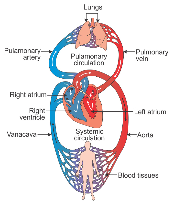
- The human circulatory system is described as closed, because the blood is
contained either within the hearts or the vessels. It is also described as double,
because the blood passes through the heart twice per circuit. The right pump sends
deoxygenated blood to the lungs where it becomes oxygenated and returns back to
the heart. The left pump sends the newly oxygenated blood around the body
- The heart sends blood away to the lungs, this is known as the pulmonary circuit. The
pulmonary circuit replenishes the blood with oxygen, as well as dropping off CO2
which has been taken from the body's tissues
- After the blood has gone around the pulmonary circuit, it is then sent
around the systemic circuit, which takes it around the rest of the body to
all of the tissues
- Because there are 2 circuits, 2 pumps are needed -
one to send blood around the systemic circuit, and
one to send blood around the pulmonary circuit.
- Every time the blood goes
through both circuits, it
goes through the heart
twice (double)
- Every time the blood goes
through both circuits, it
goes through the heart
twice (double)
- After the blood has gone around the pulmonary circuit, it is then sent
around the systemic circuit, which takes it around the rest of the body to
all of the tissues
- The right side of the heart pumps blood around the pulmonary
circuit to re-oxygenate after returning from body tissues
- Blood returns from the body into the vena cava (vein). The blood is
low in oxygen and high in CO2
- The right side pumps the blood into the pulmonary artery and then
into the pulmonary circuit
- The right side pumps the blood into the pulmonary artery and then
into the pulmonary circuit
- Blood returns from the body into the vena cava (vein). The blood is
low in oxygen and high in CO2
- The left side of the heart pumps blood round the systemic circuit to
deliver oxygen to respiring tissues of the body
- The blood from the lungs comes back through the pulmonary vein and drains into the left
side of the heart. The blood is oxygenated and needs to be sent around the body
- The left side pumps the blood into the aorta (the biggest artery in
the body) where it is then distributed around various parts of the
body
- The heart needs its own supply of oxygen and nutrients, and the coronary arteries
deliver these to the heart tissue. Cardiac veins remove the cellular wastes.
- The heart needs its own supply of oxygen and nutrients, and the coronary arteries
deliver these to the heart tissue. Cardiac veins remove the cellular wastes.
- The left side pumps the blood into the aorta (the biggest artery in
the body) where it is then distributed around various parts of the
body
- Internal structure
- 4 chambers - atria and ventricles, both left and right
- The atria are thin-walled elastic chambers which recieve blood from the veins. They can
expand and withstand rising pressure
- Deoxygenated blood enters the vena cava into the right atrium. Oxygenated blood enters
the pulmonary vein into the left atrium
- The ventricles are thick-walled chambers that pump blood out through the arteries. They need
thicker walls to pump blood to organs, not just other chambers like the atria
- The right ventricle recieves deoxygenated blood from the right atrium. The right ventricle contracts
and sends the deoxygenated blood out to the lungs via the pulmonary artery.
- The left ventricle recieves oxygenated blood from the left atrium. The ventricle contracts and sends the blood to
the rest of the body. Therefore, the left ventricle requires much more muscle in order to efficiently pump the blood
- The atria and ventricles are separated by atrio-ventricular valves that prevent blood flowing in the wrong direction
- The arteries leading from the heart are separated from the ventricles by semilunar valves
- Cardiac muscle is a specialed type of muscle found in the walls of the heart; it can
contract automatically without a signal from the brain
- Cardiac muscle contains branched fibres and myofibrils separated by intercalated discs. The
branches help to form a sheet, and each muscle cell is connected by the intercalated discs which
allow the communication of the muscle contraction to run through the whole sheet.
- The structure of cardiac muscle ensure that contraction of the heart has a pumping effect, i.e. from atria to ventricles and out again
- The structure of cardiac muscle ensure that contraction of the heart has a pumping effect, i.e. from atria to ventricles and out again
- Cardiac muscle contains branched fibres and myofibrils separated by intercalated discs. The
branches help to form a sheet, and each muscle cell is connected by the intercalated discs which
allow the communication of the muscle contraction to run through the whole sheet.
- Cardiac muscle is a specialed type of muscle found in the walls of the heart; it can
contract automatically without a signal from the brain
- The arteries leading from the heart are separated from the ventricles by semilunar valves
- The atria and ventricles are separated by atrio-ventricular valves that prevent blood flowing in the wrong direction
- The left ventricle recieves oxygenated blood from the left atrium. The ventricle contracts and sends the blood to
the rest of the body. Therefore, the left ventricle requires much more muscle in order to efficiently pump the blood
- The right ventricle recieves deoxygenated blood from the right atrium. The right ventricle contracts
and sends the deoxygenated blood out to the lungs via the pulmonary artery.
- The ventricles are thick-walled chambers that pump blood out through the arteries. They need
thicker walls to pump blood to organs, not just other chambers like the atria
- Deoxygenated blood enters the vena cava into the right atrium. Oxygenated blood enters
the pulmonary vein into the left atrium
- 4 chambers - atria and ventricles, both left and right
- The blood from the lungs comes back through the pulmonary vein and drains into the left
side of the heart. The blood is oxygenated and needs to be sent around the body
- 4 main vessels related to the heart: the vena cava which brings
deoxygenated blood from the body into the right side
- The right side sends blood out to the lungs in the pulmonary artery
- This blood comes back to the left side through the pulmonary vein
- The left side sends blood out to the body via the aorta
- The left side sends blood out to the body via the aorta
- This blood comes back to the left side through the pulmonary vein
- The right side sends blood out to the lungs in the pulmonary artery
Media attachments
Want to create your own Mind Maps for free with GoConqr? Learn more.