
Mind map
Immune Response
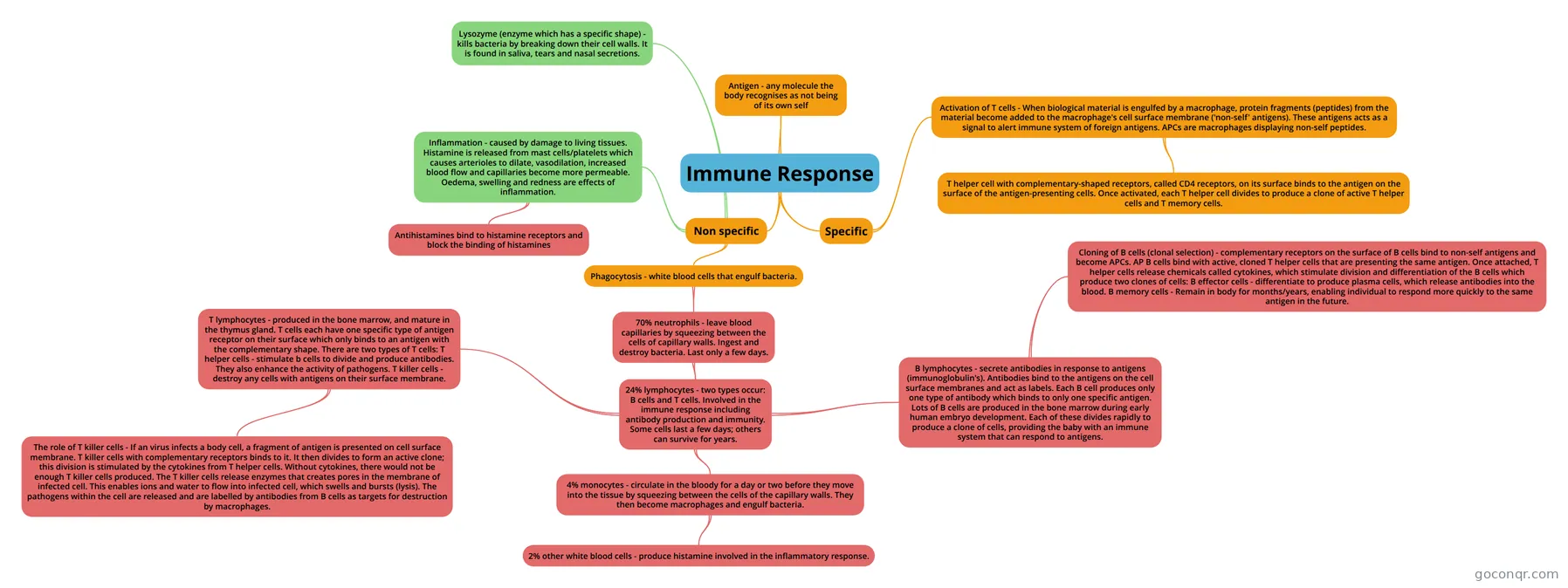
Mind map: Immune Response
Map content
Immune Response
Non specific
- Lysozyme (enzyme which has a specific shape) - kills bacteria by breaking down their cell walls. It is found in saliva, tears and nasal secretions.
- Inflammation - caused by damage to living tissues. Histamine is released from mast cells/platelets which causes arterioles to dilate, vasodilation, increased blood flow and capillaries become more permeable. Oedema, swelling and redness are effects of inflammation.
- Antihistamines bind to histamine receptors and block the binding of histamines
- Phagocytosis - white blood cells that engulf bacteria.
- 70% neutrophils - leave blood capillaries by squeezing between the cells of capillary walls. Ingest and destroy bacteria. Last only a few days.
- 24% lymphocytes - two types occur: B cells and T cells. Involved in the immune response including antibody production and immunity. Some cells last a few days; others can survive for years.
- 4% monocytes - circulate in the bloody for a day or two before they move into the tissue by squeezing between the cells of the capillary walls. They then become macrophages and engulf bacteria.
- 2% other white blood cells - produce histamine involved in the inflammatory response.
- B lymphocytes - secrete antibodies in response to antigens (immunoglobulin's). Antibodies bind to the antigens on the cell surface membranes and act as labels. Each B cell produces only one type of antibody which binds to only one specific antigen. Lots of B cells are produced in the bone marrow during early human embryo development. Each of these divides rapidly to produce a clone of cells, providing the baby with an immune system that can respond to antigens.
- Cloning of B cells (clonal selection) - complementary receptors on the surface of B cells bind to non-self antigens and become APCs. AP B cells bind with active, cloned T helper cells that are presenting the same antigen. Once attached, T helper cells release chemicals called cytokines, which stimulate division and differentiation of the B cells which produce two clones of cells: B effector cells - differentiate to produce plasma cells, which release antibodies into the blood. B memory cells - Remain in body for months/years, enabling individual to respond more quickly to the same antigen in the future.
- T lymphocytes - produced in the bone marrow, and mature in the thymus gland. T cells each have one specific type of antigen receptor on their surface which only binds to an antigen with the complementary shape. There are two types of T cells: T helper cells - stimulate b cells to divide and produce antibodies. They also enhance the activity of pathogens. T killer cells - destroy any cells with antigens on their surface membrane.
- The role of T killer cells - If an virus infects a body cell, a fragment of antigen is presented on cell surface membrane. T killer cells with complementary receptors binds to it. It then divides to form an active clone; this division is stimulated by the cytokines from T helper cells. Without cytokines, there would not be enough T killer cells produced. The T killer cells release enzymes that creates pores in the membrane of infected cell. This enables ions and water to flow into infected cell, which swells and bursts (lysis). The pathogens within the cell are released and are labelled by antibodies from B cells as targets for destruction by macrophages.
- 4% monocytes - circulate in the bloody for a day or two before they move into the tissue by squeezing between the cells of the capillary walls. They then become macrophages and engulf bacteria.
- 24% lymphocytes - two types occur: B cells and T cells. Involved in the immune response including antibody production and immunity. Some cells last a few days; others can survive for years.
- 70% neutrophils - leave blood capillaries by squeezing between the cells of capillary walls. Ingest and destroy bacteria. Last only a few days.
Specific
- Activation of T cells - When biological material is engulfed by a macrophage, protein fragments (peptides) from the material become added to the macrophage's cell surface membrane ('non-self' antigens). These antigens acts as a signal to alert immune system of foreign antigens. APCs are macrophages displaying non-self peptides.
- T helper cell with complementary-shaped receptors, called CD4 receptors, on its surface binds to the antigen on the surface of the antigen-presenting cells. Once activated, each T helper cell divides to produce a clone of active T helper cells and T memory cells.
- Activation of T cells - When biological material is engulfed by a macrophage, protein fragments (peptides) from the material become added to the macrophage's cell surface membrane ('non-self' antigens). These antigens acts as a signal to alert immune system of foreign antigens. APCs are macrophages displaying non-self peptides.
Antigen - any molecule the body recognises as not being of its own self
Information
- Description
- Mind Map on Immune Response, created by anyasan on 16/11/2015.
- Created by
- anyasan
- Views
- 0
- Rating