
Mind map
SKIN (incomplete)
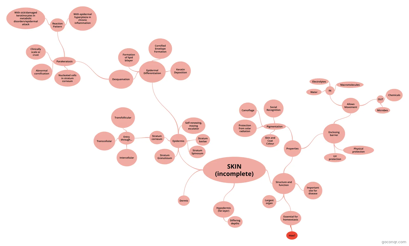
Mind map: skin structure and function
Map content
SKIN (incomplete)
Structure and function
- Largest organ
- Essential for homeostasis
- How?
- Important site for disease
- Properties
- Pigmentation
- Skin and Coat Colour
- Protection from solar radiation
- Camoflage
- Social Recognition
- Enclosing barrier
- Physical protection
- UV protection
- Allows Movement
- IN
- Water
- Electrolytes
- Macromolecules
- OUT
- Microbes
- Chemicals
- IN
- Pigmentation
Epidermis
- Stratum baslae
- Deepest layer Proliferation(here only in dog??)Stem/transient amplifying cells.Attached to other keratinocytes by desmosomes, and to lamina lucida of basement membrane via hemi-desmosomes
- Stratum Spinosum
- Superficial to stratum basale. Differentiation Attached to other keratinocytes by desmosomes. Bulk of living epidermis. These cells have popped up out of basal layer; no further cell division (in dog). Committed to terminal differentiation - WILL become external skin cells (squamous epithelium)
- Stratum Granulosum
- Superficial to stratum spinosum (Late) differentiation Attached to other karatinocytes by desmosomes. Keratohyaline granules: insoluble aggregate of enzymes involved in cross-linking keratin filaments (profillagrin) as well as proteins that form the envelope of cornified cells. Granules = scaffolding removers and packagers.
- Stratum corneum
- Most superficial layer DesquamationCells (corneocytes, squames), dead remanats of living keratinocytes, are attached by corneo-desmosomes, that are enzymatically degraded to allow shedding. A lipid seal between the squames reflects the "bricks and mortar" analogy for this key layer in skin barrier function.
- Entry through...
- Important for topical drugs
- Intercellular
- Transcellular
- Transfollicular
- Self-renewing, moving escalator
- Approx. 21 days in canine Turnover rate can increase to protect the body, but can lead to keratinisation/herplasia
- Epidermal Differentiation
- Desquamation
- Lack of breakdown between the squames. Does not bother dog, but o often not happy (aesthetics) Genetic problem - difficult to treat, but can do regular bathing with a keratolytic shampoo to help shed excess.
- Parakeratosis
- Nucleated cells in stratum corneum
- Abnormal cornification
- Clinically scale or crust
- Scale = excess stratum corneum Crust = Dried exudate
- Reaction Pattern
- With sick/damaged keratinocytes in metabolic disorders/epidermal attack
- With epidermal hyperplasia in chronic inflammation
- Formation of lipid bilayer
- Cornified Envelope Formation
- Keratin Deposition
- Desquamation
- Stratum baslae
Dermis
Hypodermis (fat layer)
- Differing depths
- Dependant on location (v. little on pinna)
- Differing depths
Information
- Description
- Mind Map on SKIN (incomplete), created by Alyson Pilgrim on 22/02/2016.
- Created by
- Alyson Pilgrim
- Views
- 3
- Rating