
Mind map
Colorectal adenocarcinoma
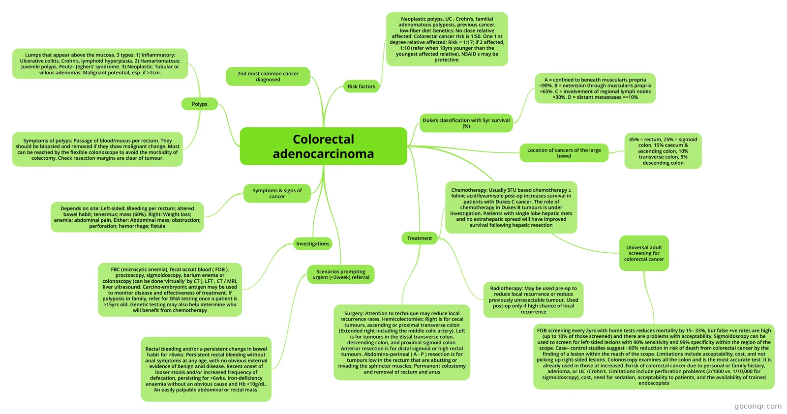
Mind map: Colorectal adenocarcinoma
Map content
Colorectal adenocarcinoma
2nd most common cancer diagnosed
Risk factors
- Neoplastic polyps, UC , Crohn’s, familial adenomatous polyposis, previous cancer, low-fiber diet Genetics: No close relative affected: Colorectal cancer risk is 1:50. One 1 st degree relative affected: Risk = 1:17; if 2 affected, 1:10 (refer when 10yrs younger than the youngest affected relative). NSAID s may be protective.
Polyps
- Lumps that appear above the mucosa. 3 types: 1) Inflammatory: Ulcerative colitis, Crohn’s, lymphoid hyperplasia. 2) Hamartomatous: Juvenile polyps, Peutz– Jeghers’ syndrome. 3) Neoplastic: Tubular or villous adenomas: Malignant potential, esp. if >2cm.
- Symptoms of polyps: Passage of blood/mucus per rectum. They should be biopsied and removed if they show malignant change. Most can be reached by the flexible colonoscope to avoid the morbidity of colectomy. Check resection margins are clear of tumour.
Symptoms & signs of cancer
- Depends on site: Left-sided: Bleeding per rectum; altered bowel habit; tenesmus; mass (60%). Right: Weight loss; anemia; abdominal pain. Either: Abdominal mass; obstruction; perforation; hemorrhage; fistula
Investigations
- FBC (microcytic anemia), fecal occult blood ( FOB ), proctoscopy, sigmoidoscopy, barium enema or colonoscopy (can be done ‘virtually’ by CT ), LFT , CT / MRI, liver ultrasound. Carcino-embryonic antigen may be used to monitor disease and effectiveness of treatment. If polyposis in family, refer for DNA testing once a patient is >15yrs old. Genetic testing may also help determine who will benefit from chemotherapy
Treatment
- Surgery: Attention to technique may reduce local recurrence rates. Hemicolectomies: Right is for cecal tumours, ascending or proximal transverse colon (Extended right including the middle colic artery). Left is for tumours in the distal transverse colon, descending colon, and proximal sigmoid colon. Anterior resection is for distal sigmoid or high rectal tumours. Abdomino-perineal ( A - P ) resection is for tumours low in the rectum that are abutting or invading the sphincter muscles: Permanent colostomy and removal of rectum and anus
- Radiotherapy: May be used pre-op to reduce local recurrence or reduce previously unresectable tumour. Used post-op only if high chance of local recurrence
- Chemotherapy: Usually 5FU based chemotherapy ± folinic acid/levamisole post-op increases survival in patients with Dukes C cancer. The role of chemotherapy in Dukes B tumours is under investigation. Patients with single lobe hepatic mets and no extrahepatic spread will have improved survival following hepatic resection
Duke’s classification with 5yr survival (%)
- A = confined to beneath muscularis propria =90%. B = extension through muscularis propria =65%. C = involvement of regional lymph nodes =30%. D = distant metastases =<10%
Location of cancers of the large bowel
- 45% = rectum, 25% = sigmoid colon, 15% caecum & ascending colon, 10% transverse colon, 5% descending colon
Scenarios prompting urgent (<2week) referral
- Rectal bleeding and/or a persistent change in bowel habit for >6wks. Persistent rectal bleeding without anal symptoms at any age, with no obvious external evidence of benign anal disease. Recent onset of looser stools and/or increased frequency of defecation, persisting for >6wks. Iron-deficiency anaemia without an obvious cause and Hb <10g/dL. An easily palpable abdominal or rectal mass.
Universal adult screening for colorectal cancer
- FOB screening every 2yrs with home tests reduces mortality by 15– 33%, but false +ve rates are high (up to 10% of those screened) and there are problems with acceptability. Sigmoidoscopy can be used to screen for left-sided lesions with 90% sensitivity and 99% specificity within the region of the scope. Case– control studies suggest ~60% reduction in risk of death from colorectal cancer by the finding of a lesion within the reach of the scope. Limitations include acceptability, cost, and not picking up right-sided lesions. Colonoscopy examines all the colon and is the most accurate test. It is already used in those at increased ;lkrisk of colorectal cancer due to personal or family history, adenoma, or UC /Crohn’s. Limitations include perforation problems (2/1000 vs. 1/10,000 for sigmoidoscopy), cost, need for sedation, acceptability to patients, and the availability of trained endoscopists
Information
- Description
- Mind Map on Colorectal adenocarcinoma, created by emailk8 on 12/01/2014.
- Created by
- emailk8
- Views
- 22
- Rating