814776
Description
Mind Map by cmharrisuk, updated more than 1 year ago
|
|
Created by cmharrisuk
over 11 years ago
|
|
Unit 9: Transport in humans
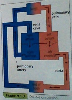
- Describe the double circulation as a low pressure flow
to the lungs and a high pressure flow to the body
tissues and relate to the functions of the two circuits
- Circulatory system
- the organ system made up of blood vessels and the
heart that transports blood. Mammals have a double
circulation with blood passing through the heart twice
in one circuit of the body
- the organ system made up of blood vessels and the
heart that transports blood. Mammals have a double
circulation with blood passing through the heart twice
in one circuit of the body
- Circulatory system
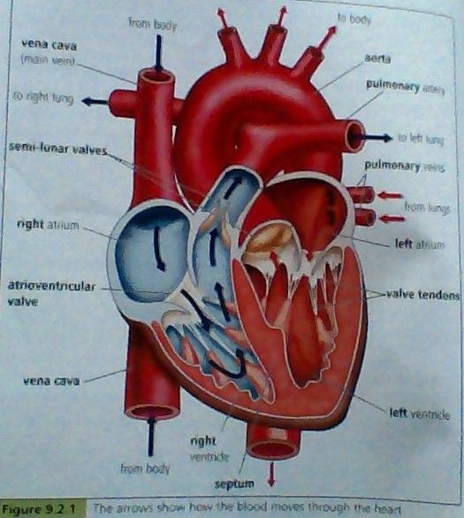
- Describe the strucure of the heart to
include muscular walls, septum and valves
- The heart consists almost entirely of cardiac muscle.
The left and right side of the heart are divided by the
septum, a wall of tissue, which prevents
deoxygenated blood on the right side mixing with
oxygenated blood on the left side. Between each
atrium and ventricle is a valve, which prevent blood
flowing back (backflow).
- Describe the heart as a pump for the
flow of blood and the role of valves to
permit only one-way flow
- the ventricles have more muscular walls than the atria because
they have to pump the blood much further than the atria - either
to the lungs or to the tissues of the rest of the body.
- the pressure of blood against the
valves make them open to let the
blood pass through, but they close
when blood flows back to fill the
pockets, so as to prevent backflow.
- the ventricles have more muscular walls than the atria because
they have to pump the blood much further than the atria - either
to the lungs or to the tissues of the rest of the body.
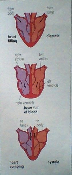
- State the sequence of events that
take place during one heart beat
- Diastole is when the heart
muscles are relaxed. Blood flows
into the atria from the veins.
- Systole is when the
heart muscles contract.
- The atia contract and force blood into
the ventricles. The valves between the
atria and ventricles open. Then the
ventricles contract to force blood out
into the arteries. The valves close.
- the right ventricle pumps
blood to the lungs in the
pulmonary artery.
- the left ventricle pumps
blood to the rest of the
body in the aorta.
- deoxygentated blood returns to the
right atrium in the vena cava.
oxygenated blood returns to the left
atrium in the pulmonary veins.
- the right ventricle pumps
blood to the lungs in the
pulmonary artery.
- Diastole is when the heart
muscles are relaxed. Blood flows
into the atria from the veins.
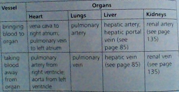
- Name the main blood vessels to and
from the heart, lungs, liver and kidney
- To
- heart
- right atrium: vena cava.
left atrium: pulmonary vein
- right atrium: vena cava.
left atrium: pulmonary vein
- lungs
- pulmonary artery
- pulmonary artery
- liver
- hepatic artery,
hepatic portal vein
- hepatic artery,
hepatic portal vein
- kidney
- renal artery
- renal artery
- heart
- From
- heart
- right ventricle: pulmonary artery.
left ventricle: aorta
- right ventricle: pulmonary artery.
left ventricle: aorta
- lungs
- pulmonary vein
- pulmonary vein
- liver
- hepatic vein
- hepatic vein
- kidney
- renal vein
- renal vein
- heart
- To
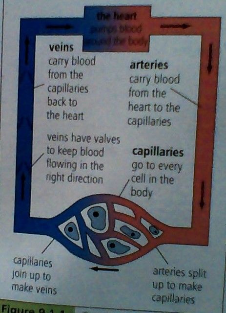
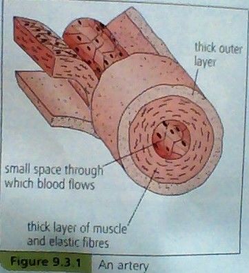
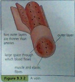
- Describe the structure and function
of arteries, veins and capillaries
- Structure
- arteries
- narrow space in centre. walls: thick
and muscular with elastic fibres.
- narrow space in centre. walls: thick
and muscular with elastic fibres.
- veins
- wider space in centre. walls: less muscular
and less elastic. has semi-lunar valves
- wider space in centre. walls: less muscular
and less elastic. has semi-lunar valves
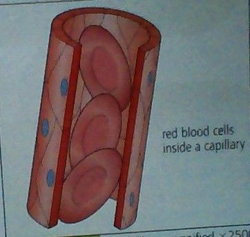
- capillaries
- are very narrow,
walls: one cell thick
- are very narrow,
walls: one cell thick
- arteries
- Function
- arteries
- the main blood vessel, carries blood
which then branches into capillaries
- the main blood vessel, carries blood
which then branches into capillaries
- veins
- carries deoxygenated blood away from organ(s)
through heart to lungs to be oxygenated again
- carries deoxygenated blood away from organ(s)
through heart to lungs to be oxygenated again
- capillaries
- carries blood. oxygen, carbon dioxide and dissolved food
substances diffuse through wall to and from blood (cells)
- carries blood. oxygen, carbon dioxide and dissolved food
substances diffuse through wall to and from blood (cells)
- arteries
- Structure
- Explain how the structure of
arteries, veins and capillaries
adapts them to their functions
- arteries
- thick muscular walls help to withstand pressure.
elastic fibres help to push blood along.
- thick muscular walls help to withstand pressure.
elastic fibres help to push blood along.
- veins
- much less pressure than in arteries. less elastic walls - pressure
from body muscles and other surrounding organs help to move
blood along. valves also help maintain a one-way flow of blood.
- much less pressure than in arteries. less elastic walls - pressure
from body muscles and other surrounding organs help to move
blood along. valves also help maintain a one-way flow of blood.
- capillaries
- one cell thick walls help with diffusion.
- one cell thick walls help with diffusion.
- arteries
- Describe how to investigate
the effect of physical activity
on pulse rate
- measure pulse rate at rest (two
fingers at throat) for 1 minute. do
light exercise for 1 minute.
measure pulse rate for 1 minute
again. do heavy exercise for 3
mins. measure pulse rate for 1
minute again... etc.
- physical activity increases pulse rate,
supplying muscles with more oxygen and
glucose and removing carbon dioxide
quicker.
- physical activity increases pulse rate,
supplying muscles with more oxygen and
glucose and removing carbon dioxide
quicker.
- measure pulse rate at rest (two
fingers at throat) for 1 minute. do
light exercise for 1 minute.
measure pulse rate for 1 minute
again. do heavy exercise for 3
mins. measure pulse rate for 1
minute again... etc.
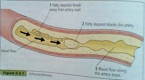
- Describe coronary heart
disease in terms of blockage
of coronary arteries
- the heart needs glucose and oxygen to keep the muscles
working. glucose and oxygen are transported to the heart in
the coronary arteries. if these arteries get blocked then the
heart muscle could become starved of oxygen and die.
- cholesterol (made in the liver) can stick to artery
walls, narrowing the artery and slowing down the
flow of blood - this is called artherosclerosis.
- the heart needs glucose and oxygen to keep the muscles
working. glucose and oxygen are transported to the heart in
the coronary arteries. if these arteries get blocked then the
heart muscle could become starved of oxygen and die.
- State the possible causes
of coronary heart disease
and ways to prevent it
- Causes
- eating a diet with too
much saturated fat
(increases
concentration of
cholesterol in blood)
- being over-weight
- smoking
- taking little or
no exercise
- stress
- eating a diet with too
much saturated fat
(increases
concentration of
cholesterol in blood)
- Ways to prevent it
- take care of
your diet - eat a
balanced diet
- take regular exercise
- don't smoke
- take care of
your diet - eat a
balanced diet
- Causes
- List the main components of blood
- plasma, white blood cells,
platelets, red blood cells
- plasma, white blood cells,
platelets, red blood cells
- Identify red and white blood
cells in diagrams, photographs
and under the microscope
- red blood cells
- have no nuclei, are red (because of
the red pigment haemoglobin)
- have no nuclei, are red (because of
the red pigment haemoglobin)
- white blood cells
- have nuclei, are colourless under a
microscope, there are two types:
phagocytes and lymphocytes
- have nuclei, are colourless under a
microscope, there are two types:
phagocytes and lymphocytes
- red blood cells
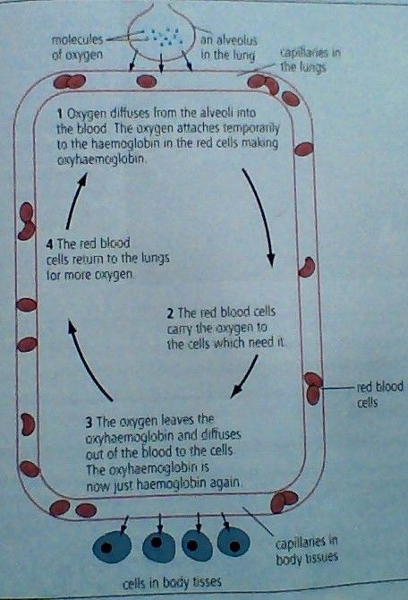
- Describe the role of red blood cells
and haemoglobin in oxygen transport
- because red blood cells have no nuclei, this leaves more
space for haemoglobin (found in cytoplasm) that combines
with oxygen to form oxyhaemoglobin. red blood cells carry
the oxygen in haemoglobin to cells around the body.
- because red blood cells have no nuclei, this leaves more
space for haemoglobin (found in cytoplasm) that combines
with oxygen to form oxyhaemoglobin. red blood cells carry
the oxygen in haemoglobin to cells around the body.
- Describe the functions of
plasma in transport
- plasma is 55% of blood volume
and consists of and transports:
- nutrients such as glucose,
amino acids, lipids,
vitamins and mineral ions
- wastes such as urea
and carbon dioxide
- blood proteins
such as albumen
and antibodies
- hormones such as
insulin, glucagon
and adrenaline
- nutrients such as glucose,
amino acids, lipids,
vitamins and mineral ions
- plasma is 55% of blood volume
and consists of and transports:
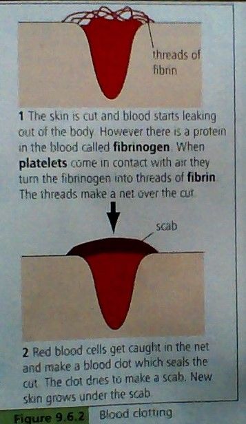
- Describe the process
of blood clotting
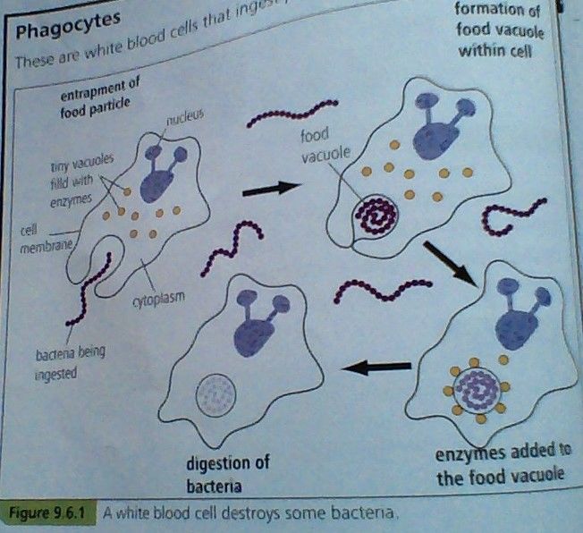
- Describe how white blood cells such
as lymphocytes and phagocytes
protect the body from disease
- phagocytes ingest pathogens,
such as bacteria - they surround
pathogens, ingest them, take
them into food vacuoles, digest
them using enzymes.
phagocytes can move through
capillary walls as well. - this
process is called phagocytosis
- lymphocytes recognise 'foreign'
bacterium or virus and make
proteins called antibodies which
attack the pathogens in a
number of ways: make them
stick together (agglutinate),
dissolve their membranes,
neutralise the toxins that some
pathogens, such as bacteria,
produce.
- phagocytes ingest pathogens,
such as bacteria - they surround
pathogens, ingest them, take
them into food vacuoles, digest
them using enzymes.
phagocytes can move through
capillary walls as well. - this
process is called phagocytosis
- Describe the roles of the immune
system in antibody production,
tissue rejection and phagocytosis
- antibody production
- there is a different type of
antibody for each type of
pathogen. after you have had a
disease, lymphocytes are ready
to produce more of the
appropriate antibodies should
the pathogen enter the body
again - this makes you immune
to thay particular disease.
- there is a different type of
antibody for each type of
pathogen. after you have had a
disease, lymphocytes are ready
to produce more of the
appropriate antibodies should
the pathogen enter the body
again - this makes you immune
to thay particular disease.
- phagocytosis
- phagoctyes ingest
and kill pathogens
- phagoctyes ingest
and kill pathogens
- tissue rejection
- drugs are given to patient to
supress immune system and
the bone marrow is treated
with radiation to stop white
blood cell production for a
period of time
- drugs are given to patient to
supress immune system and
the bone marrow is treated
with radiation to stop white
blood cell production for a
period of time
- antibody production
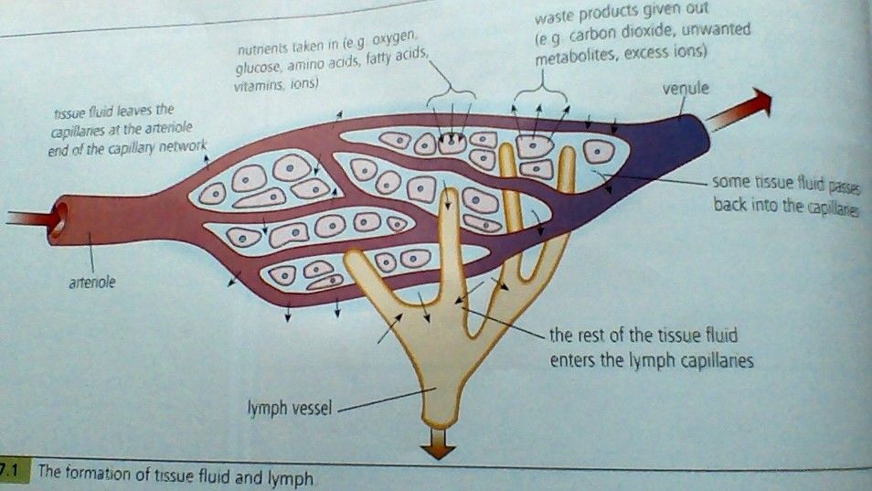
- Describe the exchange of materials
between capillaries and tissue fluid
- State that platelets cause blood to clot
- Describe the functions of the lymphatic system in
circulating lymph and producing lymphocytes
- Describe the one-way flow of blood
around the body through the pulmonary
(lungs) and systemic (rest of body) circuits
- the one-way flow of blood, means that the heart pumps
blood, giving it pressure, so that it flows inside the arteries
around the body. the pulmonary and the systemic circuits
are part of the one-way flow of blood/ double circulation
- the one-way flow of blood, means that the heart pumps
blood, giving it pressure, so that it flows inside the arteries
around the body. the pulmonary and the systemic circuits
are part of the one-way flow of blood/ double circulation
Media attachments
Want to create your own Mind Maps for free with GoConqr? Learn more.