8540059
Description
Mind Map by Rachel Hibray, updated more than 1 year ago
|
|
Created by Rachel Hibray
about 7 years ago
|
|
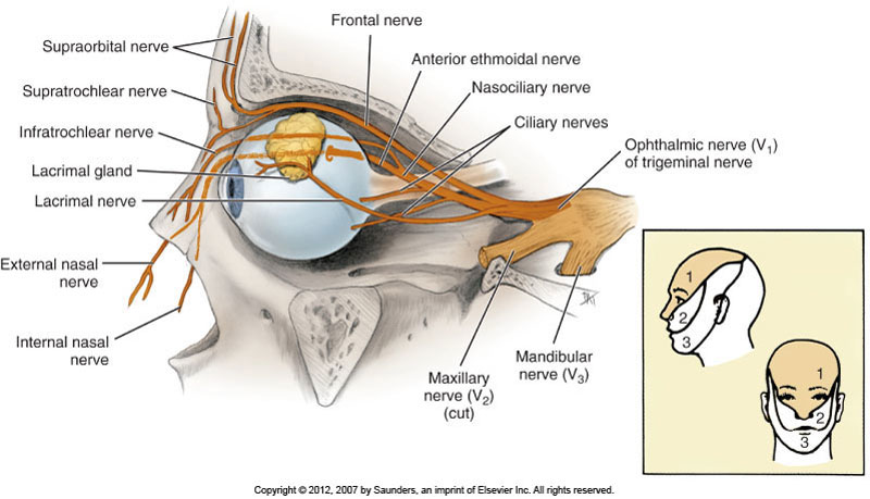
Ophthalmic nerve (V1)
- Smallest branch of
trigeminal nerve, stems
from sensory root, only
carries afferent fibers
- Branches off trigeminal ganglion
- Exits skull at superior orbital fissure then branches into..
- Frontal nerve
- Afferent branch of V1 that runs
from the superior orbital fissure
along the roof of the orbit until it
branches into..
- Supraorbital nerve
- Sensory innervation of the forehead
and anterior scalp (V1 --> frontal nerve
--> supraorbital nerve)
- Sensory innervation of the forehead
and anterior scalp (V1 --> frontal nerve
--> supraorbital nerve)
- Supratrochlear nerve
- Sensory innervation of the bridge of the
nose and medial parts of the upper eyelid
and forehead (V1 --> frontal nerve -->
supratrochlear nerve)
- Sensory innervation of the bridge of the
nose and medial parts of the upper eyelid
and forehead (V1 --> frontal nerve -->
supratrochlear nerve)
- Supraorbital nerve
- Afferent branch of V1 that runs
from the superior orbital fissure
along the roof of the orbit until it
branches into..
- Lacrimal nerve
- Afferent branch of V1 that runs
from the superior orbital fissure
along the lateral roof of the orbit.
Sensory innervation for the lateral
part of the upper eyelid,
conjunctiva, and lacrimal gland.
(V1--> lacrimal nerve)
- Also carries
parasympathetic
postganglionic fibers from
the greater patrosal nerve
of CN VII to the lacrimal
gland for tear production
- Afferent branch of V1 that runs
from the superior orbital fissure
along the lateral roof of the orbit.
Sensory innervation for the lateral
part of the upper eyelid,
conjunctiva, and lacrimal gland.
(V1--> lacrimal nerve)
- Nasociliary nerve
- Afferent branch of V1
that runs from the
superior orbital fissure
through the orbit, and
is superior to the optic
nerve (CN II). Branches
into..
- Long ciliary nerve
- Sensory innervation to
and from the eyeball.
Sensation, not vision.
(V1 --> nasociliary nerve
--> long ciliary nerve)
- Sensory innervation to
and from the eyeball.
Sensation, not vision.
(V1 --> nasociliary nerve
--> long ciliary nerve)
- Infratrochlear nerve
- Sensory
innervation to the
upper eyelid. (V1
--> nasociliary -->
infratrochlear)
- Sensory
innervation to the
upper eyelid. (V1
--> nasociliary -->
infratrochlear)
- Anterior ethmoidal nerve
- Sensory innervation to
the lacrimal sac,
superficial nose, and
anterior nasal cavity (V1
--> nasociliary nerve -->
anterior ethmoidal)
- Sensory innervation to
the lacrimal sac,
superficial nose, and
anterior nasal cavity (V1
--> nasociliary nerve -->
anterior ethmoidal)
- Posterior ethmoidal nerve
- Sensory innervation
to the paranasal
sinuses. (V1 -->
nasociliary nerve -->
posterior ethmoidal
- Sensory innervation
to the paranasal
sinuses. (V1 -->
nasociliary nerve -->
posterior ethmoidal
- Long ciliary nerve
- Afferent branch of V1
that runs from the
superior orbital fissure
through the orbit, and
is superior to the optic
nerve (CN II). Branches
into..
- Frontal nerve
Media attachments
Want to create your own Mind Maps for free with GoConqr? Learn more.