8909560
Description
Mind Map by Liffey Farrell, updated more than 1 year ago
|
|
Created by Liffey Farrell
over 8 years ago
|
|
Cell Structure
- Cells
- Organisms can be
Prokaryotes or
Eukaryotes
- All living things are
made from cells
- Eukaryotic cells are
complex and include all
animal and plant cells
- Eukaryotes are
organisms that are made
up of eukaryotic cells
- Eukaryotes are
organisms that are made
up of eukaryotic cells
- Prokaryotic cells are
smaller and simpler, e.g.
bacteria
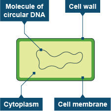
- A prokaryote is a
prokaryotic cell (it's a
single celled organism
- Bacteria are prokaryotes
- Bacterial cells are much
smaller
- It has cytoplasm, a cell
membrane and a cell wall
- Bacteria don't have
chloroplasts or
mitochondria
- Bacterial cells don't have a 'true' nucleus -
instead they have a single circular strand
of DNA that floats freely in the cytoplasm
- They may also contain one or
more small rings of DNA
called plasmids
- They may also contain one or
more small rings of DNA
called plasmids
- Bacterial cells don't have a 'true' nucleus -
instead they have a single circular strand
of DNA that floats freely in the cytoplasm
- Bacteria don't have
chloroplasts or
mitochondria
- It has cytoplasm, a cell
membrane and a cell wall
- Bacterial cells are much
smaller
- Bacteria are prokaryotes
- A prokaryote is a
prokaryotic cell (it's a
single celled organism
- All living things are
made from cells
- Plant and animal cells have
similarities and differences
- The different parts of a cell are
called subcellular structures
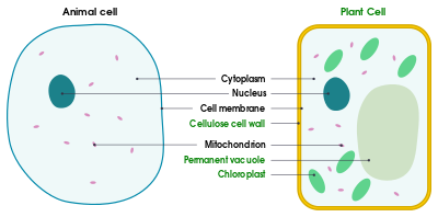
- Animal cells
- Nucleus - contains genetic material
that controls the activities of the
cell
- Cytoplasm - gel-like
substance where most of the
chemical reactions happen
- It contains enzymes that
control these reactions
- It contains enzymes that
control these reactions
- Cell membrane - holds the cell
together and controls what goes
in and out
- Mitochondria - these are where
most of the reactions for aerobic
respiration take place
- Respiration transfers energy
that the cell needs to work
- Respiration transfers energy
that the cell needs to work
- Ribosomes - these are where
proteins are made in the cell
- Nucleus - contains genetic material
that controls the activities of the
cell
- Plant cells
- They usually contains the same
subcellular structures as an animal
cell plus a few things animals cells
don't have
- Rigid cell wall
- Made of cellulose
- It supports the cell
and strengthens it
- Made of cellulose
- Permanent vacuole - contains cell
sap, a weak solution of sugar and
sals
- Chloroplasts - these are where
photosynthesis occurs, which
makes food for the plant
- They contain a green substance called
chlorophyll, which absorbs the light
needed for photosyntheis
- They contain a green substance called
chlorophyll, which absorbs the light
needed for photosyntheis
- Rigid cell wall
- They usually contains the same
subcellular structures as an animal
cell plus a few things animals cells
don't have
- Animal cells
- The different parts of a cell are
called subcellular structures
- Organisms can be
Prokaryotes or
Eukaryotes
- Microscopy
- Cells are studied using
microscopes
- Microscopes let us see
hings that we can't see
with the naked eye
- The microscopy techniques we can
use have developed over the years
as technology and knowledge have
improved
- Light microscopes use light and lenses to
form an image of a specimen and magnify
it (make it look bigger)
- They let us see individual
cells and large subcellular
structures such as nuclei
- Electron microscopes use
electrons instead of light to
form an image
- They have a higher resolution
- Resolution is the ability to distinguish
between two points, so a higher
resolution gives a sharper image
- Electron microscopes let us see much smaller
things in more detail, like the internal structure
of mitochondria and chloroplasts. They even let
us see tinier things like ribosomes and plasmids
- Electron microscopes let us see much smaller
things in more detail, like the internal structure
of mitochondria and chloroplasts. They even let
us see tinier things like ribosomes and plasmids
- Resolution is the ability to distinguish
between two points, so a higher
resolution gives a sharper image
- They have a higher resolution
- Electron microscopes use
electrons instead of light to
form an image
- They let us see individual
cells and large subcellular
structures such as nuclei
- Light microscopes use light and lenses to
form an image of a specimen and magnify
it (make it look bigger)
- The microscopy techniques we can
use have developed over the years
as technology and knowledge have
improved
- Microscopes let us see
hings that we can't see
with the naked eye
- You can calculate the
magnification of an image
using this formula:
- Magnification = image
size/real size
- They need to have the same units
- The image size or real size can
be calculated by rearranging the
equation
- Image size =
magnification x
real size
- Real size = image
size/magnification
- Image size =
magnification x
real size
- The image size or real size can
be calculated by rearranging the
equation
- Example:
- A specimen is 50um. Calculate
the width of the image of the
specimen under a
magnification of 100
- 1) Rearrange the formula
- 2) Fill in the values you know
- 3) Remember the units in your answer
- 4) Convert the units
- 4) Convert the units
- 3) Remember the units in your answer
- 2) Fill in the values you know
- Image size = 100x50
- = 5000um
- = 5mm
- To convert from micrometres to
milimetres you need to divide by
1000 e.g. 5000um / 1000 = 5mm
- To convert from micrometres to
milimetres you need to divide by
1000 e.g. 5000um / 1000 = 5mm
- = 5mm
- = 5000um
- 1) Rearrange the formula
- A specimen is 50um. Calculate
the width of the image of the
specimen under a
magnification of 100
- They need to have the same units
- Magnification = image
size/real size
- Standard form
- Because microscopes see
such tiny objects, sometimes
it's useful to write numbers
in standard form
- This is where you change
very big or small numbers
with lots of zeros into
something more
manageable e.g. 0.017 can
be written as 1.7x10⁻²
- To do this you just need to
move the decimal point
left or right
- The number of places the decimal point moves is
then represented by a power of 10, this is positive if
the decimal point's moved to the left, and the
negative if it's moved to the right
- Example:
- A mitochondria is approximately 0.0025mm
long. Write this figure in standard form
- 1) The first number needs to be
between 1 and 10 so the decimal
point needs to move after the '2'
- 2) Count how many places the
decimal point has moved - this is the
power of 10. Don't forget the minus
sign because the decimal point has
moved right
- 2) Count how many places the
decimal point has moved - this is the
power of 10. Don't forget the minus
sign because the decimal point has
moved right
- 2.5 x 10⁻³
- 1) The first number needs to be
between 1 and 10 so the decimal
point needs to move after the '2'
- A mitochondria is approximately 0.0025mm
long. Write this figure in standard form
- Example:
- The number of places the decimal point moves is
then represented by a power of 10, this is positive if
the decimal point's moved to the left, and the
negative if it's moved to the right
- To do this you just need to
move the decimal point
left or right
- This is where you change
very big or small numbers
with lots of zeros into
something more
manageable e.g. 0.017 can
be written as 1.7x10⁻²
- Because microscopes see
such tiny objects, sometimes
it's useful to write numbers
in standard form
- Cells are studied using
microscopes
- Cell Differentiation and
Specialisation
- Cells don't all look the same. They
have different structures to suit
their different functions
- Cells differentiate to become
specialised
- Differentiation is the process by
which a cell changes to become
specialised for its job
- As cells change, they develop different
subcellular structures and turn into
different types of cells
- This helps them carry
out specific functions
- Most differentiation occurs as
an organism develops
- In most animal cells, the ability to differentiate is
then lost at an early stage, after they become
specialised
- However lots of plant cells
don't ever lose this ability
- The cells that differentiate in mature animals
are mainly used for repairing and replacing cells,
such as skin or blood cells
- Some cells are undifferentiated
cells - they're called stem cells
- Some cells are undifferentiated
cells - they're called stem cells
- The cells that differentiate in mature animals
are mainly used for repairing and replacing cells,
such as skin or blood cells
- However lots of plant cells
don't ever lose this ability
- In most animal cells, the ability to differentiate is
then lost at an early stage, after they become
specialised
- Most differentiation occurs as
an organism develops
- This helps them carry
out specific functions
- As cells change, they develop different
subcellular structures and turn into
different types of cells
- Differentiation is the process by
which a cell changes to become
specialised for its job
- Examples of
specialised cells:
- Sperms cells are specialised for
reproduction
- The function of a sperm cell is
basically to get the male DNA
to the female DNA
- It has a long tail and a
streamlined head to help it swim
to the egg
- There are a lot of mitochondria
in the cell to provide the energy
needed
- It also carries enzymes in its head to
digest through the egg cell membrane
- It also carries enzymes in its head to
digest through the egg cell membrane
- There are a lot of mitochondria
in the cell to provide the energy
needed
- It has a long tail and a
streamlined head to help it swim
to the egg
- The function of a sperm cell is
basically to get the male DNA
to the female DNA
- Nerve cells are specialised
for rapid signalling
- The function of nerve cells is to carry
electrical signals from one part of the body
to another
- These cells are long (to cover more distance) and
have branched connections at their ends to connect
to other nerve cells and form a network throughout
the body
- These cells are long (to cover more distance) and
have branched connections at their ends to connect
to other nerve cells and form a network throughout
the body
- The function of nerve cells is to carry
electrical signals from one part of the body
to another
- Muscle cells are
specialised for
contraction
- The function of a muscle cell is to
contract quickly
- These cells are long so that they have
space to contract and contain lots of
mitochondria to generate the energy
needed for contraction
- These cells are long so that they have
space to contract and contain lots of
mitochondria to generate the energy
needed for contraction
- The function of a muscle cell is to
contract quickly
- Root hair cells are specialised for
absorbing water and minerals
- Root hair cells are cells on the surface of plant
roots, which grow into long "hairs" that stick
out into the soil
- This gives the plant a big surface area
for absorbing water and mineral ions
from the soil
- This gives the plant a big surface area
for absorbing water and mineral ions
from the soil
- Root hair cells are cells on the surface of plant
roots, which grow into long "hairs" that stick
out into the soil
- Phloem and Xylem cells are
specialised for
transporting substances
- Phloem and xylem cells form phloem
and xylem tubes, which transport
substances such as food and water
around plants
- To form the tubes, the cells are long
and joined end to end
- Xylem cells are hollow in the centre and
phloem cells have very few subcelluar
structures so that stuff can flow
through them
- Xylem cells are hollow in the centre and
phloem cells have very few subcelluar
structures so that stuff can flow
through them
- To form the tubes, the cells are long
and joined end to end
- Phloem and xylem cells form phloem
and xylem tubes, which transport
substances such as food and water
around plants
- Sperms cells are specialised for
reproduction
- Cells differentiate to become
specialised
- Cells don't all look the same. They
have different structures to suit
their different functions
- Binary Fission
- Prokaryotic cells can
reproduce using a type of
simple cell division called
binary fission
- Prokaryote cells replicate by
binary fission
- In binary fission, the
cell splits into two
- 1) The circular DNA and
plasmid(s) replicate
- 2) The cell gets bigger and the
circular DNA strands move to
opposite 'poles' (ends) of the
cell
- 3) The cytoplasm begins
to divide and new cell
walls begin to form
- 4) The cytoplasm divides and two
daughter cells are produced. Each
daughter cell has one copy of the
circular DNA, but can have a variable
number of copies of the plasmid(s)
- 4) The cytoplasm divides and two
daughter cells are produced. Each
daughter cell has one copy of the
circular DNA, but can have a variable
number of copies of the plasmid(s)
- 3) The cytoplasm begins
to divide and new cell
walls begin to form
- 2) The cell gets bigger and the
circular DNA strands move to
opposite 'poles' (ends) of the
cell
- 1) The circular DNA and
plasmid(s) replicate
- In binary fission, the
cell splits into two
- Bacteria can divide very
quickly if given the right
condition (e.g. a warm
environment and lots of
nutrients)
- Some bacteria, such as E.coli can take
as little as 20 minutes to replicate in
the right environment
- However if conditions become
unfavourable, the cells will stop
dividing and eventually begin to
die
- However if conditions become
unfavourable, the cells will stop
dividing and eventually begin to
die
- Some bacteria, such as E.coli can take
as little as 20 minutes to replicate in
the right environment
- Prokaryote cells replicate by
binary fission
- Prokaryotic cells can
reproduce using a type of
simple cell division called
binary fission
Media attachments
Want to create your own Mind Maps for free with GoConqr? Learn more.