
Description
|
|
Created by Emily Rowland-Rawson
over 6 years ago
|
|
Page 1
2.1 Microscopy
The first types of microscopes to be developed were light microscopes in the 16th to 17th century. Since then they have conrinued to be developed and improved. There are three well-known branches of microscopy: optical, electron, and scanning probe microscopy, along with the emerging field of X-ray microscopy.
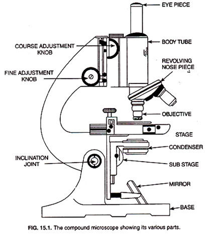
Light microscopes A compound light microscope has two lenses- the objective lens, which is placed near to the specimen, and an eyepiece lens, through which the specimen is viewed. The objective lens produces a magnified image, which is magnified again by the eyepiece lens. This objective/eyepiece lens configuration allows for much higher magnification and reduced chromatic aberration than that in a simple light microscope. Illumination is usually provided by a light underneath the sample. Opaque specimens can be illuminated from above with some microscopes.
Staining techniques Differential staining can distinguish between two types of organisms that would otherwise be hard to identify. It can also differentiate between different organelles of a single organism within a tissue sample. Gram stain technique is used to separate bacteria into two groups, Gram-positive bacteria and Gram-negative bacteria (Figure 5 ). Crystal violet is first applied to a bacterial specimen on a slide, then iodine, which fixes the dye. The slide is then washed with alcohol. The Gram-positive bacteria retain the crystal violet stain and will appear blue or purple under a microscope. Gram-negative bacteria have thinner cell walls and therefore lose the stain. They are then stained with safranin dye, which is called a counterstain. These bacteria will then appear red. Gram-positive bacteria are susceptible to the antibiotic penicillin, which inhibits the formation of cell walls. Gram-negative bacteria have much thinner cell walls that are not susceptible to penicillin. Acid-fast technique is used to differentiate species of Mycobacterium from other bacteria. A lipid solvent is used to carry carbolfuchsin dye into the cells being studied. The cells are then washed with a dilute acidalcohol solution. Mycobacterium are not affected by the acid-alcohol and retain the carbolfuchsin stain, which is bright red. Other bacteria lose the stain and are exposed to a methylene blue stain, which, unsurprisingly, is blue.
Page 2
2.2 Magnification and calibration
Magnification is how many times larger the image is than the actual size of the object being viewed. Interchangeable objective lenses on a compound light microscope allow a user to adjust the magnification. Simply magnifying an object docs not increase the amount of detail that can be seen. The resolution also needs to be increased. The resolution of a microscope determines the amount of detail that can be seen- the higher the resolution the more details are visible. Resolution is the ability to sec individual objects as separate entities. Imagine a car coming towards you at night with its headlights on. When it is a long way off you will only sec one light but. as the ca r gets close r you eventually sec that there a re, in fact, two headlights - they have been resolved. Resolution is limited by the diffraction of light as it passes through samples (and lenses). Diffraction is the tendency of light waves to spread as they pass close to physical structures such as those present in the specimens being studied. The structures present in the specimens arc very close to each other and the light reflected from individual structures can overlap due to diffraction. This means the structures arc no longer seen as separate entities and detail is lost. In optical microscopy structures that arc closer than half the wavelength of light cannot be seen separately (resolved). Resolution can be increased by using beams of electrons which have a wavelength thousands of times shorter than light (Topic 2.3, More microscopy). Electron beams arc still diffract ed but the shorter wavelength means that individual beams can be much closer before they overlap. This means objects which arc much smaller an d closer together can be seen separately without diffraction blurring the image.
Page 3
2.3 More Microscopy
In light microscopy, increased magnification can be achieved easily using the appropriate lenses, but if the image is blurred no more detail will be seen. Resolution is the limiting factor. In electron microscopy, a beam of electrons with a wavelength of less than 1 nm is used to illuminate the specimen. More detail of cell ultrastructure can be seen because electrons have a much smaller wavelength than light waves. They can produce images with magnifications of up to x500 000 and still have clear resolution. Electron microscopy Electron microscopes have changed the way we understand cells but there are some disadvantages to this technique. They are very expensive pieces of equipment and can only be used inside a carefully controlled environment in a dedicated space. Specimens can also be damaged by the electron beam and because the preparation process is very complex, there is a problem with artefacts (structures that are produced due to the preparation process). However. as techniques improve a lot of theses artefacts can be eliminated. There are two types of electron microscope: In a transmission electron microscope (TEM ) a beam of electrons is transmitted through a specimen and focused to produce an image. This is similar to light microscopy. This has the best resolution with a resolving power of 0.5 nm. In a scanning electron microscope (SEM) a beam of electrons is sent across the surface of a specimen and the reflected electrons are collected. The resolving power is from 3-1 0 nm, so the resolution is not as good as with transmission electron microscopy but stunning three-dimensional images of surfaces are produced, giving us valuable information about the appearance of different organisms.
Page 4
2.4 Eukaryotic cell structure
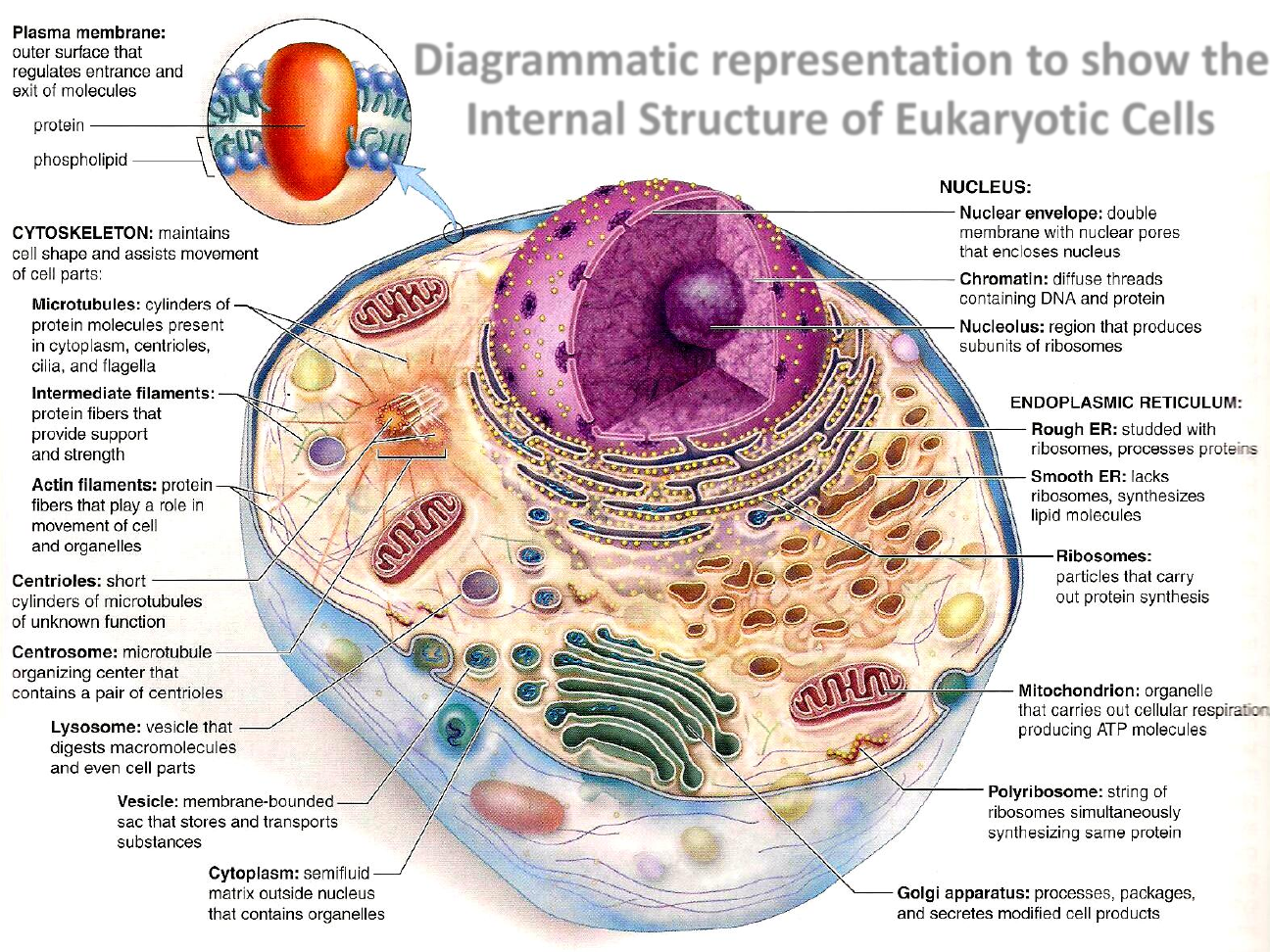
The basic unit of all living things is the cell - but not all cells are the same. There are two fundamental types of cell- prokaryotic and eukaryotic. Prokaryotes are single-celled organisms with a simple structure of just a single undivided internal area called the cytoplasm (composed of cytosol, which is made up of water, salts and organic molecules). Eukaryotic cells make up multicellular organisms like animals, plants, and fungi. Eukaryotic cells have a much more complicated internal structure, containing a membrane-bound nucleus (nucleoplasm) and cytoplasm, which contains many membrane-bound cellular components.
Page 5
2.5 The ultrastructure of plant cells
Plant cell walls Plant cell walls are made of cellulose, a complex carbohydrate. They are freely permeable so substances ca n pass into and out of the cell through the cellulose wall. The cell walls of a plant cell give it shape. The contents of the cell press against the cell wall making it rigid. This supports both the individual cell and the plant as a whole. The cell wall also acts as a defence mechanism. protecting the contents of the cell against invading pathogens. All plant cells have cellulose cell walls. Plant cell organelles Vacuoles are membrane lined sacs in the cytoplasm containing cell sap. Many plant cells have large permanent vacuoles which are very important in the maintenance of turgor, so that the contents of the cell push against the cell wall and maintain a rigid framework for the cell. The membrane of a vacuole in a plant cell is called the tonoplast. It is selectively permeable, which means that some small molecules can pass through it but others cannot. 1f vacuoles appear in animal cells, they are small and transient (not permanent). Chloroplasts are the organelles responsible for photosynthesis in plant cells. They a re found in the cells in the green parts of plants such as the leaves and the stems but not in the roots. They have a double membrane structure, similar tO mitochondria. The fluid enclosed in the chloroplast is called the stroma. They also have an internal network of membranes, which form flattened sacs called thylakoids. Several thylakoids stacked together arc called a granum (plural grana). The grana arc joined by membranes called lamellae. The grana contain the chlorophyll pigments, where light-dependent reactions occur during photosynthesis. Starch produced by photosynthesis is present as starch grains. Like mitOchondria, chloroplasts also contain DNA and ribosomes. Chloroplasts are therefore able to make their own proteins.
Page 6
2.6 Prokaryotic and eukaryotic cells
Prokaryotic cells Prokaryotic cells may have been among the earliest forms of life on Earth. They first appeared around 3.5 billion years ago when the surface of the Earth was a very hostile environment. Scientists believe that these early cells we re adapted to living in extremes of salinity, pH and temperature. These organisms are known as extremophiles and they still exist today. They can be found in hydrothermal vents and salt lakes- similar environments to those believed to have made up the early Earth. They are usually of the domain Archaea and more recently they have been found in more hospitable environments such as soil and the human digestive system. Prokaryotic organisms are always unicellular with a relatively simple structure. Their DNA is not contained within a nucleus, they have few organelles and the organelles they do have are not membrane-bound. The structure of the DNA contained within prokaryotes is fundamentally the same as in eukaryotes but it is packaged differently. Prokaryotes generally only have one molecule of DNA, a chromosome, which is supercoiled to make it more compact. The genes on the chromosome are oft en grouped in to operons, meaning a number of genes are switched on or off at the same time. The ribosomes in prokaryotic cells are smaller than those in eukaryotic cells. Their relative size is determined by the rate at which they settle, or form a sediment, in solution. The larger eukaryotic ribosomes are designated 80S and the smaller prokaryotic ribosomes, 70S. They are both necessary for protein synthesis, although the larger 80S ribosomes are involved in the formation of more complex proteins.
0 comments
Want to create your own Notes for free with GoConqr? Learn more.