Description
|
|
Created by Anneke Ernst
over 6 years ago
|
|
Page 1
Communication between Cells
Three types of signal used by cells to communicate Chemical Signals (Most Common) Mechanical Signals (Such as touch or pressure) Electromagnetic Signals (Such as light)
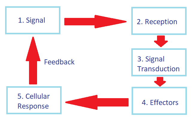
Cells depend on their ability to receive and respond to a variety of signal from their environment and from other cells. They are in constant communication. The three components of cellular communication are: Reception of a Signal Transduction of the Signal Cellular response to the Signal
Cells use signals to tell other cells: To stay alive and function To self destruct To undergo cell division for growth or repair To differentiate into a particular cell type To eliminate a pathogen that has invaded the body To activate a gene and produce the protein encoded by the gene To silence a gene To produce and enzyme or a structural protein
Variation in Signalling Molecules
Differences in chemical structures. Molecules that act as chemical signals include: Amino Acids Peptides Poly-peptides Proteins Steroids These differences mean the signalling molecules vary in size and differ in the affinity for water, some being hydrophilic and some hydrophobic.
Differences in function. Signalling molecules belong to different functional groups, examples: Animal hormones Plant hormones Neurotransmitters of nerve cells Anti-bodies of immune cells Cytokines and pheromones
Difference in scope. >Some signalling molecules exert their effect over a distance while others only act locally.
All signal molecules share these same characteristic. They bind to sites on specific receptors* on their target cells *Receptors are proteins that receive the various signals, specific receptors are for signalling molecules.
Page 2
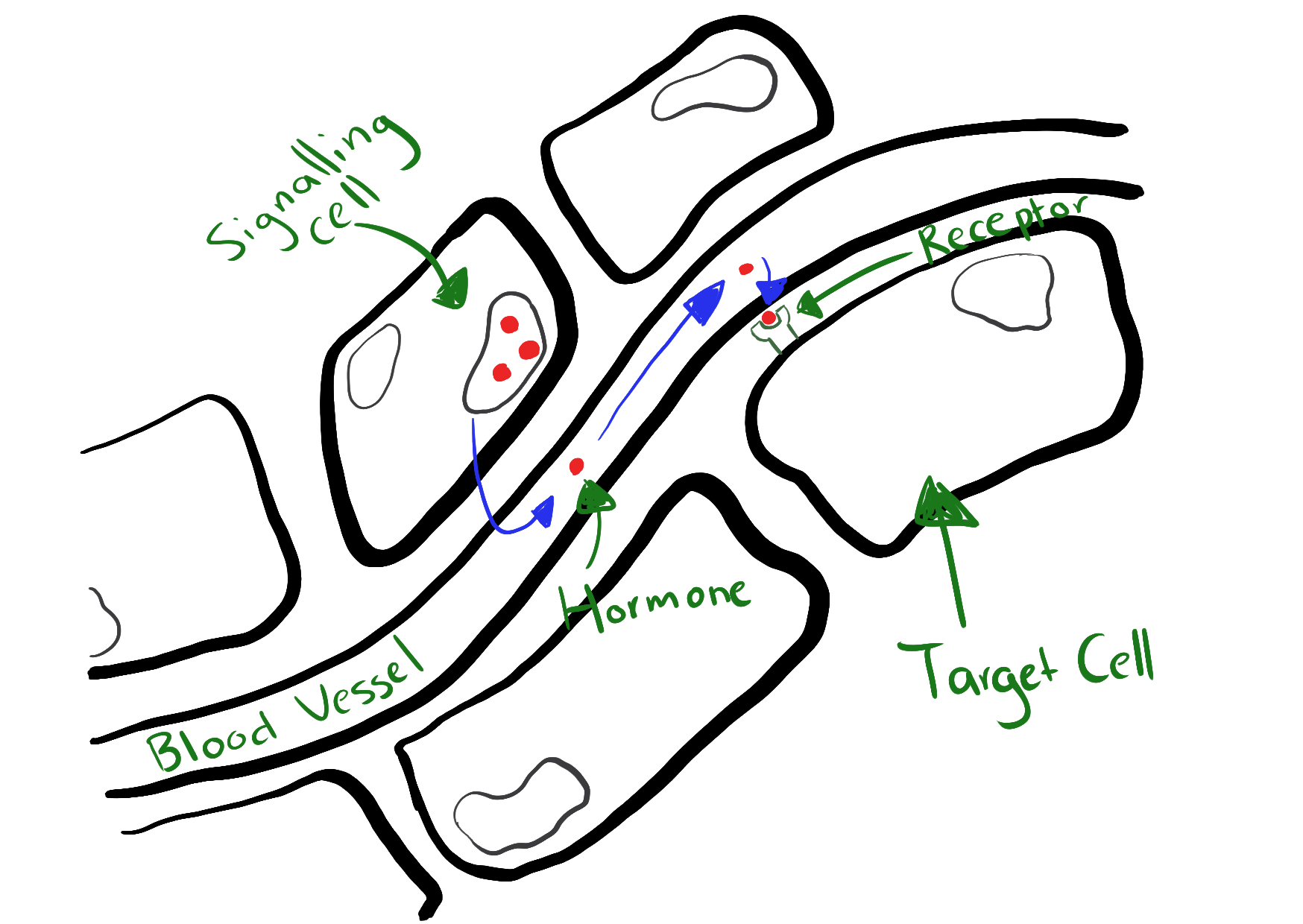
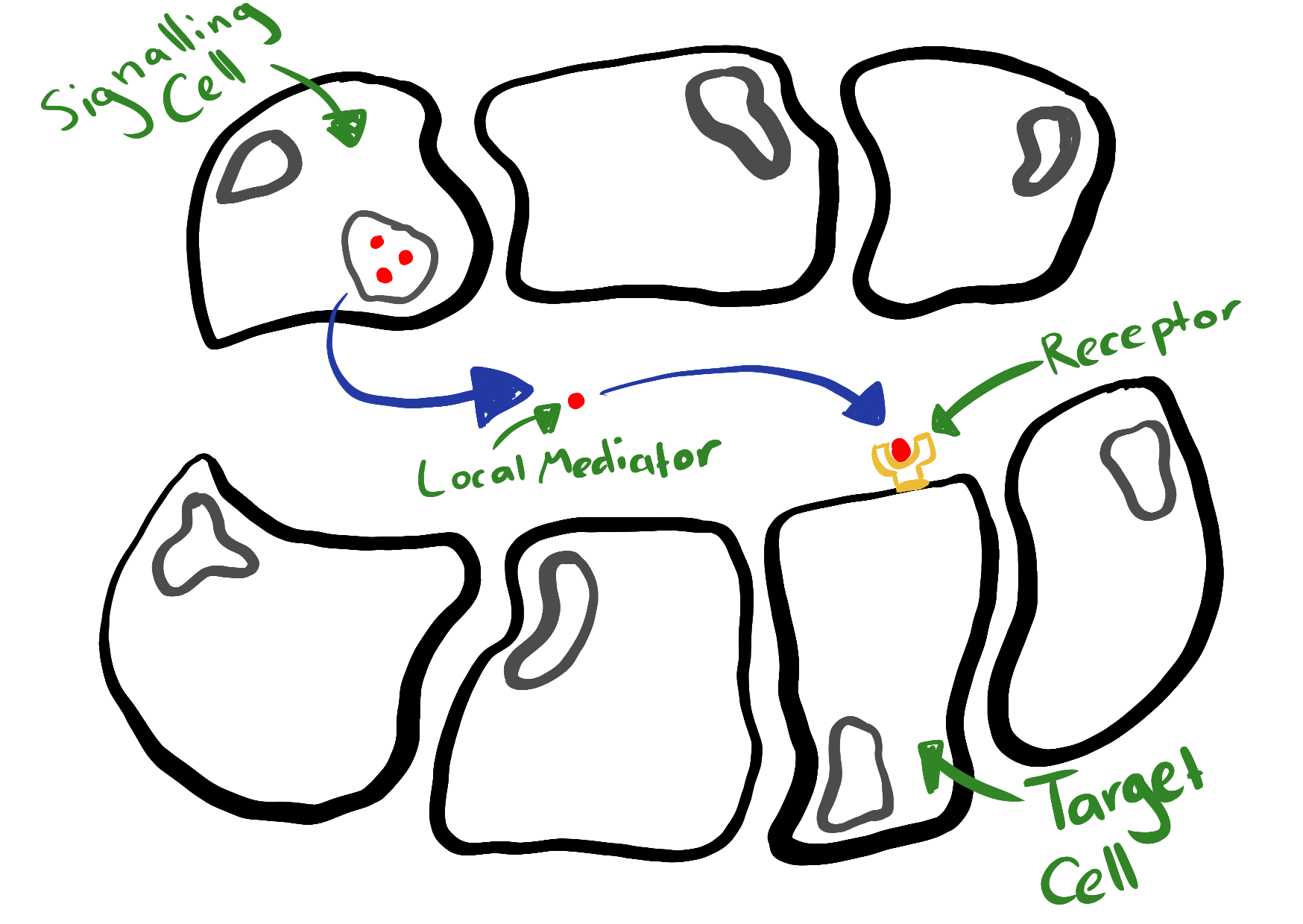
Signals moving from source to target
Signals are only effective if it binds to its specific receptor, so it must travel to reach it and this distance varies depending on what the signal and receptor These are differences between type of distance:
Long distance travel to target cells Some signalling molecules travel long distances to their specific receptors, most use the bloodstream to do this.
Travel to nearby cells Some signals travel short distances to nearby cells by diffusion through the interstitial fluid around cells. These signalling molecules are called Local Mediators.
One cell sends and receives a signal Some cells can release chemical signals that can be receive by itself.
Direct Cell to Cell contact In some cases signals can move directly from one cell to another when they come into direct contact. Some examples include: Gap Junctions "Communicating Junction". Occurs between adjacent cells in animal tissue Plasmodesma. Occurs between adjacent cells in plant cells
Page 3
Stages of Cell Signalling
Signal Reception Signal Transduction Cellular Response
Signal Reception (Stage 1)
First step of cell communication is the reception of a signalling molecule from a cell's external environment. In most cases the receptors are located to the plasma membrane of the cell, these are called 'Cell-Surface Receptors' Some cases have receptors located in the nucleus of the target cell these are called 'intracellular Receptors
Polar and hydrophilic signalling molecule cannot cross the lipid bilayer of the plasma membrane, and can only bind to cell-surface receptors. Cell-surface receptors are either trans-membrane proteins on their own or part of a complex linking the exterior and interior of the cell. The region of the protein that is exposed to the outer region of the cell is called the 'signal-binding domain'.
Hydrophobic signalling molecules can cross the lipid bilayer of the plasma membrane.
Signal Transduction (Stage 2)
Converts a signal from outside a cell into a response within the cell Signal is received in one form, is changed to another mode or molecule Transduction is activated after a signalling molecule binds to its receptor which changes the receptor's 3D shape and activates it.
TRANSDUCTION OF A HYDROPHOBIC SIGNAL Signalling molecule binds with receptor, changing its shape and revealing a new part. It becomes a complex. The complex easily moves through the cytoplasm into the nucleus. The complex binds to a specific gene, activating the gene. HYDROPHOBIC MOLECULES EASILY PASS THROUGH MEMBRANES
TRANSDUCTION OF A HYDROPHILIC SIGNAL The signalling molecule binds to the outside of the cell, changing the shape of receptor and activating it and turning into a complex The complex activates an enzyme inside the cell, embedded int he plasma membrane. The activated enzyme catalyses the production of multiple copies of a 2nd messenger. As there are multiple copies it amplifies the signal 2nd messenger molecules activates copies of key enzymes, which activates copies of another key enzymes and on and on and on, thus carrying the signal. This signal finally gets carried to the nucleus, where it activates a specific gene HYDROPHILIC MOLECULES CANNOT PASS THROUGH THE PLASMA MEMBRANE
Steroid Hormones Hydrophobic: Insoluble in water Lipophilic: soluble in lipid solvent Transported in blood, but only with the aid of carrier proteins Readily diffuse across the lipid bilayer of the plasma membrane Bind to intracellular receptors Directly regulate gene expressions No second messenger involved Longer lasting response Peptide/Protein Hydrophilic: soluble in water Lipophobic: insoluble in lipid solvents Transported in solution in blood plasma Unable to cross the lipid bilayer of the plasa membrane Bind to cell-surface receptors Indirectly act on genes Second messenger produced during signal transduction Shorter period of response
Cellular Response to Signal (Stage 3)
Effects proteins are produced by the gene activated in stage 2. These effector proteins produce the cellular response to the original signal. These do not stay activated and will switch off after a period of time to return to its resting state.
Page 4
Chemical Signal: Human Hormones
Divides into 3 groups Amino Acids derivatives: Are hydrophilic, dissolve in water Suffix ends in '-ine'. Examples: Thyrox'ine'. Epinephr'ine' Lipid-deprived Hormones: Are hydrophobic, not water soluble Suffix ends in '-ol' or '-one' Examples: Cortis'ol'. Testoster'one' Peptide and Protein Hormones Composed of small polypeptide chains (Peptides) Composed of long polypeptide chains (Proteins)
Hydrophilic hormones are transported in a solution in the blood stream Hydrophobic hormones are transported by attaching to a protein carrier through the blood stream
Release of hormones operates on a negative feedback system
Page 5
Chemical Signals: Neurotransmitters
Chemical signalling molecules of nerve cells (neurons) Neurons use two types of signals
Communication within a nerve cell. Depends on electrical signals known as action potentials that transmit a nerve impulse along the axon of a neuron.
Communication between nerve cells uses chemical signals, known as neurotransmitters that diffuse across very small gaps known as synaptic cells
Neurotransmitters transmit nerve impulses from the neurons to muscle cells, stimulating their contraction and to some glands
Junction of two Neurons Synapse is the term for the junction of two neurons and the synaptic cleft that separates them
When a neuron is stimulated, it transmits a nerve impulse in the form of an electrical signal along its axon. At the end of the axon are packages or vesicles that contain neurotransmitters. The impulse is transmitted across the synaptic cleft by the neurotransmitters molecules released from the vesicles in the pre-synaptic neuron. The neurotransmitters molecules diffuse across the synaptic cleft and bind with receptors on the surface of the post-synaptic neurons Note the presence of an enzyme that destroys the neurotransmitter, ensuring that the nerve impulse has a limited duration.
At a neuro-muscular junction, a neurotransmitter is released from the motor end plates at the terminal ends of an axon. This neurotransmitter diffuses across the same gap between the axon and the muscle and binds to the receptor on the membrane of the striated muscle The cellular response contracts the muscle Muscle tissue produces an enzyme that inactivates the transmitter substance
Page 6
Chemical Signals: Pheromones
Pheromones are produced by a variety of animal species. Pheromones are chemical signalling molecules that released by one animals that can carry a signal to a second animal of the same species (a conspecific) If this signal is received, the second anima produces a physiological or behavioural change
Detecting Pheromones Pheromones are secreted by an animal into the external environment. In addition to their olfactory system that detects smells and odors, mammals have a second system that detects pheromones, which uses a different nerve pathway to use. A structure called the vomeronasal organ (VNO) contains the detector cells of the second system.
When the animal breathes, the air is directed to the VNO. If it detects pheromones, a signal is relayed to several regions of the brain, to the hypothalamus The hypothalamus is where the physiological and reproductive responses to pheromone signals are regulated.
Pheromones in Animal World Trail Pheromones: Used to mark a trail to food Alarm Pheromones: Used to warn others when attacked Territorial Pheromones: Used to mark and keep territory/area/land Sex pheromones:Used to attract another for sexual reproduction, also to signal when ready to mate
Page 7
Chemical Signals: Cytokines
The major cells that use cytokines are immune cells (white blood cells of mammals) Cytokines are 'cell signalling molecules that aid cell-to-cell communications in immune response and stimulate the movement of cells towards sites of inflammation, infection and trauma'. Cytokines act as messengers between cells of the immune system.
Cytokines have many effects: Stimulatory Inhibitory Regulatory Cytokines are chemical mediators, they are intracellular proteins that enhance and activated the function of other proteins. Cytokines are modulators that directly influence the effects of other molecules.
Cytokines are secreted by cells in response to various stimuli They are usually briefly, locally and at very low concentrations. Cytokines must be produced a new in response to stimulus, as such cellular response to cytokine signals are slow and may take hours to appear. Once they are produced, they diffuse to nearby target cells
Cellular response produced in response to cytokines include: Promotion of cell growth and differentiation Cell proliferation Cell migration Cell activation Apoptosis Immune Response (inflammation and phagocytosis)
Cytokines in human disease Several cytokines act as mediators in the normal inflammatory response to infection Overproduction or overstimulation of pro-inflammatory cytokines can produce undesirable outcomes
Page 8
Chemical Signalling in Plants
Plant Hormones Produced mainly the cells of the growing regions (meristems) of shoots and roots, in young leaves in germinating seeds and in developing fruits Transport of plant homones is mainly in the phloem tissue. Classic plant hormones are: Auxins, which are responsible for cell division and growth in cell size. Produced in the meristem cells in the growing tips of plant shoots. Cytokinins, which are responsible for an increase in cell division. Produced in roots. Gibberelines, which are repsonsible for cell division and growth in cell size. Absinic Acid, which is responsible for dormancy Ethylene, which is responsible for ageing (senescence). Jasmonates, which are responsible for protection against herbivores.
Page 9
Apoptosis (Programmed Cell Death)
Rate of cell renewal=Rate of cell death Apoptosis is the genetically controlled and highly regulated process of self-destruction. Essential role in keeping balance in the internal systems of the body. Apoptosis plans and causes the death of: Cells at the end of their natural life Dysfunctional, damaged or diseased cells Excessive cells
Necrosis Necrosis is the unplanned death of cells Necrosis can occur when cells suffer: Mechanical damage Chemical Trauma Thermal burns Frost bite Oxygen deprivation If a large volume of cells undergo necrosis and there is no oxygen, gangrene can develop
Processes Apoptosis Cell Shrinks Plasma membrane forms 'blebs'. Breaks up into parcels called apoptotic bodies. Bodies engulfed by nearby phagocytic cells Necrosis Cell Swells Plasma membrane bursts Contents spew out into surronding tissue causing inflammation
Apoptosis Pathways
Two signal pathways: Intrinsic Pathway, initiated within a cell Extrinsic Pathway, initiated by factors outside the cell
Both pathways use enzymes called Caspases, which is the driving force of apoptosis. Caspases are present in cells in inactive form called procaspases, when activated they convert to caspases that are functional enzymes. Activating one caspases triggers a chain reaction which activates more caspases. This cannot be stopped or reversed, Caspases -3,-6 and -7 are known as executioner caspases Caspases-9 is known as a initiator caspase
Intrinsic Pathway Also known as the mitochondrial pathway Depends on factors released by the mitochondria, usually when the cell is stressed or damaged Cells can come under stress from factors such as: DNA Damage Viral Infection Hypoxia Ultraviolet radiation Deprivation of growth factors The stress activates the intrinsic pathway, which results in the cell killing itself This is beneficial, due to: Virus-infected cells remove themselves before the virus spreads and worsen, without damaging nearby cells
Extrinsic Pathway Also known as the death receptor pathway Activated when molecules outside the cell binds to the death receptors on the plasma membrane These molecules give the signal to the cell that 'it's time to die'. NOT STRESSED CELLS!!! Cells go through extrinsic pathway if they are: No longer needed Being replaced by new cells In excess of current needs
STEPS OF APOPTOSIS PATHWAYS Intrinsic Intrinsic pathway is activated Pores form on outside of mitochondria Pores release cytochrome c into cytoplasm Cytochrome c forms aggregates with other compounds Activates 'caspase-9' which activates 'caspase-3,-6 and -7' These cut up the cell into it's different parts, forming packages Phagocytic cells come and engulfs the packages Extrinsic Extrinsic pathway is activated by signalling molecules binding to death receptors on the cell's plasma membrane This leads to cutting the 'procaspases-8', which activates it turning it into 'caspase-8' 'Caspase-8' cuts 'caspases-3,-6 and -7' which activates them These cut up the cell into it's differnet parts, forming packages Phagocytic cells come and engulf the packages
Apoptosis and Human Disease If apoptosis is reduced, too little cell loss will activate. Out of date cells will accumulate abnormally. Diseases that can be cause by this are: Cancer, as mutated cells are not destroyed by apoptosis letting the cells grow blocking out normal cells. Autoimmune diseases If apoptosis is increased, too many cells would be destroyed. This means that there won't be enough cells to function. Diseases that can be caused by this are: Parkinson's disease and Alzheimer, which is caused by an abnormal loss of nerve cells in the brain Untreated acquired immune deficiency syndrome (AIDS) excessive apoptosis leads to a loss of a group of immune cells called T-helper cells with fights AIDS.
Want to create your own Notes for free with GoConqr? Learn more.