Page 1
Basic Info
Levels of Organization cell --> tissue --> organs --> organ system --> organism
Why Do Cells Reproduction Increase cell size Cell maintenance Make new cells Repair and replace
Parent vs Daughter Cells Parent Cell original cell divides to make two daughter cells Daughter Cell new cells formed from original genetically identical to the parent cell
Checkpoints Checkpoints exist to prevent mutations in the DNA. 1. End of G1 - will check for proper cell growth Checks for: Nutrients Growth Factors DNA Damage 2. End of G2 - checks for DNA malfunctions and proper cell size Checks for: Cell Size DNA Replication 3. Middle of Mitosis Checks for: Chromosome Spindle Attachment
Internal vs External Factors External Signals: if the cell starts touching other cells, the cell cycle is turned off Internal Signals: different types of growth hormones and proteins turn the cell cycle on or off
Page 2
Cell Cycle
a continuous process where cells grow, make copies of chromosomes, and divide to make daughter cells Consists of three major phases
1. Interphase
Interphase is the period of growth and DNA replication that occurs between cell divisions. This process consists of three phases. 1. Gap 1 (G1) The period of cell growth before DNA is duplicated. This period takes place in the daughter phase. 2. Synthase Phase (S) The period when DNA or chromosomes are being duplicated. 3. Gap 2 (G2) The period after DNA is duplicated and prepares for cell division.
Page 3
2. Mitosis
Mitosis: division of the nucleus
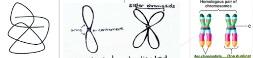
Chromatin DNA is unraveled (string-like) Sister Chromatid A single chromosome Homologous Chromosome 2 chromatids connected from each parent Homologous Pairs 2 of the same homologous chromosome
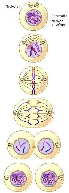
Phases of Mitosis Prophase Chromatin condenses into chromosomes Centrioles separate Spindle fibers form Nucleus breaks down Metaphase Chromosomes line up in the center of the cell Spindle fibers connect to chromosomes Anaphase Chromosomes separate at the centromere and sister chromatids move away from each other Chromosomes pulled towards opposite poles of the cell by spindle fibers Telophase Chromosomes unravel into thin strands of DNA (chromatin) Spindle fibers disappear Nucleus membrane reappears as nuclei are formed Results in two identical cells Cytokinesis The actual division of the cytoplasm Different in plant and animal cells Animal cells - pinches the cells apart Plant cells - a cell plate is formed, this becomes the new cell wall
Page 4
3. Meiosis
Meisosis: cell divison to make gametes Haploid: cell has half the number of chromosomes (n) In humans, the haploid amount is 23 (n) Diploid: cell has a full set of chromosomes (2n) In humans, the diploid amount is 46 (2n)
Meiosis 1 Interphase 1 DNA is in chromatin form During the S phase, DNA replicates Cells grow in prep for the division Prophase 1 Chromatin condenses into a homologous chromosome Nucleus disappears Spindle fibers form Tetrad occurs Crossing over occurs: exchange of genetic material between a homologous pair Metaphase 1 Tetrads (homologous pairs) line up in the middle of the cell Anaphase 1 Tetrads separate and each homologous chromosome moves to opposite ends of the cell Telophase 1 Nucleus appears Spindle fibers disappear Cytokinesis 1 Cytoplasm divides into 2 cells 2 haploid daughter cells
Meiosis 2 Interkinesis Resting phase No DNA replication Prophase 2 Chromosomes form again in both cells Nucleus disappears Spindle fibers form Crossing over does not occur Metaphase 2 Homologous chromosomes line up in the middle of the cell Anaphase 2 Chromosomes pull apart at the centromere Sister chromatids move to opposite ends of the cell Telophase 2 Nucleus reappears Spindle fibers disappear Chromosomes unravel Cytokinesis 2 Cells divide into 4 cells All cells are now haploid gametes (sex cells) All cells are genetically different
Page 5
Karyotype
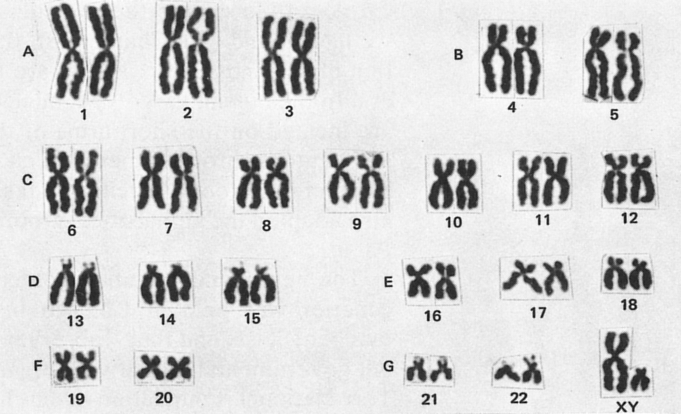
A karyotype is a picture of all chromosomes in an organism. It is used to help identify genetic disease due to abnormal numbers or damaged chromosomes. Nondisjunction: results in too few or too many chromosomes in a cell Can lead to: Monosomy: having only 1 chromosome instead of 2 Trisomy: having 3 chromosomes instead of 2
Page 6
Cancer
Cancer is an error in the DNA replication process that can alter genes that regulate cell growth. Cancer: cells that do not “listen” to checkpoints and signal from the body to stop dividing (continuous uncontrolled cell division) Cancer is caused by mutations and carcinogens, which are environmental factors that cause mutations in DNA.
Want to create your own Notes for free with GoConqr? Learn more.