
Fluxograma
Stomach
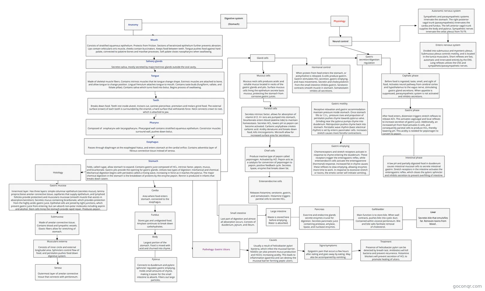
Flowchart: Stomach digestive system anatomy
Conteúdo do fluxograma
Anatomy
Digestive system
(Stomach)
Physiology
Mouth
Consists of stratified squamous epithelium. Protects from friction. Sections of keratinized epithelium further prevents abrasion. Lips contain orbicularis oris muscle, cheeks contain buccinators. Keeps food between teeth. Tongue pushes food against hard palate, connected to palatine bones and maxillae processes. Soft palate closes nasopharynx when swallowing.
Tongue
Made of skeletal muscle fibers. Contains intrinsic muscles that let tongue change shape. Extrinsic muscles are attached to bone, and allow tongue to change position. Lingual frenulum attaches tongue to mouth. Contains taste buds (fungiform, vallate, and foliate pillae). Contains saliva which turns food into bolus. Begins process of swallowing.
Salivary glands
Secretes saliva, mostly secreted by major/extrinsic glands outside the oral cavity.
Pharynx
Composed of oropharynx adn laryngopharynx. Pharyngeal wall contains stratified squamous epithelium. Constrictor muscles surround wall, pushes down bolus.
Esophagus
Passes through diaphragm at the esophegeal hiatus, and enters stomach at the cardial orifice. Contains adventitia layer of fibrous connective tissue instead of serosa.
Stomach
Folds, called rugae, allow stomach to expand. Contains gastric juice composed of HCL, intrinsic factor, pepsin, mucus, and bicarbonate. Gastric pits provide the opening for gastric glands. Utilizes two types of digestion: mechanical and chemical. Mechanical digestion begins with peristalsis called a mixing wave, increasing in force as it reaches the pylorus. The major chemical digestion in the stomach is the breakdown of proteins by the enzyme pepsin. Rennin is produced in infants that breaks down casein.
Liver
Secretes bile that emulsifies fat. Removes toxins from blood .
Gallbladder
Main function is to store bile. When wall contracts, pushes bile into cystic duct. Contained within visceral peritoneum. Bile and bile salts facilitate removal of cholesterol.
Pancreas
Exocrine and endocrine glands secrete enzymes crucial for digestion. Secretes pancreatic juice containing protease, amylase, lipase, and nuclease enzymes.
Small intestine
Last part of digestion and almost all absorption occurs. Consists of duodenum, jejnum, and illeum.
Large intestine
Waste is stored here before emptying. Water is absorbed.
Teeth
Breaks down food. Teeth rest inside alveoli. Incisors cut, canines pierce/tear, premolars and molars grind food. The external surface (crown) of each tooth is surrounded by the enamel, a hard surface that withstands force. Neck connects crown to root, which is attached to jaw.
Cephalic phase
Before food is ingested, taste, smell, and sight of food. Activates neural pathway from cerebral cortex and hypothalamus to the vagus nerve, stimulating gastric gland secretions. When appetite is suppressed, parasympathetic system is not activated and inhibits secretions.
Gastric phase
After food enters, distension triggers stretch reflexes to release ACh. This activates vagovagal and local reflexes to increase secretion of gastric juice. Additionally, an increased pH from food activates G cells, and consequently parietal cells to produce HCL, thereby lowering pH. This acidity is needed for pepsinogen to convert to pepsin.
Intestinal phase
A low pH and partially digested food in duodenum causes intestinal mucosal cells to secrete intestinal gastrin. Stretch receptors in the intestine activates the enterogastric reflex, which closes the pyloric sphincter and inhibits secretion to prevent overfilling of intestine.
Neural control
Gastric secretion/digestion regulation
Gland cells
Mucous cells
Mucous neck cells produces acidic and soluble mucus located in necks of the gastric glands and pits. Surface mucous cells lining the epithelium secrete basic mucous, protecting the stomach from corrosive gastric juices.
Parietal cells
Secretes intrinsic factor, allows for absorption of vitamin b12. H+ ions are pumped into stomach, bicarbonate enters blood (alakline tide) to maintain homeostasis. Secretes HCL, lowers pH so pepsin can become activated. Carbonic anyhydrase creates carbonic acid. Acidity denatures and breaks down food, kills microrganisms. Microvilli allow for increased surface area for secretions.
Chief cells
Produce inactive type of pepsin called pepsinogen. Activated by HCl. Pepsin acts as a catalysis for conversion of pepsinogen to pepsin, positive feedback cycle. Secretes lipase, enzyme that breaks down fat.
Enteroendocrine cells
Releases histamine, serotonin, gastrin, and somatostatin. Histamine triggers parietal cells to secrete HCL.
Enteric nervous system
Divided into submucous and myenteric plexus. Submucous plexus controls motility, and is located in the tunica muscularis. Short reflexes are fast, automatic and innervated entirely by the ENS. Long reflexes utilizes the CNS and sympathetic/parasympathetic nerves.
Autonomic nervous system
Sympathetic and parasympathetic systems innervate the stomach. The right posterior vagal trunk (parasympathetic) innervates the cardia and fundus. The left anterior vagal trunk supplies the body and pylorus. Sympathetic nerves innervate the celiac plexus from T6-T9.
Hormonal control
When protein from food enters the stomach, or acetycholine is released, G-cells produce gastrin. Gastrin stimulates HCL secretion, gastric emptying, and mass movements. Secretin and cholecystokinin from the small intestine inhibits gastrin. Serotonin contracts smooth muscle in stomach. Somatostatin inhibits all secretions.
Gastric motility
Receptive relaxation and gastric accommodation maintain pressure inside stomach. Once stomach fills to 1.5 L, pressure rises and propulsion of peristalsis pushes chyme towards pylorus valve. Grinding near the pylorus pumps chyme into duodenum. Retropulsion pushes chyme back into stomach. Peristaltic wave rhythm (basic electrical rhythm) is set by enteric pacemaker cells. Increases stretch causes more forceful contractions.
Gastric emptying
Chemoreceptors and stretch receptors activate in response to chyme entering the duodenum. These receptors trigger the enterogastric reflex, while enteroendocrin cells activate the enterogastrone (hormonal) response. Increased fat in chyme causes these reflexes to slow emptying, allowing enzymes more time to work. In response to excessive stretch or toxins, the emetic center will intitiate vomiting.
Gastric mucosa
Innermost layer. Has three layers: simple columnar epithelium (secretes mucus), lamina propria (loose areolar connective tissue, capillaries that supply epithelium, and lymphoid follicles provide protection) and muscularis mucosae (smooth muscle that assists in absorption/secretion). Secretes mucus containing bicarbonate, which provides protection from the highly acidic gastric juice. Epithelial cells are joined by tight junctions, which prevent gastric juice from entering, but can absorb non-polar molecules including aspirin and alcohol. Stem cells lining the stomach provide rapid repair. Produces pepsin.
Cardia
Area where food enters stomach, connected to the esophagus.
Histology
Submucosa
Made of areolar connective tissue. Contains blood and lympathic vessels. Elastic fibers allow for stretching of stomach.
Muscularis externa
Consists of inner circle and external longitudal area. Sphincters control flow of food, and peristalsis pushes food down digestive system.
Serosa
Outermost layer of areolar connective tissue that connects with peritoneum.
Fundus
Stores gas and undigested food. Amylase continues to break down carbohydrates.
Body
Largest portion of the stomach. Food is mixed with acid and churned into chyme.
Pylorus
Connects to duodenum and pyloric sphincter regulates gastric emptying. Holds small amounts of chyme, making it easier for the small intestine to absorb. Filters out large particles.
Pathology: Gastric Ulcers
Causes
Usually a result of helicobacter pylori bacteria, which infect the mucosal barrier. NSAIDs can also prevent mucus production and HCO3, increasing acidity. This leads to inflammation (gastritis) and can destroy the mucosal barrier forming peptic ulcers.
Signs/symptoms
Epigastric pain that occurs a few hours after eating and goes away by eating. May also be accompanied by vomiting.
Treatment
Presence of helicobacter pylori can be detected by breath test. Antibiotics will kill bacteria and prevent recurrence. Histamine blockers will prevent secretion of HCL to promote healing of ulcers.
Informações
- Descrição
- Fluxograma sobre Stomach, criado por Matt Wyatt em 13-01-2019.
- Criado por
- Matt Wyatt
- Visualizações
- 8
- Avaliação
Mais sobre Stomach and Gastric Secretion