Mapa mental
The musculoskeletal system (mod 2, mod 10 (wk6), mod 11 (wk2)
Conteúdo do mapa
The musculoskeletal system (mod 2, mod 10 (wk6), mod 11 (wk2)
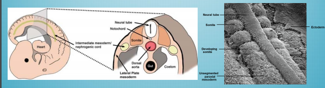
Embryology of the musculoskeletal system
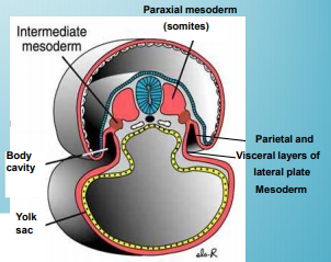
- Originates predominantly from the mesoderm with some components from the ectoderm (neural crest cells) that become nervous and blood tissue incorporated into the skeletal system The ectodermal components form the bones of the face
- Mesoderm
- Paraxial
- Somites
- 38 pairs form cranially and caudally bilaterally along the neural tube ad notochord
- Sclerotomes
- Vertebral columns and ribs
- Dermomyotomes
- Muscle cells and dermatomes
- Somites
- Lateral
- Forms the lining of the primitive body cavity and organs, peritoneal, pleural and pericardial cavities
- Somatic
- Forms into the Bone, skeletal muscle, connective tissue of limbs, pelvis and sternum Dermis of skin in body wall and limbs
- Splanchnic
- Forms into the Smooth muscle and cardiac
- Paraxial
- Ectoderm
- Ossification / osteogenesis
- Occurs in-utero in the foetus and continues into childhood
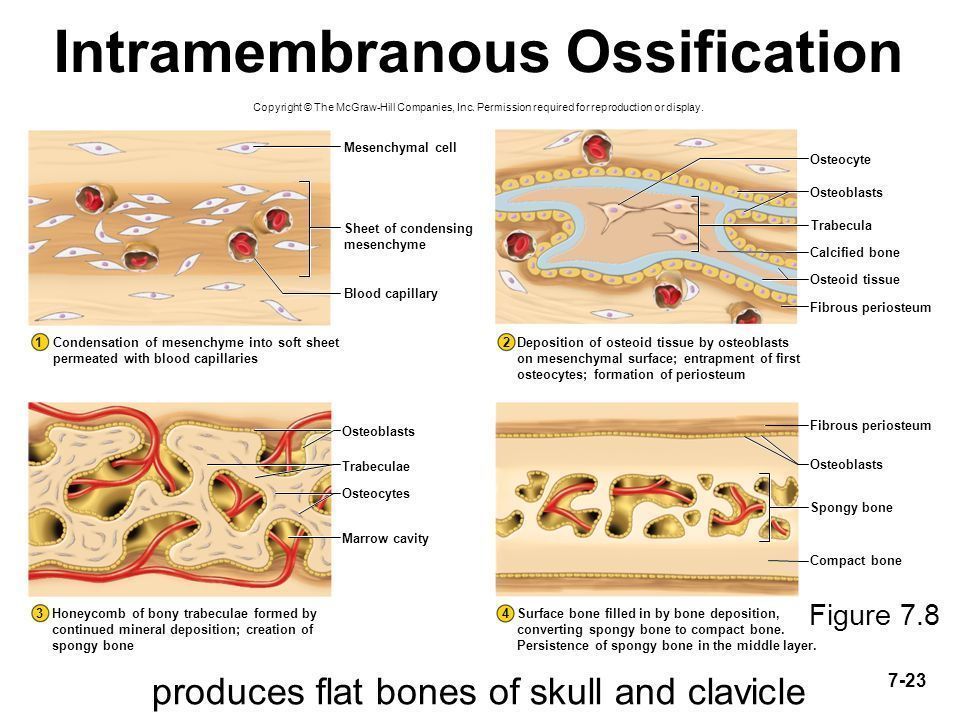
- Intra-membranous ossification
- Produces flat bones of the skull and clavicle
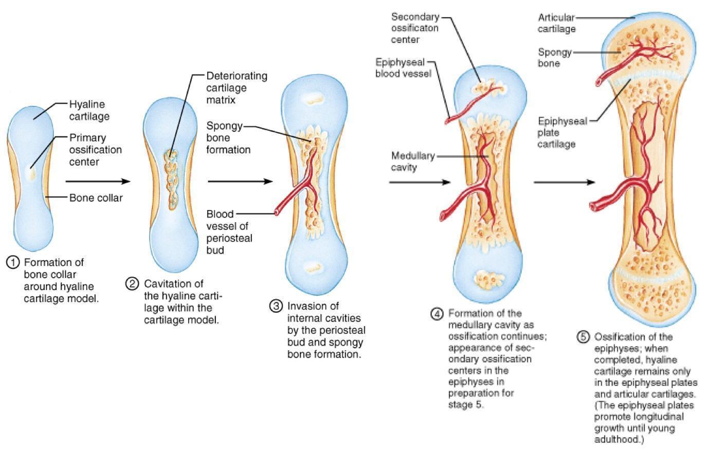
- Endochondral ossification
- This process of bone development utilises a template of hyaline cartilage that gradually becomes replaced by osseous tissue Occurs in week 6 of foetal life and continues into the 20s Mode of development of most bones of the body
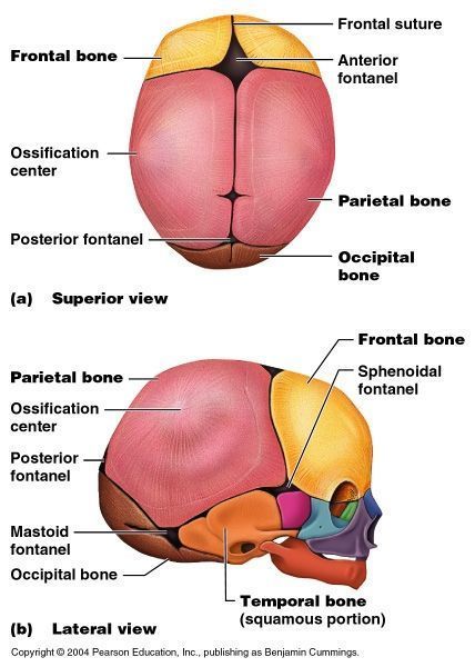
- The skull in utero and in childhood
- In utero, the cranial part of the skull is not a whole bone, but divided into separate bones by fontanelles, which fuse later in life to become sutures This is to allow molding through the vaginal and pelvic cavity during child birth, as the flat bones slide over each other during birth
- The fontanelles: The anterior The posterior The sphenod The mastoid
The Human Skeleton
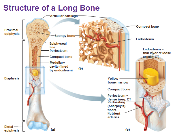
- Anatomy of bone
- Diaphysis
- Epiphysis
- The site of attachment for tendons and ligaments
- Articular cartilage
- Covers the ephiphyseal parts of the bone Site of joint formation Made of hyaline cartilage Produces synovial fluid to lubricate the joint surfaces during movement to prevent wear and tear
- Periosteum
- Provides a strong attachment and continuation for muscles to tendon to bone Plays a vital role in bone growth and fracture healing
- Epiphyseal plate
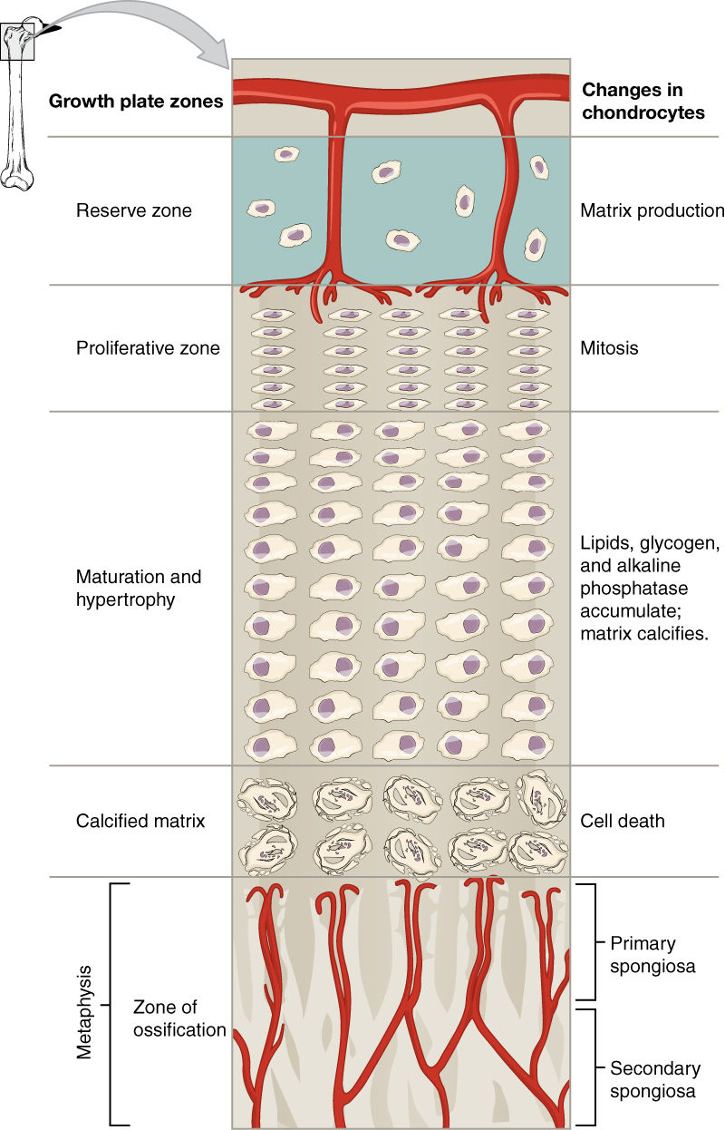
- Found in children and adolescents A plate of hyaline cartilage that separates the epiphysis from the diaphysis The zone of bone growth in length Seen as a translucent line on X-Ray
- Metaphysis
- The metaphysis is the area connecting the epiphyseal head to the diaphysis, with the epiphyseal plate (in under 20s) or line (over 20s) runnng through it This is the site of bone growth (transitional zone) where cartilage turns into bone
- Function of the skeleton
- Support
- Protection
- Storage and balance of electrolytes
- Acid-Base balance
- Haematopoeisis
- Locomotion
- Types of bone
- Irregular
- Vertebral bodies Ethmoid, sphenoid bones of the skull, etc.
- Flat bones
- Cranial bones the sternum Scapula Ribs Hip bones
- Long bones
- Arm: Humerus, Radius, Ulna Legs: Tibia, Fibula, Femur Hands and feet: metacarpals, metatarsals, phalanges of the hands and feet
- Short bone
- Carpals and tarsals of the wrists and ankles Patella of the knee
- Irregular
- Structure of the skeleton
- The skeleton is 75% compact bone and 25% spongy bone
- Axial skeleton
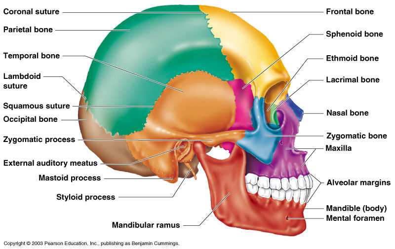
- Skull
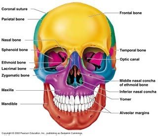
- Facial bones
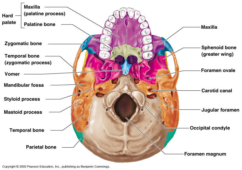
- The maxillae
- intraorbital foramen
- infraorbital erves and bessels
- Incisive foramen
- Naso-palantine nerves
- intraorbital foramen
- The palantine bones
- Greater palantine foramen
- Palantine nerves
- Greater palantine foramen
- The zygomatic bones
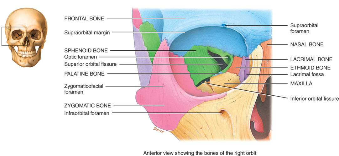
- Zygomaticofacial foramen
- Zygomaticofacial nerve
- Zygomaticotemporal foramen
- Zygomaticotemporal nerve
- Zygomaticofacial foramen
- The lacrimal bones
- Lacrimal foramen
- Tear duct leading to the nasal cavity
- Lacrimal foramen
- The nasal bones
- Inferior nasal conchae
- The vomer
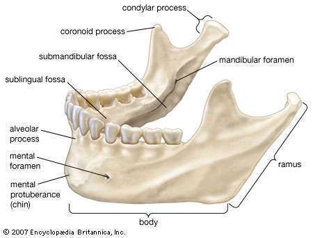
- The mandible
- Mental foramen
- mental nerve and vessels
- Mandibula foramen
- inferoior alveolar nerves and vessels to lower teeth
- Mental foramen
- Maxilla sphenoid region
- Infraorbital fissure
- Infraorbital nerve Zygomatic nerve Infraorbital nerve
- Infraorbital fissure
- The maxillae
- Cranial bones
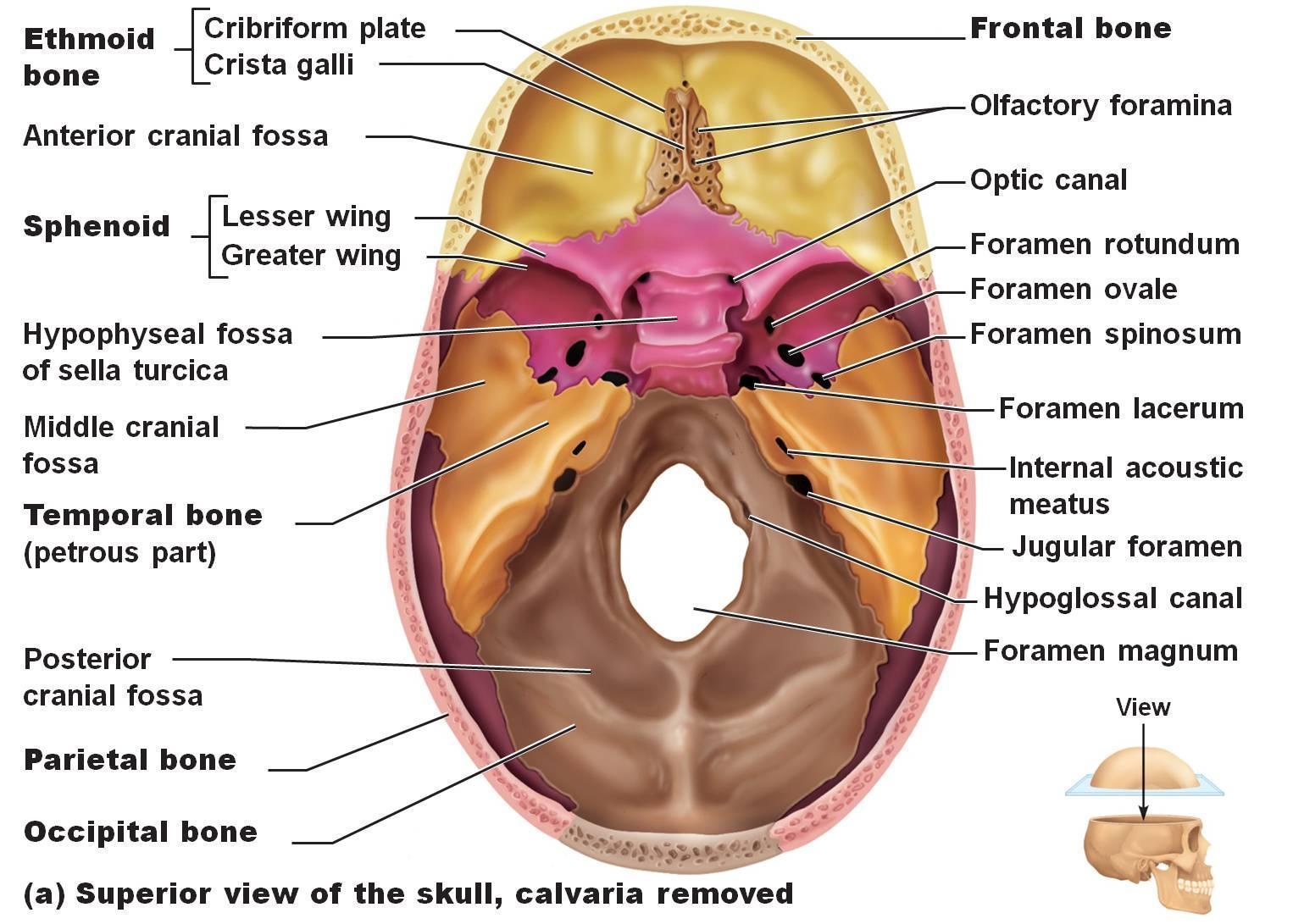
- Made up of 2 pairs of cranial fossa, divided into 3
- Cranial floor
- The anterior cranial fossa: frontal lobe Medial cranial fossa: temporal lobe Posterior cranial fossa: cerebellum
- Frontal bone
- Supraorbital foramen
- parietal bones
- Made up of the sagittal surture, corononal suture, lambdoid suture ad squamous suture
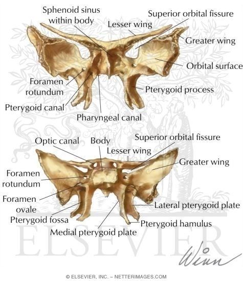
- Sphenoid bones
- foramen ovale
- Mandibular branch of the trigeminal nerve (CN V) Accessory meningeal artery
- Foramen rotundum
- Maxillary division of CN V (trigeminal nerve)
- Foramen spinosum
- Middle meningeal artery Spinosal nerve Some branch of the trigeminal nerve
- optic canal
- Optic nerve Ophthalmic artery
- Superior orbital fissure
- Oculomotor nerve (CN III) trochlear (CN IV) abducens nerve (CN VI) Ophthalmic division of trigeminal nerve (CN V)
- foramen ovale
- Occipital bone
- Foramen magnum
- Spinal cord Accessory nerve (CN XI) Vertebral arteries
- Hypoglossal canal
- Condylar canal
- Vein from transverse sinus
- Foramen magnum
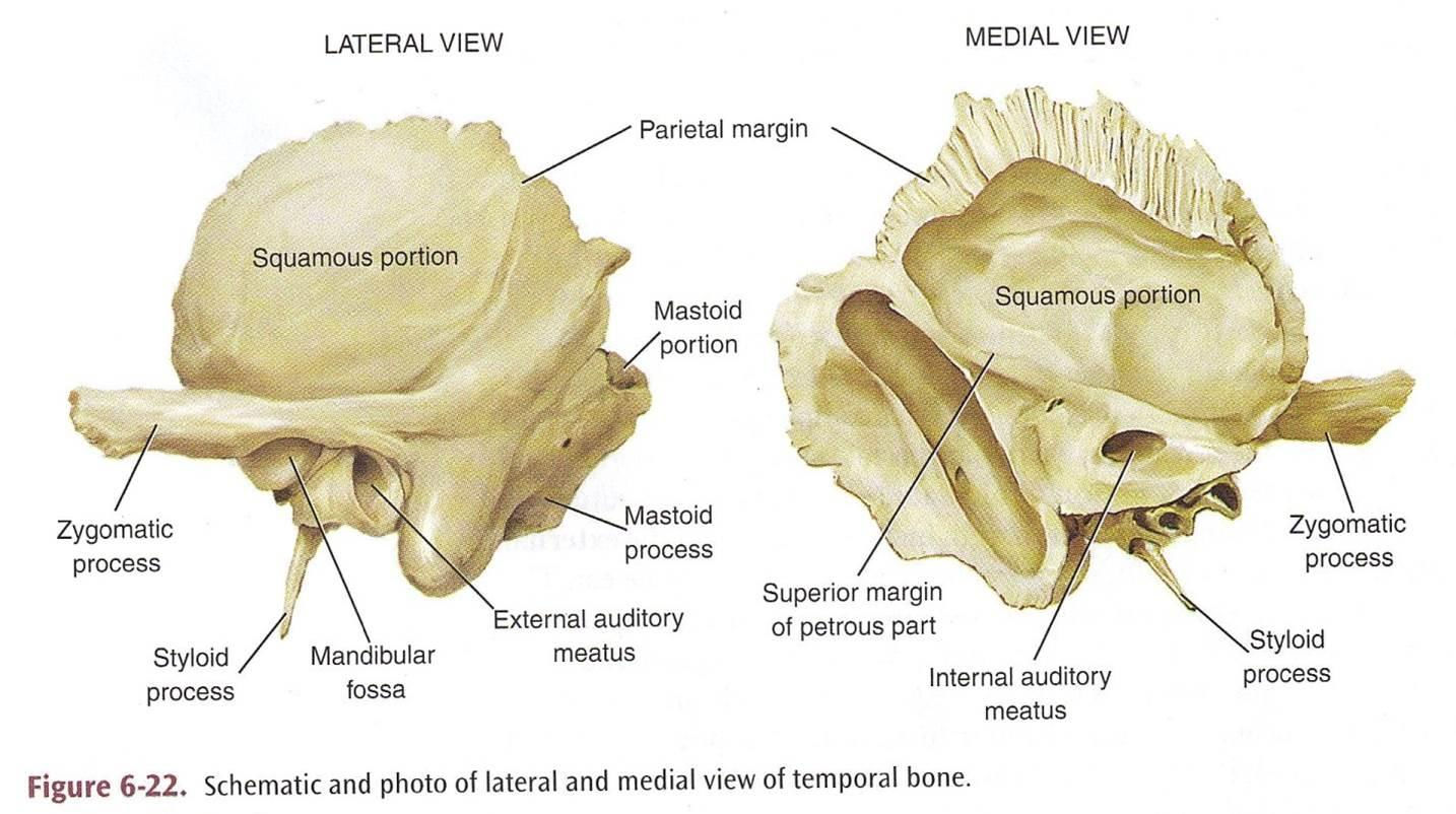
- Temporal bones
- Styloid process
- Attachment point for muscles of the tongue, pharynx and hyoid bone
- Carotid canal
- internal carotid artery
- External acoustic meatus
- Internal acoustic meatus
- vestibulocochlear nerve (CN VIII)
- Stylomastoid foramena
- Facial nerve (CN VII)
- Mastoid foramen
- Meningeal artery, sigmoid sinus vein branch
- Styloid process
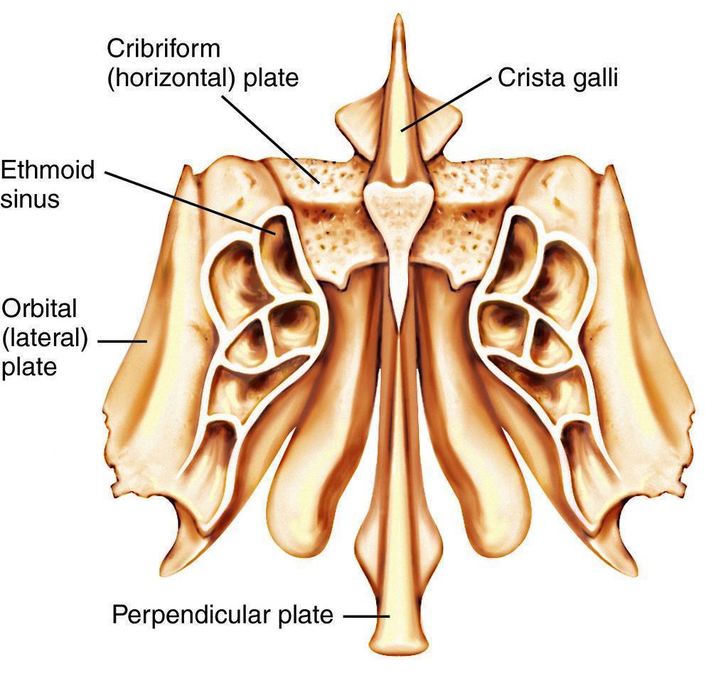
- Ethmoid bone
- Cribriform foramen
- Olfactory nerve (CN I)
- Cribriform foramen
- Cavities of the brain
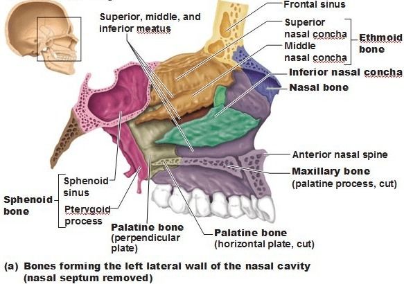
- Nasal cavity
- The bones are designed to create air turbulence to allow contact with the membrane to warm, moisturise and humidify the air before reaching the lungs
- Also involved carrying the sensory cells of smell
- Oral cavity
- Auditory cavities
- Para-nasal sinuses
- Connected by the nasal cavity, and are filled with air making the skull light-weight, help in producing vocal sounds
- Ethmoid sinus
- Maxillary sinus
- Sphenoid sinus
- Frontal sinus
- Orbital cavity
- Nasal cavity
- Auditory ossicles
- Malleus Incus Stapes
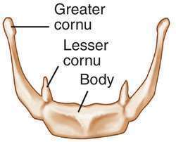
- Hyoid bone
- Facial bones
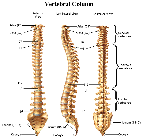
- The spinal column
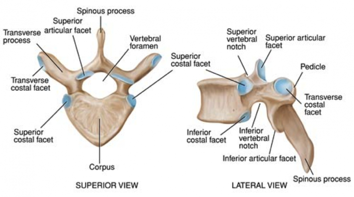
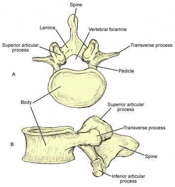
- The vertebral column: Protects the spinal cord Absorbs the stresses from locomotion and external forces Attachment for the ribs, skull, pelvic and shoulder girdle 33 vertebral bones
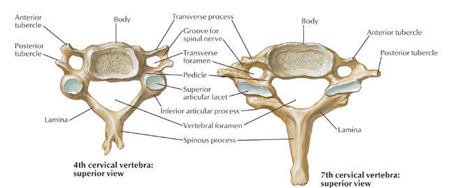
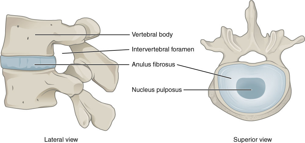
- Basic structure The spinal cord runs through the vertebral foramen The space between 2 pedicles of 2 vertebrae: intervertebral foramen--> the passage of pinal nerves
- Vertebrae
- Cervical
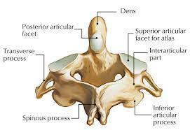
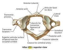
- C1 - C7 C1: the atlas --> flexion and extension C2: the axis --> rotation
- Thoracic
- T1-T12 The site of attachment of the 12 pairs of ribs to enclose the lungs, heart, and hepatobiliary and gastroenterological structures
- Lumbar
- L1-5
- Sacral
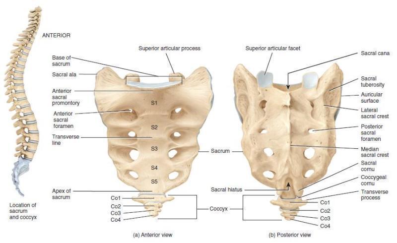
- Forms the posterior wall of the pelvic gurdle Begin as 5 separate bones They contain foramena, passages for nerves and blood vessels to the pelvis Forms the sacroiliac joint
- Coccygeal
- The coccyx is the attachment point for muscles of the pelvic floor Consists of 4 fused bones
- Cervical
- Intervertebral discs
- Between each vertebrae is cushioning tissue: intervertebral discs They bind adjacent vertebrae together Support the weight of the body bsorb shock
- Herniated disc
- The thoracic cage
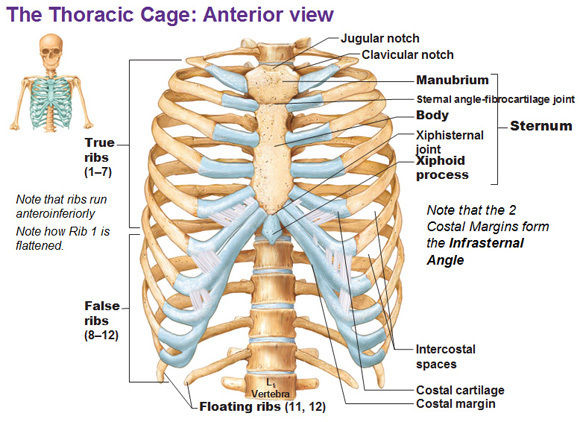
- Formed by the thoracic verebrae, sternum and ribs The enclosure of the heart and the lungs, the abdominal organs (spleen, liver, pancreas, kidneys) Also involved in breathing by helping to create the intra-thoracic pressures needed for exhalation ad inhalationThe attachment point for the pectoral girdle and upper limb
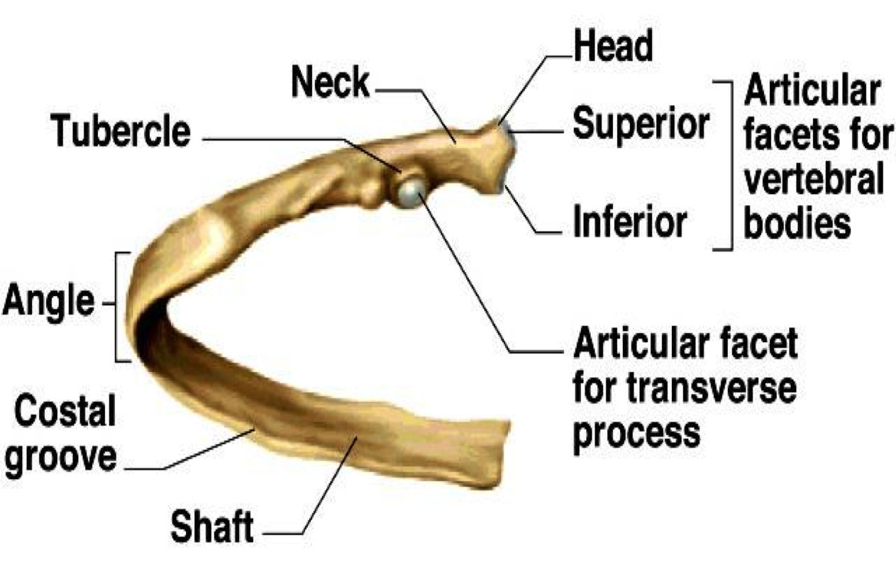
- The ribs
- Attach to the sternum anteriorly distally and the thoracic vertebrae posteriorly proximally, Anteriorly, they have the costal cartilage
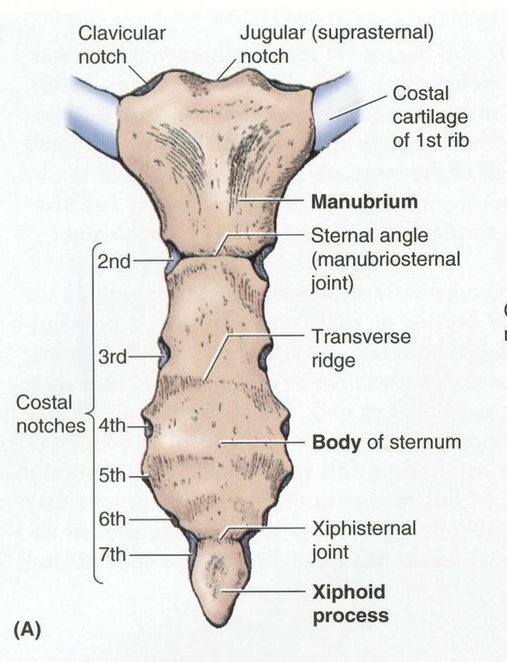
- The sternum
- The body of the sternum extends from T5 to T9 The xiphoid process is the point of attachment of some of the abdominal muscles The landmark for CPR (2 fingers away from it)
- Skull
- Appendicular skeleton
- Shoulder gurdle
- Also known as the pectoral gurdle acts as the attachmet of the upper limbs to the axial skeleton
- It is formed by the Sternoclavicular joint Acromioclavicular joint Glenohumeral joint
- The clavicles
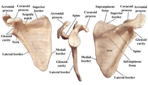
- The scapula
- The coracoid process
- The attachment of the tendons of the biceps brachii
- The glenoid cavity
- The coracoid process
- Pelvic gurdle
- Upper Limbs
- The brachium
- Antebrachium
- The carpus
- Lower limb
- Shoulder gurdle
- Histology of bone
- Bone cells
- Osteoblasts
- Bone-forming cells: lay down the mineral deposits of bone to reinforce and rebuild bone Formed from osteogenic cells Stress and fractures stimulate increased osteogenic cells mitosis and differentiation into osteoblasts Also secrete osteocalcin that stimulates insulin secretion, and increases insulin sensitivity of adipocytes and limits their growth
- Osteoclasts
- Bone-dissolving cells Different cell lineage from the other bone cells Large cells, formed from multiple stem cells (3-50!) Bone resorption
- Osteocytes
- Old osteoblasts Resorb bone matrix, deposit bone matrix Play a role in bone density and blood concentrations of calcium and phosphate Strain sensors --> stress induces bone remodelling of shape and density
- Osteogenic cells
- Stem cells from embryonic mesenchymal cells Continuously undergo mitosis, some differentiate into osteoblasts
- Osteoblasts
- Histological structure
- Compact bone
- Layers of matrix: lamellae Arranged around central canals: Haversian canal Connected by canaliculi to make Haversian systems
- Spongy bone
- Bone marrow
- Red marrow
- In children, all their bone contains red marrow Red marrow is haemapoietic, containing myeloid tissue In adults, the red marrow is only found in the axial skeleton, part of the pelvic gurdle and the proximal heads of the humerus and femur
- Yellow marrow
- Replaces most of the red marrow in adults Consists of adipose tissue In severe chronic anaemia, it can revert back to red marrow!
- Red marrow
- Compact bone
- Bone matrix
- 85% hydroxyapatite (crystallized calcium phosphate salt) 10% CaCO3 Mg, Na, K+,Fl, SO4(2-), HCO3-, OH- ions
- Bone cells
- Bone physiology
- Bone growth and remodelling
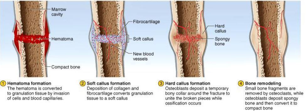
- Fractures and repair
- Bone elongation
- Occurs at the epiphyseal plate, the growth zone of bones, resulting in increase in height The epiphyseal plate contains hyaline cartilage in the middle with transitional zones at each end Starts from the cartilage cells (zone 1), which are derived from the reserve zone Zone 5 is nearer to the bone marrow and the epiphyseal head. Growth starts in zone 1, 2 and 3 leading to interstitial growthFailure of growth from the cartilage results in dwarfism
- Bone widening and thickening
- Occurs in appositional growth, growth of new tissue on the surface
- paget's disease of the bone
- Bone remodelling
- Continues throughout life Occurs by absorption of old bone and deposition of new bone Annually, we replace 10% of our bone to repair micro-fractures, release minerals into the blood and reshape bones in response to use and disuse. Increased stress in a bone over time increases its density Occurs via interaction of the osteoclasts and osteoblasts Disuse: increased osteoclasts activity to decrease mass Increased use: thickened bone
- Osteoporosis
- Puberty
- Oestrogen, testosterone and GH are secreted at the start of puberty and are what initiate bone ossification of the epiphysial plates Thus it is the last thing that occurs in children (GnRH secretion by the hypothalamus) to allow the child to attain their potential height
- The sex steroids have an influence on osteogenic cells, chondrocytes and osteoblasts
- Oestrogen
- Has a more significant effect on bone mineralization on girls than testosterone
- Testosterone
- Precocious puberty
- Leads to rapid growth, but causes early fusion of the epiphyseal plates leading to stunted growth
- Delayed puberty
- Leads to prolonged bone growth, slow attainment of their adult height, and bone age less than their chronological age
- Mineral deposition and resorption
- Mineral deposition
- Performed by osteoblasts by absorbing PO4(3-), Ca2+, etc. from the blood and layering them into the bone Involved in ossification Can occur abnormally in organs and tissue such as the brain, kidneys, ureters, bladder, joints, arteries (arteriosclerosis), tendons, etc.
- Mineral resorption
- Performed by osteoclasts releasing minerals into the blood They have receptors to detect calcium levels in the blood and dissolve bone in response to low levels (using HCl- and acid phosphatase)
- Mineral deposition
- Calcium homeostasis
- Calcium plays a role in action potential in neurones, activation of enzymes and intracellular metabolic processes, blood clotting, muscle contraction and exocytosis
- The skeleton acts a storage for calcium (~1kg) 1% flows in blood, 99% in bones 18% is exchanged with the blood per year Blood serum levels: 9.2-10.4mg/dl (2.25-2.6 mmol/L) 45% is ionised and can pass through membranes, the rest is bound to albumin and other plasma proteins
- Hypocalcaemia
- Signs
- Tetany (prolonged held contraction) Muscle tremors and spasms Trousseau's sign: strong spasmodic flexion of the wrist and thumb and extension of the fingers on compression of the brachial nerve by a blood pressure cuff Rarely, laryngospasm
- Causes
- Vitamin D deficiency Diarrhoea, Thyroid tumours (suppress parathyroid glands) Hypoparathyroidism Pregnancy and lactation due to increased demand of calcium from the foetus Iatrogenic: accidental parathyroidectomy secondary to a thyroidectomy in hyperthyroidism or in a head & neck surgery (vascular damage)
- Osteomalacia
- Rickets
- Signs
- Hypercalcaemia
- SIgns
- Depression of the nervous system Emotional disturbances Muscle weakness Sluggish reflexes Cardiac arrest
- SIgns
- Skin
- The skin utilises UV B light to turn cholesterol into cholecalciferol (VD3)
- Liver
- The liver turns the VD3 into calcidiol
- Kidneys
- The kidneys turn VD2 into VD1 (calcitriol)
- Bowel
- The bowel uses vitamin D1 to absorb calcium into the blood system
- Bone
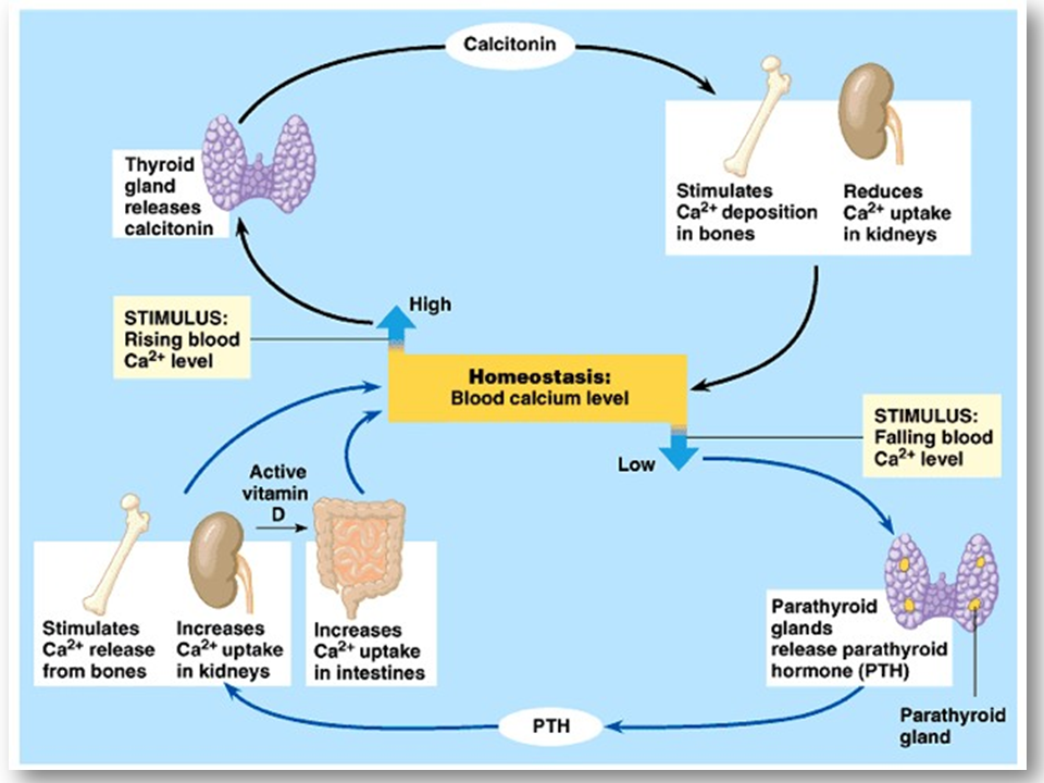
- The bone absorbs calcium in high levels with stimulation In low levels, the PTH stimulates osteoclasts to resorb calcium and release it into the blood In high levels, calcitonin is released by the thyroid glands to inhibit osteoclast actitivity and increase osteoblast actitvity
- Parathyroid gland
- Thyroid
- Calcitonin
- Secreted by c cells of the thyroid gland when serum calcium is too high
- Acts by inhibiting osteoclast activity and stimulating osteoblast activity Essential in pregnant and lactating women
- Calcitonin
- Parathyroid hormone
- PTH is secreted in low calcium levels It acts on the bones, kidneys and gut
- Bones: binds to osteoblasts to stimulate RANKL secretion to bind to RANKR on osteoclasts to activate their differentiation and activity --> bone resorption Also inhibits bone deposition by osteoblasts
- Increases calcium resorption by the kidneys from urine and increases phosphate excretion in the urine
- Promotes vitamin D activation by the kidneys to increase calcium absorption by the gut
- Thyroid
- Calcitriol
- Calcitriol on the gut: increases absorption of calcium and phosphate
- On the bones: increases resorption by triggering osteoblasts to secrete RANKL which bind to RANK receptors on the osteoclasts to activate them to resorb bone and release calcium and phosphate into the blood
- Kidneys: acts to increase reabsorption of Ca2+ from the urine
- Calcidiol
- Cholecalfiferol
- Phosphate homeostasis
- Phosphate is vital for enzyme activation, cell receptors, DNA, RNA, ATP and many other proteins in the body Phosphate ions are used to buffer in acidic conditions
- Bone growth and remodelling
- Anatomy of bone
Anatomy and physiology of muscles
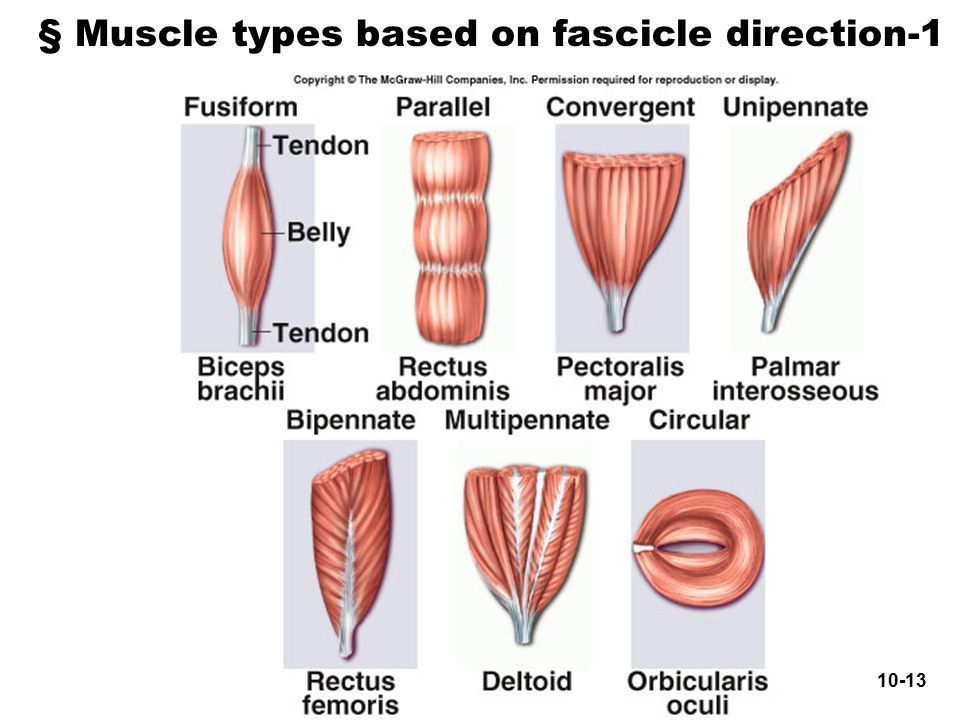
- Structure of muscles
- Macroscopic
- Fascia
- Compartmentalises muscles
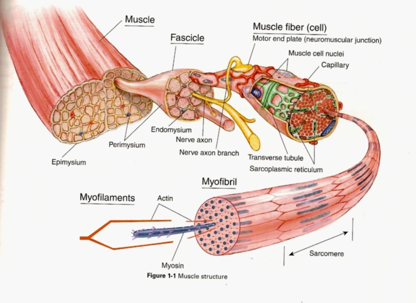
- Epimysium
- Perimysium
- Bundles muscle fibres together to form fascicles: the parallel strands of muscles Carries the larger nerves and blood vessels and stretch receptors (muscle spindles)
- Endomysium
- Surrounds each muscle fibre, and creates room for capillaries and nerve fibres to each fibre, space for extra-cellular chemical environment and nerve endings
- Perimysium
- Fascia
- Microscopic
- Macroscopic
- Anatomy of the muscular system
- Pelvic muscles
- Abdominal muscles
- Abdominal muscles
- Upper limb muscles
- Lower limb muscles
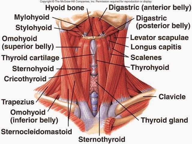
- Head and neck muscles
- Thoracic muscles
- Physiology of muscles
- Functions of muscles
- Movement
- Locomotion Movement of body contents: air, fluid, blood, foetus, food, faeces, urine Communication
- Stability
- Control of body openings and passages between the body and environment
- Heat production
- Glycaemic control
- Movement
- Structure of muscles