Cells are studied using microscopes
Microscopes let us see things that we cant see with the naked eye. The microscopy techniques we can use have developed over the years as technology and knowledge have improved.
Light microscopes use light and lenses to form an image of a specimen and magnify it. They let us see individual cells and large subcellular structures, like nuclei.
Electron microscopes use electrons instead of light to form an image. They have a much higher magnification than light microscopes.
They also have higher resolution. Resolution is the ability to distinguish between two points, so a higher resolution gives a sharper image.
Electron microscopes let us see much smaller things in more detail, like the internal structure of mitochondria and chloroplasts. They even let us see tinier things like ribosomes and plasmids.
Slide 2
Using a light microscope
Preparing a slide:
Place the specimen onto the slide
Add drops of stain (iodine for a plant and methylblue for animal) to the specimen. Stains like iodine make features such as the nucleus easier to see.
Place a cover slip over the specimen and make sure air bubbles are removed
Place the slide on the stage and adjust the objective lens and focus
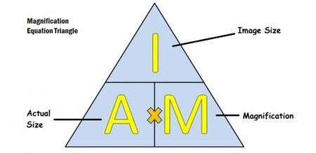
Calculating magnification:
I = image size I = A x M
A = actual size A = ! / M
M = magnification M = I / A
1. A red blood cell is 7.5 micrometres in diameter. It is magnified 2000 times. Calculate the diameter of the image seen through the microscope in mm.
Answer:
I = A x M
I = 7.5 x 2000
I = 15,000 mm
2. A sperm cell has a tail 40 micrometres long and a student draws it 40 mm long. Calculate the magnification.
Answer:
M = I / A M = 40 mm / 40 micrometres
M = 4000 / 4
M = 1000
Slide 4
A light microscope uses a series of lenses to produce a magnified image of an object:
The object is placed on a rectangular glass slide
The slide is placed on a stage with a light source below
Light shines through the object and into the objective lens
The light passes through the eyepiece lens and from there into your eye.
The Light Microscope and Observing Cells
When you observe cells, it is usual to make a drawing of what you see. Very often there is so much to see that you can only aim to draw part of it.
Use a pencil rather than pen or colours
Outline the features as accurately as you can
Use as little shading as possible
Label your drawing with the name of the sample and the total magnification you used
