
Karteikarten
Eye
Karten in diesem Stapel (89)
- 1What are common complaints or concerns?Pain
Redness
Discharge
Photophobia
Foreign body
Visual disturbances - 2How do you test for visual acuity?Pick a chart (Snellen or Rosenbaum). Test with and without corrective lenses. Cover one eye. Smallest line read with >50% accuracy.
- 3What is the top number in visual acuity mean?Distance between patient and chart
- 4What does the bottom number in visual acuity mean?Distance at which a normal eye can read letters
- 5What is the visual acuity for legal blindness?20/200
- 6OS - oculus sinisterleft eye
- 7OD - oculus dexterright eye
- 8OU - oculus uterqueboth eyes
- 9During pinhole testing, what does improvement of vision mean?Implies a functional problem (ex: refractive error)
- 10During pinhole testing, what does a lack of improvement in vision mean?Implies a structural problem (ex: optic nerve damage)
- 11MyopiaNearsightedness - difficulty seeing far away
- 12HyperopiaFarsightedness - difficulty seeing close up
- 13Presbyopiainability to focus on near objects (aging vision)
- 14Diplopiadouble vision
- 15Scotomasblind spot in visual field
- 16Photophobiapain with vision/light
- 17Give an example of confrontation testing of visual fields.static finger wiggle test. Put them one up high and one low on either side of pt.
- 18—Bitemporal Hemianopsia

- 19—Homonymous Hemianopsia

- 20Homonymous Hemianopsiathe loss of half of the field of view on the same side in both eyes
- 21Bitemporal Hemianopsialoss of vision in the outer (temporal or lateral) half of both the right and left visual fields
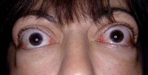
- 22Exophthalmosprotruding of eyeball
- 23Enophthalmosrecession of eyeball
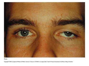
- 24PtosisDropping of the upper eyelid. Interruption of CN III nerve supply.
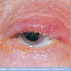
- 25BlepharitisInflammation of lid margin with redness, thickening and scales of crust
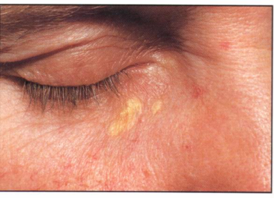
- 26XanthelasmaCholesterol deposits
- 27—Blepharitis
- 28—Xanthelasma
- 29—Exophthalmos
- 30—Ptosis
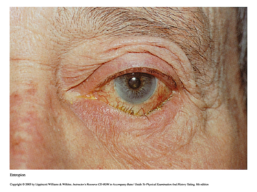
- 31EntropionLid droops inward, lower lash may irritate conjunctiva and cornea
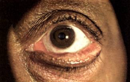
- 32EctropionLid droops outward exposing palpebral conjunctiva - when punctum gets blocked it causes poor drainage and tearing
- 33—Entropion
- 34—Ectropion
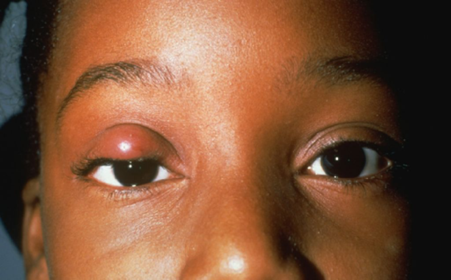
- 35ChalazionBeady nodule inside the eyelid due to a blocked sebaceous gland
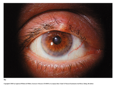
- 36HordeolumSty - painful, tender infection around hair follicle lid margin
- 37—Chalazion
- 38—Hordeolum
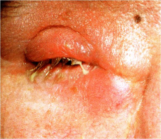
- 39DacrocystitisInflammation of the lacrimal sac (painful, red and tender)
- 40—Dacrocystitis
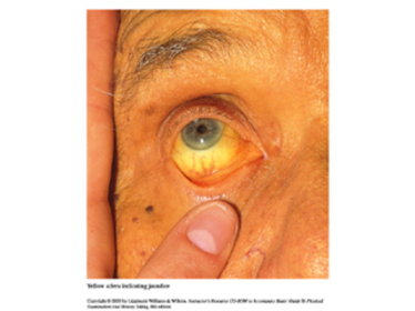
- 41—Scleral Icterus (jaundice in eye)
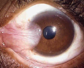
- 42—Pterygium
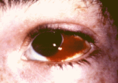
- 43—Subconjunctival Hemorrhage
- 44PterygiumA triangular thickening of the bulbar conjunctiva that grows slowly across the outer surface of the cornea (usually from the nasal side)
- 45ConjunctivitisDiffuse dilatation of conjunctival vessels with redness that tends to be maximal peripherally. Can be viral, allergic, bacterial.
- 46StrabismusA deviation of the eyes from the normally conjugate position
- 47NystagmusFine, rhythmic oscillations of the eyes, normal for a few beats seen at terminal range of eye movement. Sustained indicates neurological condition. Lid lag can indicate hyperthyroidism
- 48Miosisconstriction of the pupils
- 49Mydriasisdilation of pupils
- 50AnisocoriaUnequal pupils
- 51AccommodationAbility of pupils to constrict when focus shifts from distance to close object
- 52ConvergenceAbility of eyes to move toward the nose symmetrically when following an object moving toward them.
- 53PERRLAPupils Equal, Round, Reactive to Light and Accommodation
- 54What muscle moves the eyes to the left and right?Lateral Rectus & Medical Rectus
- 55What muscle moves the eye up and down?Superior Rectus and Inferior Rectus
- 56Name the 6 cardinal gazes1. Medial Rectus
2. Lateral Rectus
3. Superior Rectus
4. Inferior Rectus
5. Superior Oblique
6. Inferior Oblique - 57What is the nerve innervation of lateral rectus?Abducens nerve (CN VI)
- 58What is the nerve innervation of superior oblique?Trochlear nerve (CN IV)
- 59What is the nerve innervation for the majority of the ocular muscles?Oculomotor nerve (CN III)
- 60FundusPosterior aspect of the eye seen through the ophthalmoscope
- 61Retinavisual screen and inner layer of the eye
- 62What does the optic disc contain?optic nerve (CN II), retinal vein and arteries
- 63FoveaA small depression in the retinal surface where largest concentration of cones are contained therefore the central point of vision
- 64Maculayellow oval around the fovea - acts as a natural blocker of UV light and sunblock for the fovea
- 65Physiologic cupdepression in the optic disc
- 66What is the red reflex?During a fundoscopic exam, it is the light reflecting off the retina. (absence of red reflex can mean cataracts or vitreous hemorrhage)
- 67What are the two types of glaucoma?1. open angle glaucoma (2nd leading cause of blindness in Americans)
2. acute closed angle glaucoma - 68A-V taperingThe vein appears to taper down on either side of the artery.
- 69A-V nickingvein abruptly stops on either side of the artery
- 70—A-V tapering
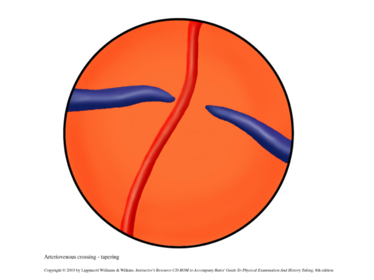
- 71—A-V nicking
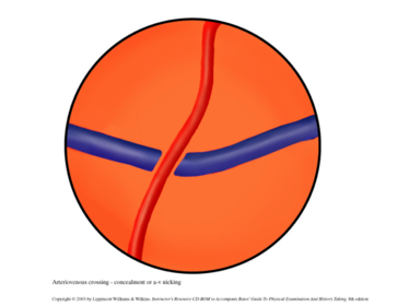
- 72—Cotton-wool patches
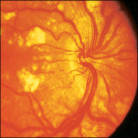
- 73—Retinal Detachment
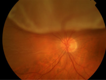
- 74What is a sit lamp examination and what does it examine?Magnifying device to examine anterior portion of the eye.
- cornea
- anterior chamber
- iris
- lens - 75Conjunctivaclear membrane covering the eye and the underside of the eyelids
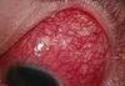
- 76—Scleritis
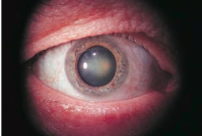
- 77Nuclear Cataractlooks gray when seen by a flashflight
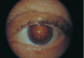
- 78Peripheral cataractProduces spokelike shadows that point gray against black, as seen with a flashlight, or black against red with an ophthalmoscope
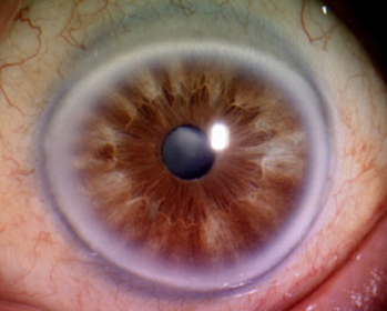
- 79Arcus senilisRing around the iris. Common in elderly, From cholesterol deposits, may indicate hyperlipidemia.
- 80—Arcus Senilis
- 81—Peripheral Cataract
- 82—Nuclear Cataract
- 83Angle Closure Glaucomaa sudden elevation in intraocular pressure that occurs when the iris blocks the eye's drainage channel—the trabecular meshwork
- 84Extraocular MovementsTesting the function of each ocular muscle and the cranial nerve that supplies it by asking the patient to move the eye in the direction controlled by that muscle (H)
- 85Consensual reactionpupillary constriction in the opposite eye
- 86What does the swinging flashlight test examine for?Used to test for impairment of optic nerves II and III
- 87How do pupils constrict?Reflex arc is initiated by reaction to light: CN2 (afferent) and CN3 (efferent)
CN3: parasympathetic innervation, pupil constricts
Sympathetic: dilate - 88Name possible retinopathies for diabeticsHard exudates
Cotton wool patches
Deep hemorrhages - 89Name possible retinopathies for hypertensivesAV tapering, nicking, banking
Copper wiring
Hard exudates
Papilledema
Informationen
- Beschreibung
- PD Module 1 Karteikarten am Eye, erstellt von Adriana Morales am 22/10/2016.
- Erstellt von
- Adriana Morales
- Aufrufe
- 1
- Verlauf
-
- Erstellt von Chelsea Cheeley · 16 Okt 2016
- Kopiert von Adriana Morales · 22 Okt 2016
- Bewertung