
Karteikarten
Cardiac Physiology PA School
Karten in diesem Stapel (56)
- 1Arteries* Deliver oxygenated blood to the tissues
* Thick-walled. with elastic tissue and smooth muscle
* Under high pressure
* Blood volume is called stressed volume - 2Arterioles* Smallest branches of the arteries
* Site of highest resistance in the cardiovascular system
* Resistance regulated by the ANS
* Alpha1 adrenergic receptors found on arterioles of the skin, splanchnic and renal circulations
* Beta2 adrenergic receptors found on arterioles of skeletal muscle - 3Capillaries* Have largest total cross-sectional and surface area
* Single layer of endothelial cells surrounded by basal lamina
* Thin-walled
* Site of exchange of nutrients, water, and gases - 4Venules* Formed from merged capillaries
- 5Veins* The largest vein is the vena cava which returns blood to the heart
* Thin-walled
* Low pressure
* Contain highest proportion of blood in the cardiovascular system
* Blood volume contained is called unstressed volume
* Have Alpha1 adrenergic receptors - 6Velocityv = Q/A
Q = blood flow
A = cross-sectional area
* directly proportional to blood flow
* inversely proportional to the cross-sectional area - 7Velocity is _________ in the aorta than in the sum of all the capillaries.Blood velocity is higher in the aorta (small-cross-sectional area) than in the sum of all the capillaries (large cross-sectional area).
The lower velocity in the capillaries optimizes conditions for exchange of substances across the capillary wall. - 8Cardiac Output =Q = ΔP/R
CO = (MAP - Right arterial pressure)/Total peripheral resistance
* Blood flow is inversely proportional to the resistance of the blood vessels - 9Resistance =R = (8ηl)/(πr^4)
η = viscosity of blood
l = length of vessel
* Resistance is directly proportional to viscosity and the length of the vessel
* It is inversely proportional to the 4th power of the vessel radius. Ex: If vessel decreased by a factor of 2 then resistance increases by a factor of 16 and blood flow decreases by a factor of 16. - 10Parallel Resistance* Systemic circulation: each organ is supplied by an artery that branches off the aorta.
R(total) = 1/R1 + 1/R2 + 1/R3 + 1/R4, etc.
* Total resistance is less than the resistance of any of the individual arteries.
* The pressure in each parallel artery is the same. - 11Series Resistance* Illustrated by the vessels within an organ. Each organ is supplied by a large artery, smaller arteries, arterioles, capillaries, and veins.
R(total) = R(artery) + R(arterioles) + R(capillaries)
* The largest portion of resistance is from the arterioles.
* As blood flows through the series of blood vessels, pressure decreases. - 12Reynold's Number* Predicts whether blood flow will be laminar or turbulent.
* The higher the number, the greater the chance for turbulent flow.
* Increased by a decrease in blood viscosity
* Increased by an increase in blood velocity - 13Capacitance (Compliance)* Describes the dispensability of blood vessels
* C = V/P (volume/pressure)
* Is much greater for veins than arteries
* A decrease in venous capacitance decreases venous volume and increases arterial volume
* Capacitance of the arteries decreases with age - 14The largest decrease in pressure...Occurs across the arterioles because they are the site of highest resistance.
- 15Mean pressures in the: Aorta Arterioles Capillaries Vena cavaAorta: 100
Arterioles: 50
Capillaries: 20
Vena cava: 4 - 16Systolic Pressure* Highest arterial pressure during a cardiac cycle
* Measured after the heart contracts (systole) and blood is ejected into the arterial system - 17Diastolic Pressure* Lowest arterial pressure during a cardiac cycle
* Measured when the heart is relaxed (diastole) and blood is returned to the heart via the veins - 18Pulse Pressure* Difference between the systolic and diastolic pressures
- 19The most important determinant of pulse pressure isStroke volume.
As blood is ejected from the left ventricle into the arterial system, arterial pressure increases because of the relatively low capacitance of the arteries. - 20Decreases in capacitance, cause ________ in pulse pressure.Increases
- 21Mean Arterial Pressure (MAP)Average arterial pressure with respect to time.
~ diastolic pressure + 1/3 pulse pressure - 22P wave* Represents atrial depolarization
- 23PR interval* The interval from the beginning of the P wave to the beginning of the Q wave
* Varies with conduction velocity through the AV node. If conduction decreases, the PR interval increases - 24An increase in the conduction velocity by the ___________ nervous system, ___________ the PR interval.An increase in the conduction velocity by the sympathetic nervous system, decreases the PR interval.
- 25A decrease in the conduction velocity by the ___________ nervous system, ___________ the PR interval.A decrease in the conduction velocity by the parasympathetic nervous system increases the PR interval.
- 26QRS complexRepresents depolarization of the ventricles.
- 27QT interval* The interval from the beginning of the Q wave to the end of the T wave
* Represents the entire period of depolarization and repolarization of the ventricles. - 28ST segment* From the end of the S wave to the beginning of the T wave
* Represents the period when the ventricles are depolarized
* Is isoelectric - 29T waveRepresents ventricular repolarization
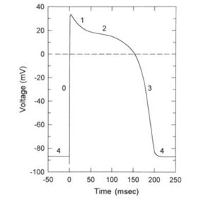
- 30Phase O:* Caused by the transient increase in the Na+ conductance which results in an inward Na+ current that depolarizes the membrane.
* At the peak of the action potential, the membrane potential approaches the Na+ equilibrium potential. - 31Phase 1* Brief period of initial repolarization
* Caused by an outward current, in part by the movement of K+ ions out of the cell and a decrease in Na+ conductance.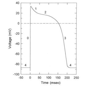
- 32Phase 2* Plateau of the action potential
* Caused by a transient increase in Ca++ conductance which results in an inward Ca++ current and by an increase in K+ conductance.
* Outward and inward currents are approximately equal to membrane potential is stable.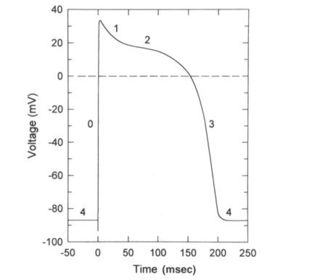
- 33Phase 3* Repolarization
* Ca++ conductance decreases, K+ conductance increases
* High K+ conductance results in large outward K+ current which hyperpolarizes the membrane back toward the K+ equilibrium potential.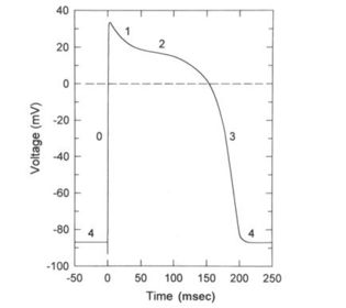
- 34Phase 4* The resting membrane potential
* Period during which the inward and outward currents are equal and the membrane potential approaches the K+ equilibrium potential.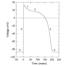
- 35Sinoatrial (SA) node* The pacemaker of the heart
* Has an unstable resting potential - 36AV node* The upstroke of the action potential in the AV node is the result of an inward CA++ current.
- 37Conduction Velocity* Reflects the time required for excitation to spread throughout cardiac tissue
* The larger the inward current during the upstroke, the higher the velocity - 38Conduction velocity is fastest in thePurkinje system
- 39Conduction velocity is slowest in theAV node (PR interval) allowing time for ventricular filling. If conduction velocity through the AV node is increased, ventricular filling may be compromised.
- 40ExcitabilityThe ability of cardiac cells to initiate action potentials in response to inward, depolarizing current
- 41Absolute refractory periodReflects the time during which no action potential can be initiated regardless of how much inward current is supplied
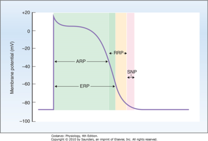
- 42Effective refractory periodReflects the time during which a conducted action potential cannot be elicited.
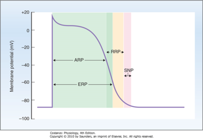
- 43Relative refractory periodPeriod during which an action potential can be elicited, but more than the usual inward current is required.
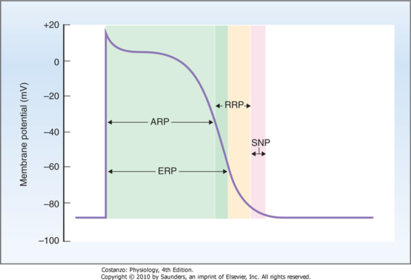
- 44Chronotropic Effects* Produce changes in heart rate
* A negative effect decreases heart rate by decreasing the firing rate of the SA node
* A positive effect increases heart rate by increasing the firing rate of the AV node - 45Dromotropic Effects* Produce changes in conduction velocity primarily through the AV node
* A negative effect decreases conduction velocity --> slowing the conduction of action potentials from the atria to the ventricles and increasing the PR interval. - 46The SA node, atria, and AV node have __________ _________ innervation, but the ventricles do not.The SA node, atria, and AV node have parasympathetic vagal innervation, but the ventricles do not.
- 47The parasympathetic neurotransmitter, ACh* Acts on muscarinic receptors
* Has a negative chronotropic effect: decreases HR
* Has a negative dromotropic effect: decreases conduction velocity through the AV node and increases the PR interval. - 48The sympathetic nervous system neurotransmitter is __________ and acts on ___________ receptors.The sympathetic nervous system neurotransmitter is norepinephrine and acts on Beta-1 receptors.
- 49Norepinephrine has ____________ chronotropic and ___________ dromotropic effects on the heart.* Positive chronotropic effect: Increase HR
* Positive dromotropic effect: Increase conduction velocity, decrease PR interval - 50Which organs have ONLY sympathetic innervation?Sweat glands, vascular smooth muscle, pilomotor muscles of skin, liver, adipose tissue, and kidneys (adrenal medulla)
- 51Where are α1 adrenergic receptors located and what is their mechanism of action?*Vascular smooth muscle (Skin, renal, splachnic)
*GI tract sphincter muscles
*Badder sphincter
*Radial muscle (iris)
*MOA: IP3 increases intracellular Ca++ - 52Where are the α2 adrenergic receptors and what is their mechanism of action?*GI wall muscles
*Presynaptic adrenergic neuron
*MOA: Inhibit adenylyl cyclase, decreasing cAMP - 53Where are the β1 adrenergic receptors located and what is their mechanism of action?*Heart
*Salivary glands
*Adipose tissue
*Kidney
*MOA: Stimulates adenylyl cyclase, increase cAMP - 54Where are the β2 adrenergic receptors located and what is their mechanism of action?*Vascular smooth muscle
*GI tract wall muscle
*Bladder wall muscle
*Bronchioles
*MOA: Stimulates adenylyl cyclase, increases cAMP - 55Where are the nicotinic (colinergic) receptors located and what is their mechanism of action?*Post-ganglionic neurons of the SNS/PNS
*Adrenal medulla
*Skeletal muscle
*MOA: Opening Na+ and K+ channels leading to depolarization - 56Where are the muscarinic (cholinergic) receptors located and what is their mechanism of action?*Effector organs of PNS
*Sweat glands of SNS
*MOA: IP3 increases intracellular Ca++
Inhibits adeylyl cyclase, decreasing cAMP
Direct K+ channel activation
Informationen
- Beschreibung
- 1 PA School Karteikarten am Cardiac Physiology PA School, erstellt von Elle Ashe am 05/02/2017.
- Erstellt von
- Elle Ashe
- Aufrufe
- 93
- Tags
- Bewertung