
Mindmap
RADIOCHEMISTRY
Inhalt der Mindmap
RADIOCHEMISTRY
RADIATION : The energy radiated or transmitted in the form of rays, waves, or particles. A stream of particles or electromagnetic waves that is emitted by the atoms and molecules of a radioactive substance as a result of nuclear decay.
NUCLEAR REACTIONS
- Spontaneous disintegration
- nothing initiates; nothing stops energy range: 15 keV < x < 20 MeV
- Artificial mutation
- interaction of two nuclei producing other nuclei
- Nuclear fission
- heaviest nuclei split into 2 or more lighter nuclei
- Nuclear fusion
- light nuclei combined to form heavier nuclei
- Spontaneous disintegration
Why is Radiation a Useful Analytical Tool?
- characterisedby the type and energy of the radiation they emit
- Easily detected and quantified
- Generally have low background
- Measure concentrations as low as 10^-14 M
Main Radioanalytical Methods
- Measuring Naturally Occuring Radiation
- e.g. Measuring radon in homes Dating artifacts or sediments
- Tracer Methods
- Radioactivity is physically introduced to the sample by adding a measured amt of radioactive sp. (tracer) most important - ISOTOPE DILUTION METHOD (a weighed quantity of radioactive tagged analyte having a known activity is added to a measured amt of the sample. mixed- a fraction of the component of interest is isolated & purified.
- Activation Analysis
- Measuring Naturally Occuring Radiation
Language
- Units
- Define these as measure of the activityof a substance In general activities in the range of 0.1 –20 kBq(3-500 nCi) are sufficient for analytical applications.
- Becquerel (Bq)= 1 decay per second
- Curie (Ci)= the activity of 1 g of 226Ra = 3.70 x 10^10Bq
- Electron Volt = 1 MeV = 1.6 ×10⁻¹³J per particle≈105 MJ/mol
- Half Life
- Time required for one half of the number of radioactive atoms in a sample to undergo decay
- Positron ẞ+
- - forms when the number of protons in nucleus reduced by 1 - has mass of electron- transitory existence –reacts with electron to form γrays
- Negatron ẞ-
- high energy electron that is formed when a neutron is converted to a proton
- Electron capture
- electron is captured by the nucleus and combines with proton to form a neutron. Neutrino is emitted.
- Decay Processes
- α decay
- in isotopes mass >150 particles either monoenergetic or distributed among few discrete energies lose energy as result collisions as pass through matter low penetrating power easily measured
- β decay
- - atomic number changes but mass number does not - lose energy by interacting with electrons in orbitals - particles have continuos spectrum of energies - penetrating power greater because smaller particles
- negatron formation
- positron formation
- electron capture
- γ-ray emission - (produced by nuclear relaxation)
- - occurs when nucleus left in excited state by α or β emission process - returns to ground state in one or more quantized steps with release of monoenergetic γ-rays - X-rays result of electronic relations = γ-ray wavelength generally 1/100thX-ray = γ-rays produced by nuclear relaxations - γ-ray emission spectrum characteristic for each nucleus highly energetic = highly penetrating
- Lose E via:
- photoelectric effect (γ-ray photon disappears)
- electrons released on exposure of (metal) surface by overcoming electron binding energy (low E)
- Compton effect (photon recoils, repeat the rxn)
- electrons ejected from atoms but with only part of the photon’s energy; photon recoils with reduced energy to act again (Relatively energetic)
- pair production (creates positron & electron)
- photon totally absorbed in creating a positron and an electron in the field near the nucleus (High Energy)
- photoelectric effect (γ-ray photon disappears)
- α decay
- Units
Measuring Radioactivity
- Spontaneous disintegration
- Stability of nucleus
- - complicated function of atomic number and atomic mass - trend for stable nuclei to have more neutrons than protons
- Probability
- - cannot tell when an individual atom will disintegrate but the average behaviour of a large number of nuclei can be predicted precisely
- Characteristic spectra
- each type of nucleus has a characteristic pattern of disintegration i.e. emit specific kinds of radiation with individual energy patterns
- Disintegration rates
- rates are always first order (straight line - easy to predict)
- Stability of nucleus
- Radioactive decay rates
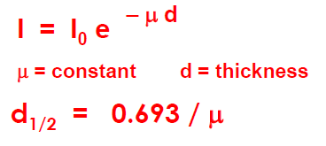
- –dN/dt = λN
- N - # radioactive nuclei t- time λ - decay constant
- If we look at an interval from t0 to t then: –( ln N –ln N0) = λdt ln (N / N0) = –λt N = N0e –λt
- Activity (A) : A = A0e –λt (A: disintegration rate)
- A = c N c = detection coefficient of detector This means:–dA/dt = –dN/dt and hence also ln (A / A0) = –λt
- Half Life t(1/2) = (0.693/ λ)
- half life = time taken for N = N0/2
- –dN/dt = λN
- Radioactive equilibrium
- - Steady state in which all radioactive ‘child’ products are decaying at the same rate as they are being formed by decay of their radioactive ‘parents’. - useful for determining the half life of a very long-lived radioactive element such as uranium.
- λ1N1= λ2N2
- Spontaneous disintegration
DETECTORS
- depends on the ionization of matter caused by radioactivity
- Photographic emulsions
- Photoelectrons knocked off halide ions cause reduction of Ag+ Ag atom then act as a focus for further reduction Produce black spot on the photographic film
- Cloud chambers
- Air supersaturated with water (or other vapour like ethanol) Produces ‘tracks’ along the path of radiation Droplets condense around ions / electrons produced by radiation.
- Gas-filled detectors
- QUANTITATIVE
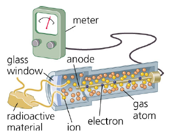
- Inert gas filled - ionized (Ar+) and electron = (ion pairs)
- e- move towards anode and ions move twrds cathode. - e- movement - charged electric current twrds the meter - reading
- detector behaviour depends on the voltage applied
- too low, the ion pairs simply recombine and are not detected. high enough, collisions between accelerated electrons and gas molecules cause secondary ion pair production, i.e. amplification.
- Ionisation chamber ==> (# electrons reach anode = total # produced by radiation)
- e.g. smoke detector
- Proportional ==> (# electrons increases with voltage because increase ions)
- Geiger ==> (enormous amplification but current limited by tube design)
- * most sensitive
- 'dead time' = Ionizations in the chamber take a finite time to dissipate, during which no further response can occur.
- Scintillation counters
- QUANTITATIVE
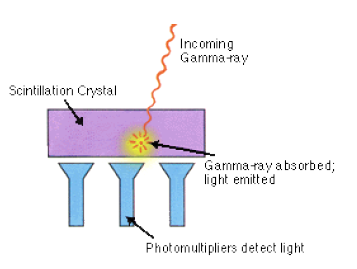
- Based on radiation-induced luminescence
- i.e. electronic excitation rather than ionization
- Excited atom relaxes, emitting a flash of light (at longer wavelength than γ)
- Structure
- - scintillation crystal - doped with organic substance (usually w benzene ring) to absorb the gamma radiation - use crystal (more dense) and gamma radiation tend to react w that
- Semiconductor detectors
- QUANTITATIVE Lithium-drifted silicon detectors Si(Li) Lithium-drifted germanium detectors Ge(Li)
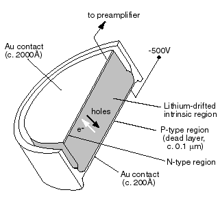
- Diode-like devices based on lithium-doped germanium and silicon
- gamma / X-rays
- gamma rays react with LI- pop the electron from Li. the electron migrate to form the actual compound
- structure
- Li vapour-deposited onto p-type Ge/Si crystal and heated Li atoms diffuse into the crystal where they act like ionizable atoms in a gas-filled detector
- Better than a proportional gas-filled counter because there is no secondary ionization so dead times are small.
- disadvantage- must be cooled by liquid N2
- - prevent Li diffusion out of the Ge/Si - decrease noise to an acceptable level - modern examples only need cooling when in use
- not portable
- Ge used instead of Si when wavelength < 0.3 Angstroms must be cooled at all times
- Counting Corrections
- Background radiation
- cosmic effects surroundings laboratory contamination
- Counting geometry
- radiation is emitted in all directions orientation and distance of counter is important
- Back-scattering
- - radiation directed away from the counter can be reflected back into it by objects behind sample - use reproducible sample holders made of absorbing material
- Self-scattering
- - radiation is deflected and absorbed by the sample itself so counts = f (sample thickness) - make samples as thin and as reproducible as possible
- Decay
- samples with short half-lives decay during counting need to correct if counting period > 10% of t½
- Dead-time corrections
- - all detectors take finite time to recover after sensing the arrival of a radioactive particle - other particles arriving in this time interval are not detected- also called ‘co-incidence corrections’
- Geiger tube50 –200 μs Scintillation0.25 μs Know dead time R* = R/(1-Rτ)
- R* = true count rate R = obs count rate τ = dead time
- Determining Dead Time
- Background radiation
RADIOANALYTICAL METHODS
- Chemical tagging
- Introduce a ‘hot’ (radioactively visible) isotope into a process containing a ‘cold’ isotope, to follow the latter
- Analytical procedures
- Used to determine % efficiencies (errors from co-ppt, occlusion, etc. are thus eliminated)
- Industrial processes
- Trace disposed waste / transport of pollutants
- Biological processes
- Trace biochemical pathways e.g. in citric acid cycle
- Determining solubilities
- Can measure the activity of a saturated solution without evaporation or weighing.
- Advantages
- -Selectivity (Independent of quantitative isolation) -Simplicity of equipment
- Disadvantages
- Exchangeability
- If isotope occurs in a compound it can sometimes ‘exchange’ e.g. tritium+ and H+
- Differences due to atomic mass
- Isotopes do not always mimic each other exactly e.g. diffusion of deuterium and tritium is much slower than that of hydrogen
- Exchangeability
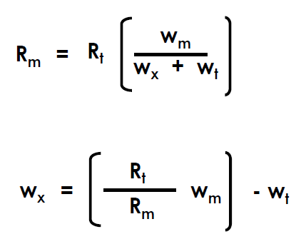
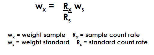
- Isotope Dilution
- Rm= activity of isolated mass wm= weight of isolated mass wx= weight of unknown wt= weight of tracer Rt= count of tracer
- Specific activity of a radionuclide is reduced when it is mixed with its stable counterpart.
- The extent of reduction in activity provides a measure of the amount of stable isotope with which the radio-isotope is mixed.
- Activation Analysis
- making sample radioactive by irradiating with neutrons/charged particles
- Sources : reactors radionuclides particle accelerators
- thermal neutrons
- Energetic neutron passed through moderating material to dissipate energy and create
- fast neutrons
- For light elements e.g. F, O, N use fast neutrons(14 eV)
- detection limit
- Detection limits for many elements are < 1 μg; for some elements as low as picogram level
- Neutron captured by analyte nucleus causing excited state and γray emmission
- Capture cross-sections
- measures of probability that interaction will occur. The values are obtained from tables. Units are in barns (1 b = 10-24cm2)
- Number of nuclei that are produced during irradiation (N* = N φσS)
- S = saturation factor
- activity is directly proportional to number of radionuclides
- Advantages
- high sensitivity, minimal sample preparation, easy to calibrate, often non-destructive,
- Disadvantages
- Expensive equipment and special facilities required OHS considerations when dealing with radionuclides Time for analysis when using long-lived radionuclides
- Chemical tagging
OHS and Radioanalytical Chemistry
- Dosimetry
- Special steps need to be taken when working with radioactive sources e.g. personal radiation monitors for monitoring dose
- Thermoluminescent dosemeter (TLD)
- Electrons in the crystal structure of the TLD material excited to higher energy levels as a result of irradiation and are trapped in the crystal structure. Response of TLD dependent on the energy and type of radiation
- Units
- Different units are used because the effects depend on the type of radiation, type of material and other circumstances
- Roentgen (ion dose)
- unit of exposure defined in terms of amount of charge generated when radiation is stopped in air Now, 1 R = 2.58 ×10⁻⁴Ci / kg For a standard human, I R ≈10-5J
- RAD (energy dose)
- unit of absorbed dose defined as amount of radiation that liberates 10-2J/kg of irradiated material independent of the type of radiation but function of material
- Gray (Gy)
- SI unit for energy dose 1 Gy = 1 J/kg = 100 rad
- REM (equivalent dose)
- measure of biological damage, defined in terms of a factor called the Relative Biological Effect(RBE)which takes account of different circumstances 1 REM = dose in RADS ×RBE RBE= dose of γrays that produces the same effect as 1 RAD of the particular type of radiation (so standardising REM)
- RBE
- Sievert (Sv)
- dose in Gy multiplied by an effectiveness factor 1 mGy dose of αrays = 20 mSv of equivalent dose 1 mGy dose of βrays = 1 mSv equivalent dose In most cases the effectiveness factor is unity and the dose in grays is equal to the dose in sieverts
- Dosimetry
Bilder und Dateien der Mindmap
Informationen
- Beschreibung
- undergraduate degree analytical chemistry Mindmap am RADIOCHEMISTRY, erstellt von chaz_id89 am 05/11/2013.
- Erstellt von
- chaz_id89
- Aufrufe
- 74
- Tags
- Bewertung