
Mindmap
Cell Division
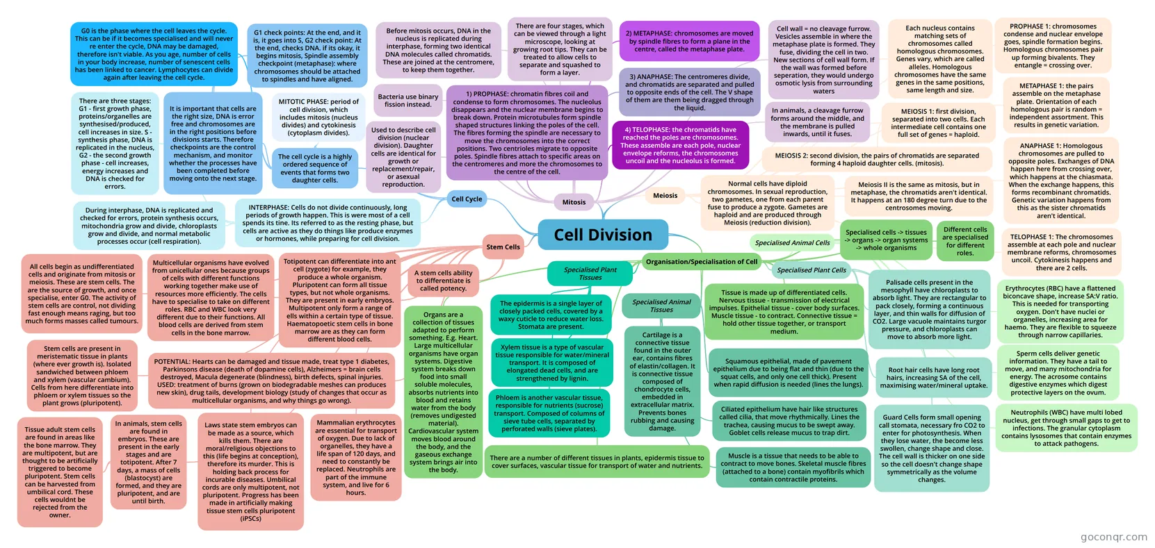
Mind map: Cell division
Inhalt der Mindmap
Cell Division
Cell Cycle
- The cell cycle is a highly ordered sequence of events that forms two daughter cells.
- MITOTIC PHASE: period of cell division, which includes mitosis (nucleus divides) and cytokinesis (cytoplasm divides).
- G0 is the phase where the cell leaves the cycle. This can be if it becomes specialised and will never re enter the cycle, DNA may be damaged, therefore isn't viable. As you age, number of cells in your body increase, number of senescent cells has been linked to cancer. Lymphocytes can divide again after leaving the cell cycle.
- It is important that cells are the right size, DNA is error free and chromosomes are in the right positions before divisions starts. Therefore checkpoints are the control mechanism, and monitor whether the processes have been completed before moving onto the next stage.
- G1 check points: At the end, and it is, it goes into S, G2 check point: At the end, checks DNA. if its okay, it begins mitosis, Spindle assembly checkpoint (metaphase): where chromosomes should be attached to spindles and have aligned.
- INTERPHASE: Cells do not divide continuously, long periods of growth happen. This is were most of a cell spends its tine. Its referred to as the resting phase, but cells are active as they do things like produce enzymes or hormones, while preparing for cell division.
- During interphase, DNA is replicated and checked for errors, protein synthesis occurs, mitochondria grow and divide, chloroplasts grow and divide, and normal metabolic processes occur (cell respiration).
- There are three stages: G1 - first growth phase, proteins/organelles are synthesised/produced, cell increases in size. S - synthesis phase, DNA is replicated in the nucleus, G2 - the second growth phase - cell increases, energy increases and DNA is checked for errors.
- The cell cycle is a highly ordered sequence of events that forms two daughter cells.
Mitosis
- Used to describe cell division (nuclear division). Daughter cells are identical for growth or replacement/repair, or asexual reproduction.
- Bacteria use binary fission instead.
- Before mitosis occurs, DNA in the nucleus is replicated during interphase, forming two identical DNA molecules called chromatids. These are joined at the centromere, to keep them together.
- There are four stages, which can be viewed through a light microscope, looking at growing root tips. They can be treated to allow cells to separate and squashed to form a layer.
- 1) PROPHASE: chromatin fibres coil and condense to form chromosomes. The nucleolus disappears and the nuclear membrane begins to break down. Protein microtubules form spindle shaped structures linking the poles of the cell. The fibres forming the spindle are necessary to move the chromosomes into the correct positions. Two centrioles migrate to opposite poles. Spindle fibres attach to specific areas on the centromeres and more the chromosomes to the centre of the cell.
- 2) METAPHASE: chromosomes are moved by spindle fibres to form a plane in the centre, called the metaphase plate.
- 3) ANAPHASE: The centromeres divide, and chromatids are separated and pulled to opposite ends of the cell. The V shape of them are them being dragged through the liquid.
- 4) TELOPHASE: the chromatids have reached the poles are chromosomes. These assemble are each pole, nuclear envelope reforms, the chromosomes uncoil and the nucleolus is formed.
- In animals, a cleavage furrow forms around the middle, and the membrane is pulled inwards, until it fuses.
- Cell wall = no cleavage furrow. Vesicles assemble in where the metaphase plate is formed. They fuse, dividing the cell in two. New sections of cell wall form. If the wall was formed before seperation, they would undergo osmotic lysis from surrounding waters
Meiosis
- Normal cells have diploid chromosomes. In sexual reproduction, two gametes, one from each parent fuse to produce a zygote. Gametes are haploid and are produced through Meiosis (reduction division).
- Each nucleus contains matching sets of chromosomes called homologous chromosomes. Genes vary, which are called alleles. Homologous chromosomes have the same genes in the same positions, same length and size.
- MEIOSIS 1: first division, separated into two cells. Each intermediate cell contains one full set of genes = haploid.
- MEIOSIS 2: second division, the pairs of chromatids are separated forming 4 haploid daughter cells. (mitosis).
- PROPHASE 1: chromosomes condense and nuclear envelope goes, spindle formation begins. Homologous chromosomes pair up forming bivalents. They entangle = crossing over.
- METAPHASE 1: the pairs assemble on the metaphase plate. Orientation of each homologous pair is random = independent assortment. This results in genetic variation.
- ANAPHASE 1: Homologous chromosomes are pulled to opposite poles. Exchanges of DNA happen here from crossing over, which happens at the chiasmata. When the exchange happens, this forms recombinant chromatids. Genetic variation happens from this as the sister chromatids aren't identical.
- TELOPHASE 1: The chromosomes assemble at each pole and nuclear membrane reforms, chromosomes uncoil. Cytokinesis happens and there are 2 cells.
- Meiosis II is the same as mitosis, but in metaphase, the chromatids aren't identical. It happens at an 180 degree turn due to the centrosomes moving.
Organisation/Specialisation of Cell
- Different cells are specialised for different roles.
- Specialised cells -> tissues -> organs -> organ systems -> whole organisms
- Specialised Animal Cells
- Erythrocytes (RBC) have a flattened biconcave shape, increase SA:V ratio. This is needed for transporting oxygen. Don't have nuclei or organelles, increasing area for haemo. They are flexible to squeeze through narrow capillaries.
- Neutrophils (WBC) have multi lobed nucleus, get through small gaps to get to infections. The granular cytoplasm contains lysosomes that contain enzymes to attack pathogens.
- Sperm cells deliver genetic information. They have a tail to move, and many mitochondria for energy. The acrosome contains digestive enzymes which digest protective layers on the ovum.
- Specialised Plant Cells
- Palisade cells present in the mesophyll have chloroplasts to absorb light. They are rectangular to pack closely, forming a continuous layer, and thin walls for diffusion of CO2. Large vacuole maintains turgor pressure, and chloroplasts can move to absorb more light.
- Root hair cells have long root hairs, increasing SA of the cell, maximising water/mineral uptake.
- Guard Cells form small opening call stomata, necessary fro CO2 to enter for photosynthesis. When they lose water, the become less swollen, change shape and close. The cell wall is thicker on one side so the cell doesn't change shape symmetrically as the volume changes.
- Tissue is made up of differentiated cells. Nervous tissue - transmission of electrical impulses. Epithelial tissue - cover body surfaces. Muscle tissue - to contract. Connective tissue = hold other tissue together, or transport medium.
- Specialised Animal Tissues
- Squamous epithelial, made of pavement epithelium due to being flat and thin (due to the squat cells, and only one cell thick). Present when rapid diffusion is needed (lines the lungs).
- Ciliated epithelium have hair like structures called cilia, that move rhythmically. Lines the trachea, causing mucus to be swept away. Goblet cells release mucus to trap dirt.
- Cartilage is a connective tissue found in the outer ear, contains fibres of elastin/collagen. It is connective tissue composed of chondrocyte cells, embedded in extracellular matrix. Prevents bones rubbing and causing damage.
- Muscle is a tissue that needs to be able to contract to move bones. Skeletal muscle fibres (attached to a bone) contain myofibrils which contain contractile proteins.
- There are a number of different tissues in plants, epidermis tissue to cover surfaces, vascular tissue for transport of water and nutrients.
- Specialised Plant Tissues
- The epidermis is a single layer of closely packed cells, covered by a waxy cuticle to reduce water loss. Stomata are present.
- Xylem tissue is a type of vascular tissue responsible for water/mineral transport. It is composed of elongated dead cells, and are strengthened by lignin.
- Phloem is another vascular tissue, responsible for nutrients (sucrose) transport. Composed of columns of sieve tube cells, separated by perforated walls (sieve plates).
- Organs are a collection of tissues adapted to perform something. E.g. Heart. Large multicellular organisms have organ systems. Digestive system breaks down food into small soluble molecules, absorbs nutrients into blood and retains water from the body (removes undigested material). Cardiovascular system moves blood around the body, and the gaseous exchange system brings air into the body.
Stem Cells
- All cells begin as undifferentiated cells and originate from mitosis or meiosis. These are stem cells. The are the source of growth, and once specialise, enter G0. The activity of stem cells are control, not dividing fast enough means raging, but too much forms masses called tumours.
- A stem cells ability to differentiate is called potency.
- Totipotent can differentiate into ant cell (zygote) for example, they produce a whole organism. Pluripotent can form all tissue types, but not whole organisms. They are present in early embryos. Multipotent only form a range of ells within a certain type of tissue. Haematopoetic stem cells in bone marrow are as they can form different blood cells.
- Multicellular organisms have evolved from unicellular ones because groups of cells with different functions working together make use of resources more efficiently. The cells have to specialise to take on different roles. RBC and WBC look very different due to their functions. All blood cells are derived from stem cells in the bone marrow.
- Mammalian erythrocytes are essential for transport of oxygen. Due to lack of organelles, they have a life span of 120 days, and need to constantly be replaced. Neutrophils are part of the immune system, and live for 6 hours.
- In animals, stem cells are found in embryos. These are present in the early stages and are totipotent. After 7 days, a mass of cells (blastocyst) are formed, and they are pluripotent, and are until birth.
- Tissue adult stem cells are found in areas like the bone marrow. They are multipotent, but are thought to be artificially triggered to become pluripotent. Stem cells can be harvested from umbilical cord. These cells wouldnt be rejected from the owner.
- Stem cells are present in meristematic tissue in plants (where ever growth is). Isolated sandwiched between phloem and xylem (vascular cambium). Cells from here differentiate into phloem or xylem tissues so the plant grows (pluripotent).
- POTENTIAL: Hearts can be damaged and tissue made, treat type 1 diabetes, Parkinsons disease (death of dopamine cells), Alzheimers = brain cells destroyed, Macula degenerate (blindness), birth defects, spinal injuries. USED: treatment of burns (grown on biodegradable meshes can produces new skin), drug tails, development biology (study of changes that occur as multicellular organisms, and why things go wrong).
- Laws state stem embryos can be made as a source, which kills them. There are moral/religious objections to this (life begins at conception), therefore its murder. This is holding back process for incurable diseases. Umbilical cords are only multipotent, not pluripotent. Progress has been made in artificially making tissue stem cells pluripotent (iPSCs)
Informationen
- Beschreibung
- A level Biology (6 - CELL DIVISION) Mindmap am Cell Division, erstellt von Molly Butcher am 09/04/2017.
- Erstellt von
- Molly Butcher
- Aufrufe
- 1
- Bewertung
Mehr zu Phases of the Cell Cycle