
Mindmap
Receptors
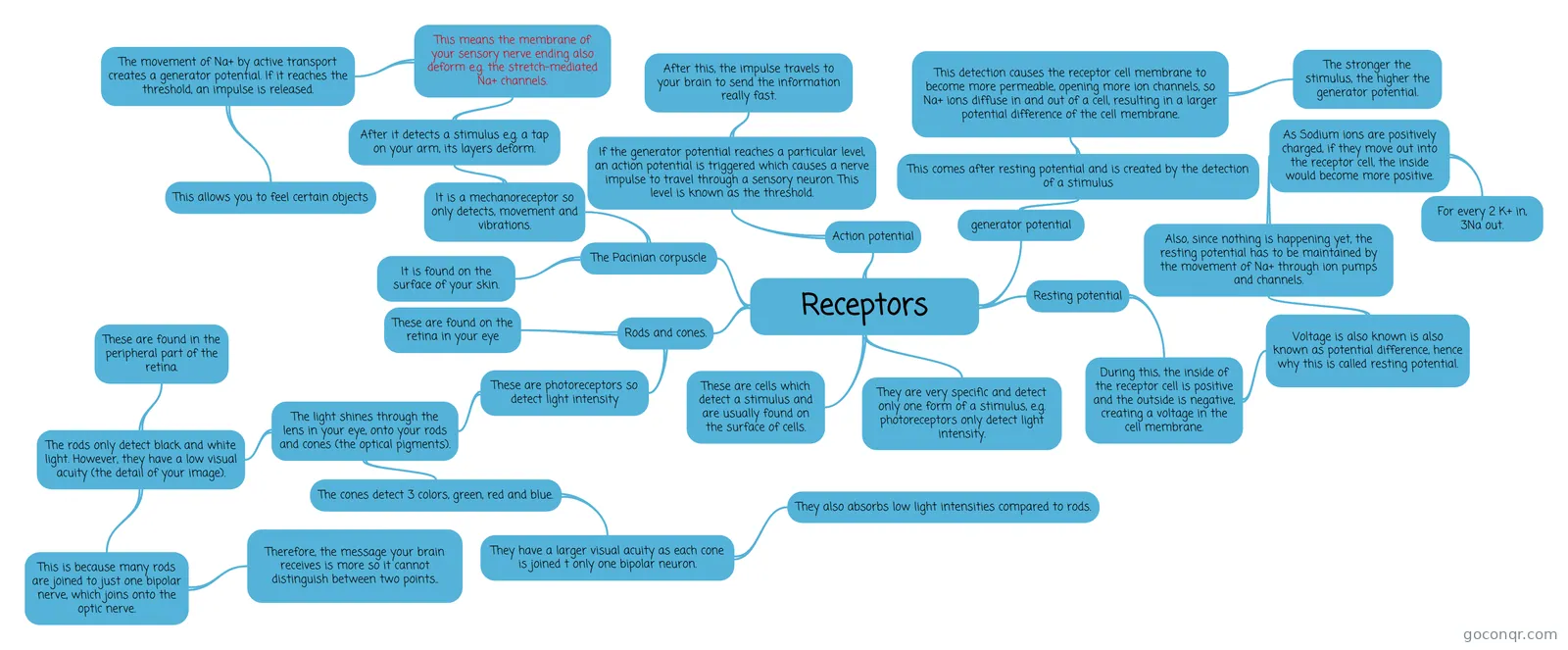
Mind map: Receptors biology
Inhalt der Mindmap
Receptors
These are cells which detect a stimulus and are usually found on the surface of cells.
They are very specific and detect only one form of a stimulus, e.g. photoreceptors only detect light intensity.
Resting potential
- During this, the inside of the receptor cell is positive and the outside is negative, creating a voltage in the cell membrane.
- Voltage is also known is also known as potential difference, hence why this is called resting potential.
- Also, since nothing is happening yet, the resting potential has to be maintained by the movement of Na+ through ion pumps and channels.
- As Sodium ions are positively charged, if they move out into the receptor cell, the inside would become more positive.
- For every 2 K+ in, 3Na out.
- As Sodium ions are positively charged, if they move out into the receptor cell, the inside would become more positive.
- Also, since nothing is happening yet, the resting potential has to be maintained by the movement of Na+ through ion pumps and channels.
- Voltage is also known is also known as potential difference, hence why this is called resting potential.
- During this, the inside of the receptor cell is positive and the outside is negative, creating a voltage in the cell membrane.
generator potential
- This comes after resting potential and is created by the detection of a stimulus
- This detection causes the receptor cell membrane to become more permeable, opening more ion channels, so Na+ ions diffuse in and out of a cell, resulting in a larger potential difference of the cell membrane.
- The stronger the stimulus, the higher the generator potential.
- This detection causes the receptor cell membrane to become more permeable, opening more ion channels, so Na+ ions diffuse in and out of a cell, resulting in a larger potential difference of the cell membrane.
- This comes after resting potential and is created by the detection of a stimulus
Action potential
- The potential differences are measured in milivolts (mV).
- If the generator potential reaches a particular level, an action potential is triggered which causes a nerve impulse to travel through a sensory neuron. This level is known as the threshold.
- After this, the impulse travels to your brain to send the information really fast.
The Pacinian corpuscle
- It is found on the surface of your skin.
- It is a mechanoreceptor so only detects, movement and vibrations.
- After it detects a stimulus e.g. a tap on your arm, its layers deform.
- This means the membrane of your sensory nerve ending also deform e.g. the stretch-mediated Na+ channels.
- The movement of Na+ by active transport creates a generator potential. If it reaches the threshold, an impulse is released.
- This allows you to feel certain objects
- The movement of Na+ by active transport creates a generator potential. If it reaches the threshold, an impulse is released.
- This means the membrane of your sensory nerve ending also deform e.g. the stretch-mediated Na+ channels.
- After it detects a stimulus e.g. a tap on your arm, its layers deform.
Rods and cones.
- These are found on the retina in your eye
- These are photoreceptors so detect light intensity
- The light shines through the lens in your eye, onto your rods and cones (the optical pigments).
- The rods only detect black and white light. However, they have a low visual acuity (the detail of your image).
- This is because many rods are joined to just one bipolar nerve, which joins onto the optic nerve.
- Therefore, the message your brain receives is more so it cannot distinguish between two points..
- These are found in the peripheral part of the retina.
- This is because many rods are joined to just one bipolar nerve, which joins onto the optic nerve.
- The cones detect 3 colors, green, red and blue.
- They have a larger visual acuity as each cone is joined t only one bipolar neuron.
- They also absorbs low light intensities compared to rods.
- They have a larger visual acuity as each cone is joined t only one bipolar neuron.
- The rods only detect black and white light. However, they have a low visual acuity (the detail of your image).
- The light shines through the lens in your eye, onto your rods and cones (the optical pigments).
Informationen
- Beschreibung
- A level Biology Mindmap am Receptors, erstellt von Jumael Zafar am 21/08/2017.
- Erstellt von
- Jumael Zafar
- Aufrufe
- 84
- Tags
- Bewertung