
Notizen
Application of the Nursing process of clients experiencing Musculoskeletal Trauma
Inhalt der Notizen (19)
Seite 1
Soft Tissue Injuries
Including sprains, strains, dislocations, and subluxations, usually result from trauma, and sports related injuries. Excess myogloblin will appear in the urine as dark red and will imply compartment syndrome. Resulting in insufficient blood supply to tissue. Patients in the ER will require consent for surgery and a Tetnus sho t(Good for 10yrs) to reduce the risk of infection from skin cuts and open wounds such as a open fracture. Bone penetrating antibiotics such as cephalosporins are administered prior to surgery. Open fractures should be covered with sterile dressings to prevent contamination.
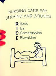
RICE (Rest, Ice, Compression, Elevation) may decrease local inflammation and pain for most musculoskeletal injuries.
Seite 2
Sprains & Strains
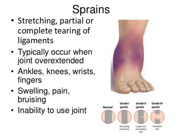
A sprain is an injury to the ligaments surrounding a joint, usually caused by a wrenching or twisting motion. Most sprains occur in the ankle, wrist, and knee joints
A strain is an excessive stretching of a muscle, its fascial sheath, or a tendon. Most strains occur in the large muscle groups, including the lower back, calf, and hamstrings.
Nursing Management: Sprains and Strains:
(1) Health Promotion
Warming up muscles before exercising and vigorous activity, followed by stretching, may significantly reduce the risk of sprains and strains. Strength, balance, and endurance exercises are also important. Strengthening exercises 1464that involve working against resistance build muscle strength and bone density. Balance exercises, which may overlap with some strengthening exercises, help to prevent falls. Endurance exercises should start at a low level of effort and progress gradually to a moderate level.
(2) Acute Care
If an injury occurs, immediate care focuses on (1) stopping the activity and limiting movement, (2) applying ice packs to the injured area, (3) compressing the involved area, (4) elevating the extremity, and (5) providing analgesia as needed.
Causes:
Falls
• Direct blows
• Crush injury
• Motor vehicle crashes
• Sports injuries
Assessments:
• Edema
• Bruising (contusion)
• Pain, tenderness
• Decreased sensation with severe edema
• Decreased pulse, coolness, capillary refill >2 sec
• Decreased movement
• Pallor
• Shortening or rotation of extremity
• Inability to bear weight with lower extremity involvement
• Limited or decreased function with upper extremity involvement
• Muscle spasms
Interventions:
Initial [PRIORITY]
• Ensure airway, breathing, and circulation.
• Perform neurovascular assessment of involved limb.
• Elevate involved limb. Elevate the injured part above heart level, even during sleep, to help mobilize excess fluid from the area and prevent further edema.
• Apply compression bandage unless dislocation present.
• Apply ice packs to affected area. Cold is most useful when applied immediately after the injury has occurred. Apply ice no more than 20 to 30 minutes at a time; avoid applying ice directly to the skin.
After the acute phase (usually 24 to 48 hours), apply warm, moist heat to the affected part to reduce swelling and provide comfort. Heat applications should not exceed 20 to 30 minutes, allowing a “cool-down” time between applications.
• Immobilize affected extremity in the position found. Do not attempt to realign or reinsert protruding bones.
• Anticipate x-rays of injured extremity.
• Give analgesia as necessary.
• Administer tetanus and diphtheria prophylaxis if there is an open fracture.
• Administer antibiotic prophylaxis for open fracture, large tissue defects, or mangled extremity injury.
Ongoing Monitoring
• Monitor for changes in neurovascular condition.
• Implement weight-bearing restrictions as ordered for lower extremity involvement.
• Anticipate compartment pressure monitoring if neurovascular assessment changes and compartment syndrome suspected.
Seite 3
Seite 4
Dislocation
Considered a orthopedic emergency!
A patient who has dislocated a joint may be at greater risk for repeated dislocations because of loose ligaments.
Definition: Separation of two bones from their normal position within a joint.
The longer the joint remains unreduced, the greater the possibility of avascular necrosis = Loss of blood supply in the bone.
The first goal of management of a dislocation is to realign the dislocated portion of the joint to its original anatomic position. Nursing management of dislocation is directed toward pain management and support and protection of the injured joint.
Closed reduction (no incision) may be performed under local or general anesthesia or IV moderate to deep sedation (formerly called conscious sedation). Anesthesia is often needed to relax the muscle so that the bones can be manipulated. In some situations, open reduction (joint visualized through surgical incision) may be needed. After reduction, the extremity is immobilized by a brace, splint, or sling, or by taping to allow torn ligaments and surrounding tissue to heal.
Seite 5
Carpal Tunnel Syndrome (CTS)
Carpal tunnel syndrome (CTS) is caused by compression of the median nerve, which enters the hand at the wrist through the narrow carpal tunnel
CTS is the most common compression neuropathy in the upper extremity. It is associated with hobbies or occupations that require continuous wrist movement (e.g., musicians, carpenters, computer operators).
Women are more likely than men to develop CTS, possibly because of a smaller carpal tunnel.
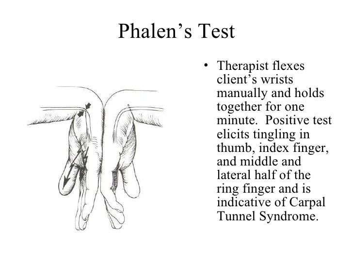
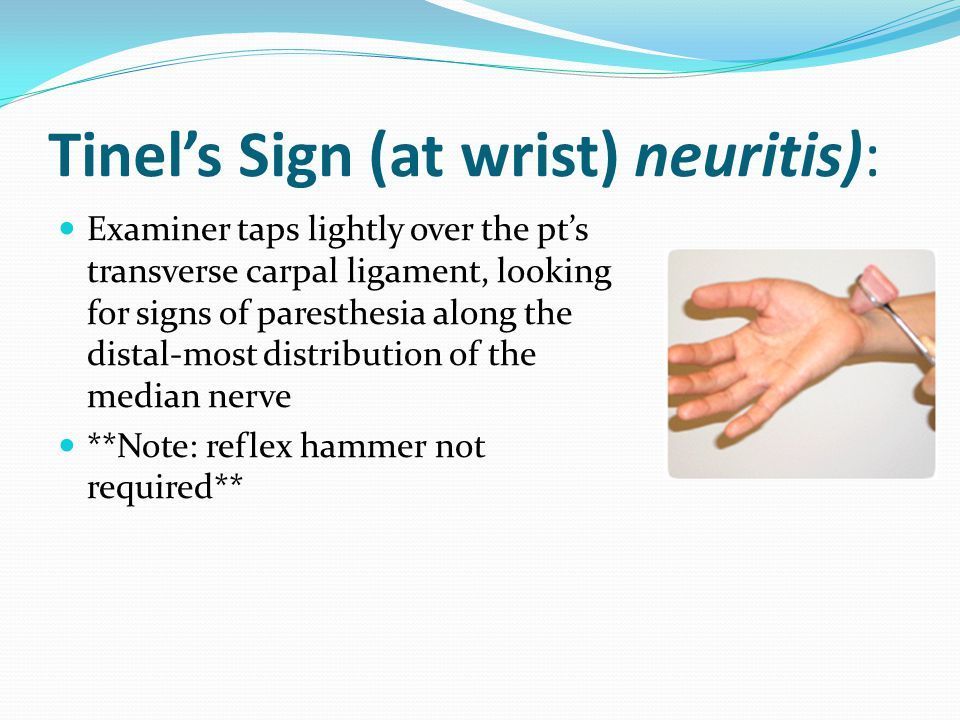
Manifestations of CTS include a positive Tinel's sign and Phalen's sign. Tinel's sign can be elicited by tapping over the median nerve as it passes through the carpal tunnel in the wrist. A positive response is a sensation of tingling in the distribution of the median nerve over the hand. Phalen's signcan be elicited by allowing the wrists to fall freely into maximum flexion and maintain the position for longer than 60 seconds. A positive response is a sensation of tingling in the distribution of the median nerve over the hand. In late stages, atrophy of the thenar muscles around the base of the thumb results in recurrent pain and eventual dysfunction of the hand.
Additional manifestations of CTS are weakness, pain, numbness, or impaired sensation in the distribution of the median nerve
Adaptive devices such as wrist splints may be worn to hold the wrist in a slight extension and relieve pressure on the median nerve
Injection of a corticosteroid drug directly into the carpal tunnel may provide short-term relief.
Special keyboard pads and computer mice that help prevent repetitive pressure on the median nerve are available for computer users. Other ergonomic changes include workstation modifications, change in body positions, and frequent breaks from work-related activities.
Interprofessional care of the patient with CTS is directed toward relieving the underlying cause of the nerve compression. Early symptoms of CTS can usually be relieved by stopping the aggravating movement and by resting the hand and wrist by immobilization in a hand splint. Splints worn at night help keep the wrist in a neutral position and may reduce night pain and numbness. Physical therapy with hand and wrist exercises may lessen symptom severity. Injection of a corticosteroid drug directly into the carpal tunnel may provide short-term relief. The patient may need to consider a change in occupation because of discomfort and sensory changes.
Seite 6
Meniscus Injury
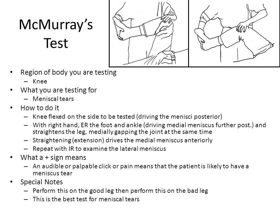
he menisci are crescent-shaped pieces of fibrocartilage in the knee.Injuries are closely associated with ligament sprains common among athletes in sports such as basketball, football, soccer, and hockey.
Meniscus injuries alone do not usually cause significant edema
Pain is elicited by flexion, internal rotation, and then extension of the knee (McMurray's test).
Because meniscus injuries are commonly caused by sports-related activity, teach athletes to do warm-up exercises. The acutely injured knee should be examined within 24 hours of injury. Initial care of this injury involves ice application, immobilization, and use of crutches with weight bearing as tolerated. Most meniscus injuries are treated in an outpatient setting. Using a knee brace or immobilizer during the first few days after the injury protects the knee and offers some pain relief.
After acute pain has decreased, physical therapy can help the patient regain knee flexion and muscle strength to assist in returning to full function. In older adults with degenerative meniscus tears, progressive exercise therapy may improve neuromuscular function and muscle strength.
Teach athletes to do warm up activities.
Knee braces and other immobilizers used.
Popping sensation when the meniscus is torn
Seite 7
Anterior Cruciate Ligament Injury (ACL)
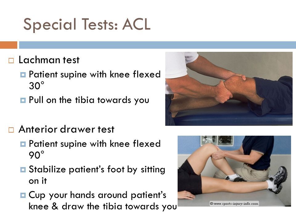
Conservative treatment for an intact ACL injury includes rest, ice, NSAIDs, elevation, and ambulation as tolerated with crutches.
Reconstructive surgery is usually recommended for physically active patients who have sustained severe injury to the ACL and meniscus. In reconstruction, the torn ACL tissue is removed and replaced with autologous or allograft tissue. ROM is encouraged soon after surgery, and the knee is placed in a brace or immobilizer. Rehabilitation with physical therapy is critical, with progressive weight bearing determined by the degree of surgical repair. A safe return to the patient's prior level of physical functioning may take 6 to 8 months.
Common knee injury
Positive Lachman's test will suggest an ACL tear.
MRI is often used to diagnose an ACL tear
Seite 8
Anthroplasty
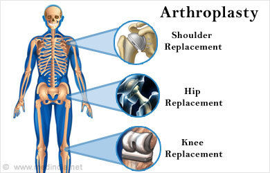
Arthroplasty is the reconstruction or replacement of a joint to relieve pain, improve or maintain ROM, and correct deformity.
Total Hip Anthroplasty (THA) - Arthroplasty is most commonly performed on patients with OA (osteoarthritis) , RA (Rheumatoid Arthritis), avascular necrosis, congenital deformities or dislocations, and other systemic problems. In THA, the prosthesis (implant) replaces the ball-and-socket joint and upper shaft of the femur
Seite 9
Hip Fractures
Hip fractures are common in older adults, with 95% of these fractures resulting from a fall.
Hip fractures refer to a fracture of the proximal (upper) third of the femur, which extends 5 cm below the lesser trochanter.
Seite 10
Nursing Management of A Hip Fracture
PREOPERATIVE CARE
Appropriate analgesics or muscle relaxants, comfortable positioning (unless contraindicated), and properly applied traction (if used) can help to manage the spasms.
When possible, teach the patient the method for exercising the unaffected leg and both arms.
POSTOPERATIVE CARE
Hospitalization averages 3 or 4 days.
In the initial postoperative period, assess vital signs, intake, and output; monitor respiratory function and encourage deep breathing and coughing; administer pain medication; and observe the dressing and incision for signs of bleeding. (eNursing Care Plan 62-2 for the orthopedic surgical patient is presented on the website for this chapter.)
Assess the patient's extremity for (1) color, (2) temperature, (3) capillary refill, (4) distal pulses, (5) edema, (6) sensation, (7) motor function, and (8) pain. Edema is alleviated by elevating the leg when the patient is in bed or in a chair. The pain resulting from surgical repair of the affected extremity can be reduced by maintaining limb alignment with pillows between the patient's knees when turning the patient to the nonoperative side. Avoid turning the patient to the affected side unless approved by the surgeon.
Inform the patient and caregiver about positions and activities that place the patient at risk for dislocation (more than 90 degrees of flexion, adduction across the midline [crossing of legs and ankles], internal rotation of hip). Many daily activities may reproduce these positions: (1) putting on shoes and socks, (2) crossing the legs or feet while seated, (3) assuming the side-lying position incorrectly, (4) standing up or sitting down while the hip is flexed more than 90 degrees relative to the chair, and (5) sitting on low seats, especially low toilet seats. Teach the patient to avoid these activities until the soft tissue capsule around the hip has healed enough to stabilize the prosthesis (usually at least 6 weeks)
Seite 11
Hip Replacement Reminders
Hip Replacement Patient Teaching
After a hip replacement by posterior surgical approach, include the following instructions when teaching a patient and caregiver.
Do
• Use an elevated toilet seat.
• Place chair inside shower or tub and remain seated while washing.
• Use pillow between legs for first 6 wk after surgery when lying on nonoperative side or when supine.
• Keep hip in neutral, straight position when sitting, walking, or lying.
• Notify surgeon immediately if severe pain, deformity, or loss of function occurs.
• Discuss personal risk factors for prosthetic joint infection with surgeon and dentist before dental work.
Do Not
• Flex hip greater than 90 degrees (e.g., sitting in low chairs or toilet seats).
• Adduct hip (i.e., bring legs together at knees).
• Internally rotate hip (i.e., turn toward planted foot on affected side).
• Cross legs at knees or ankles.
• Put on own shoes or stockings without adaptive device (e.g., long-handled shoehorn or stocking-helper) until 4-6 wk after surgery.
• Sit on chairs without arms. The arms of chairs will help the patient rise to a standing position.
Taking a tub bath and driving a car are not allowed for 4 to 6 weeks.
Seite 12
Hip Fractures with The Elderly
Factors that increase the risk of a hip fracture in older adults include:
(1) a tendency to fall due to an altered center of gravity and the inability to correct a postural imbalance,
(2) decreased fat and muscle to act as local tissue shock absorbers, and
(3) reduced skeletal strength.
Other factors that increase the older adult's risk of falling include
(1) gait and balance problems,
(2) altered vision and hearing,
(3) slowed reflexes,
(4) orthostatic hypotension, and
(5) medication use.
Homes can be made safer by:
(1) eliminating tripping hazards (e.g., loose rugs, uneven surfaces), (2) adding grab bars inside and outside the tub or shower, and beside the toilet, (3) adding railings on both sides of the stairs, and (4) installing better lighting
Seite 13
Amputations
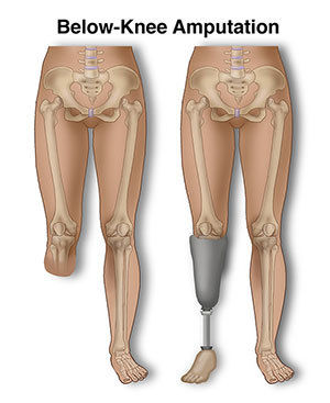
An amputation is the removal of a body extremity by trauma or surgery.
Older people have the highest incidence of amputation due to peripheral vascular disease (PVD), atherosclerosis, and vascular changes related to diabetes mellitus.
Amputation in young people is usually secondary to trauma (e.g., motor vehicle crashes, land mines, farm-related injury).
Other common reasons for amputation are trauma, thermal injuries, tumors, osteomyelitis, and congenital limb disorders. Although pain is often present, it is not usually the primary reason for amputation.
Vascular tests such as arteriography, Doppler studies, and venography provide information about the circulation of the extremity.
Nursing Diagnoses Related to Amputations:
Nursing diagnoses for the patient with an amputation may include, but are not limited to, the following:
• Disturbed body image related to loss of body part and impaired mobility
• Impaired skin integrity related to immobility and improperly fitted prosthesis
• Chronic pain related to phantom limb sensation or residual limb pain
• Impaired physical mobility related to amputation of lower limb
Preoperative:
Tell the patient that the amputated limb may feel like it is still present after surgery = Phantom Limb Sensation Mirror therapy reduces phantom limb sensation and pain in some patients. The mirror is thought to provide visual information to the brain, replacing sensory feedback expected from the missing limb.
Teach the patient to perform upper extremity exercises such as push-ups in bed or the wheelchair to promote arm strength essential for crutch walking and gait training.
Postoperative:
Teach the patient to perform upper extremity exercises such as push-ups in bed or the wheelchair to promote arm strength essential for crutch walking and gait training.
Proper bandaging ensures the residual limb is shaped and molded for eventual prosthesis fitting. The surgeon usually orders a compression bandage to be applied immediately after surgery to support soft tissues, reduce edema, hasten healing, minimize pain, and promote residual limb shrinkage and maturation.
If an immediate postoperative prosthesis has been applied, carefully observe the surgical site. If excessive bleeding occurs, notify the surgeon immediately. A surgical tourniquet must always be available for emergency use.
The delayed prosthetic fitting may be the best choice for patients who have had amputations above the knee or below the elbow, older adults, and those with infection.
Not all patients are candidates for prostheses. The seriously ill or debilitated patient may not have the upper body strength and energy needed to use a lower extremity prosthesis. Mobility with a wheelchair may be the most realistic goal for this patient.
Seite 14
Patient & Caregiver Teaching
Following Lower Extremity Amputation
After lower extremity amputation, include the following instructions when teaching the patient and caregiver.
1. Inspect the residual limb daily for signs of skin irritation, especially redness, abrasion, and odor. Pay particular attention to areas prone to pressure.
2. Discontinue use of the prosthesis if irritation develops. Have the area checked before resuming use of the prosthesis.
3. Wash the residual limb thoroughly each night with warm water and bacteriostatic soap. Rinse thoroughly and dry gently. Expose the residual limb to air for 20 min.
4. Do not use lotions, alcohol, powders, or oil on residual limb unless prescribed by the HCP.
5. Wear only a residual limb sock in good condition and supplied by the prosthetist.
6. Change residual limb sock daily. Launder in mild soap, squeeze, lay flat to dry.
7. Use prescribed pain management techniques.
8. Perform ROM to all joints daily. Perform general strengthening exercises (including for upper extremities) daily.
9. Do not elevate residual limb on a pillow.
10. Lay prone with hip in extension for 30 min three or four times daily.
Seite 15
Neurovascular Assessment for Fractures
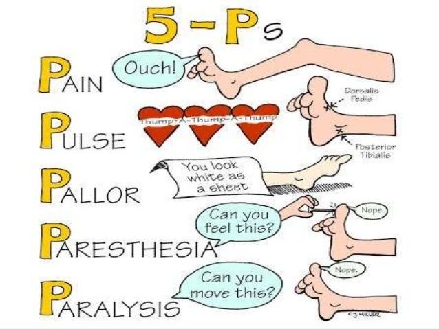
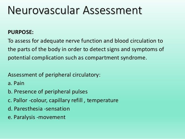
Assessment of neurovascular status is monitoring the 5 P's: pain, pallor, pulse, paresthesia, and paralysis.
Assess the neurovascular condition of the affected fracture extremity at least every 2 hours during the first 48 hours.
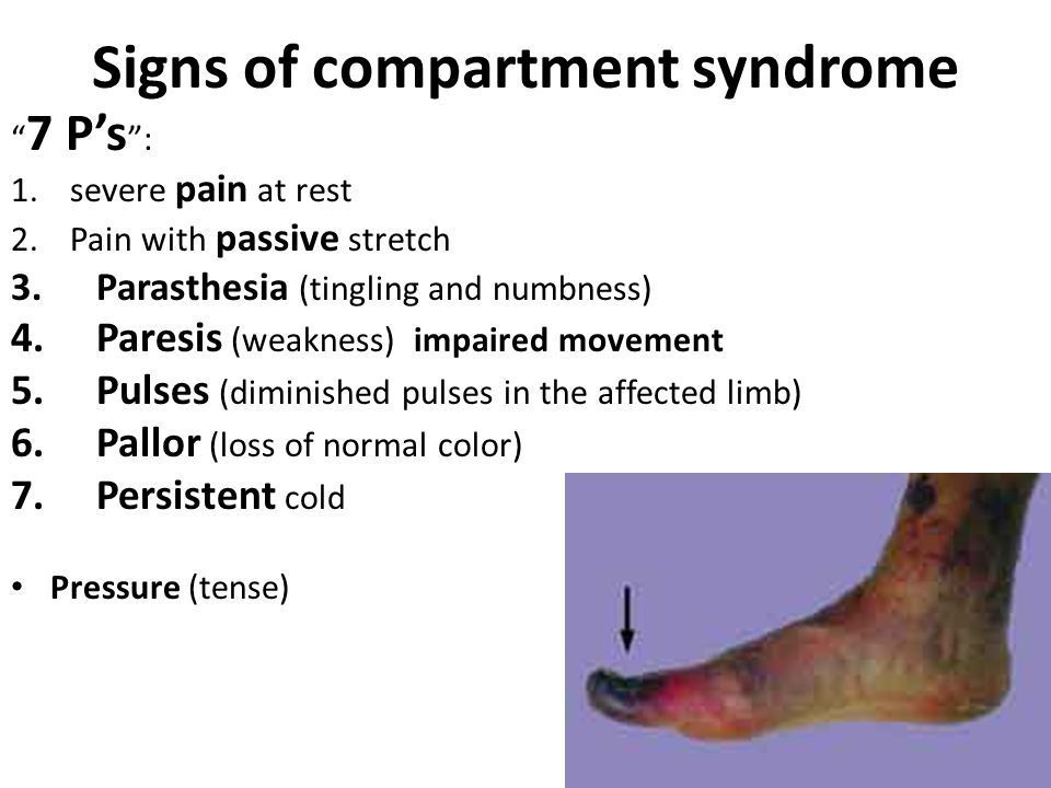
Compartment syndrome can be identified through neurovascular assessment in patients following extreme trauma. It tests blood flow to the extremity, distal to the injury, and nerve function.
Compartment syndrome is a painful condition that occurs when pressure within the muscles builds to dangerous levels. This pressure can decrease blood flow, which prevents nourishment and oxygen from reaching nerve and muscle cells. Compartment syndrome can be either acute or chronic.
Seite 16
Fat Embolism
Seite 17
Venous thromboembolism (VTE)
Seite 18
Pulmonary Embolism (PE)
pg. 529
Seite 19
Compression Syndrome
There are 36 compartments within the body.
Can happen with any condition that increases pressure within a limited tissue space.
Ex.) Cellulitis or burns result in edema fluid collecting in the compartments.
Ex.) Trauma causes bleeding within a compartment
Informationen
- Beschreibung
- Nursing Notiz am Application of the Nursing process of clients experiencing Musculoskeletal Trauma, erstellt von Kalyn S am 05/03/2018.
- Erstellt von
- Kalyn S
- Aufrufe
- 143
- Tags
- Bewertung