
Notizen
B3a: Molecules of life
Inhalt der Notizen (5)
Seite 1
Cells
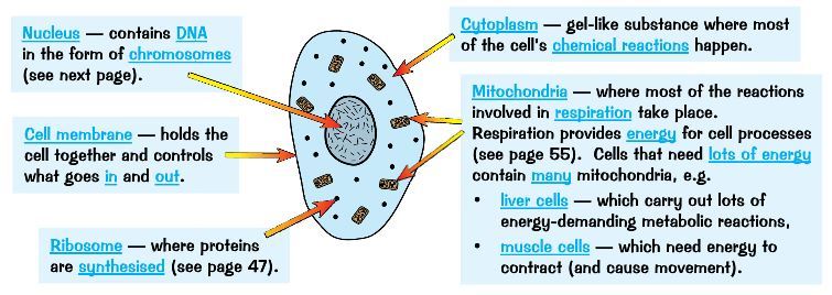
This is a diagram of an animal cell. There are labelled cell structures that make animal cells unique:
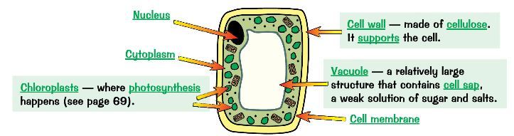
This is a diagram of a plant cell. It has all the structures of an animal cells (ie. ribosome and mitochondria) and a few extra:
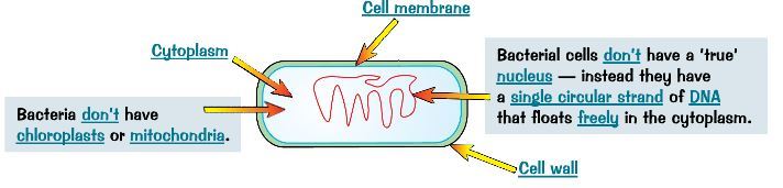
This is a diagram of a bacterial cell. It is much simpler and smaller than plant and animal cells.
Seite 2
Mitochondria and ribosomes
What are mitochondria?
Mitochondria are tiny structures found in cells (one is called a mitochondrion).
What happens in the mitochondria?
Respiration,
the chemical reaction that releases energy from glucose, happens in
mitochondria. This provides energy for life process such as movement and
growth.
Why do liver and muscle cells have larger numbers of mitochondria?
Active cells, such as liver and muscle cells, has to have a higher respiration rate. This is so that they have more energy to function as they are constantly used.
What are ribosomes?
They are found in the cytoplasm and are the site of protein synthesis.
Why can't we see them?
Ribosomes are too small to be seen with the light microscope. They can only be seen using an electron microscope.
Seite 3
Watson and Crick
Scientists struggled for decades trying to work out the structure of DNA.
Francis Crick and James Watson were the first scientists to build a model of the DNA in 1953.
They used data from other scientists to help them understand the structure. This included:
X-ray data, from Maurice Wilkins and Rosalind Franklin, showing that DNA is a double helix.
Other data showing that bases occurred in pairs.
They were awarded The Nobel Prize in Physiology or Medicine 1962. They were awarded 9 years later as new discoveries such as Watson and Crick's aren't widely accepted and other scientist had to repeat the work to make sure the results are reliable.
Seite 4
DNA
What is DNA?
DNA controls the production of different proteins. These are needed for the growth and repair of cells.
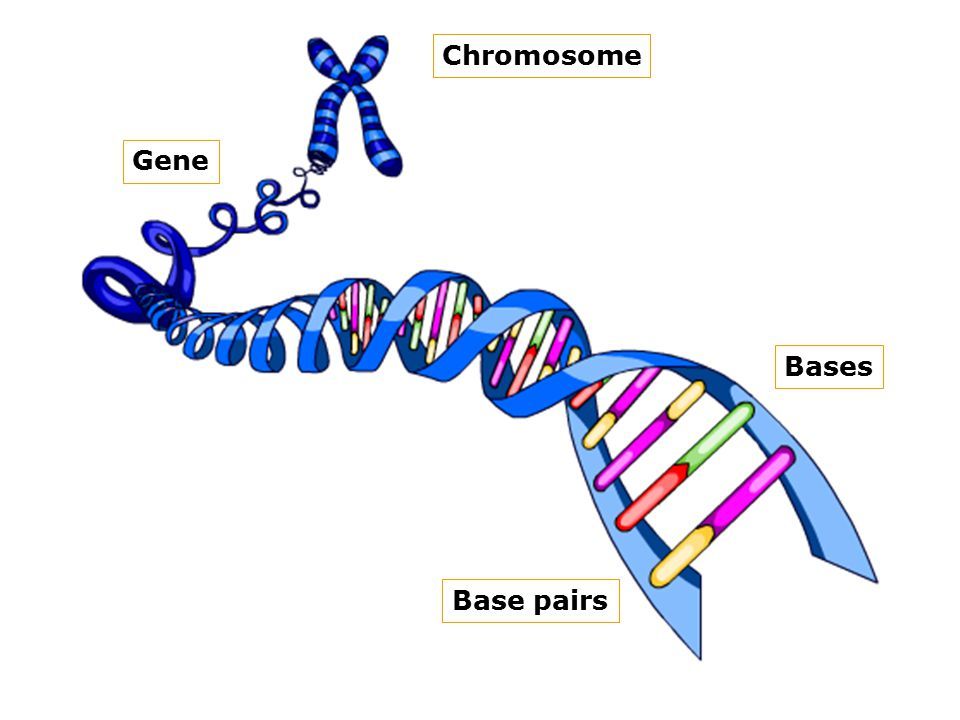
Gene
Section of a DNA
Each gene is a container of a different sequence of bases coding for a particular protein - genetic code
Chromosomes
Contained in the cell's nucleus
Long, coiled molecules of DNA
Divided up into regions called genes.
DNA Helix
Double strands of nucleotides (bases) coiled to form double helix
Cross-link between the two strands, formed by pairs of bases
Base pair
A, T
C, G
These are called complimentary base pairs
Backbone
Bases are attached to this
Sugar phosphate structure
How does DNA replicate itself?
DNA copies itself every time a cell divides - so each new cell has a full amount of DNA.
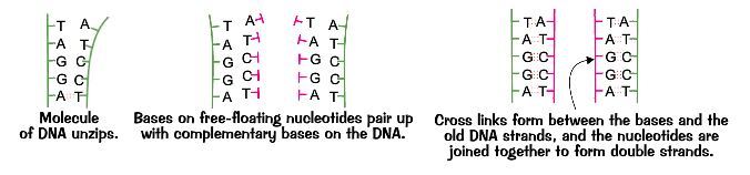
To copy itself, the DNA double helix 'unzips' - to form 2 single strands.
New nucleotides (floating freely in nucleus) the join on using complimentary base-pairing - making an exact copy of the DNA on the other strand.
The results is 2 double stranded molecules of DNA that are identical to the original molecule of DNA.
Seite 5
Protein Synthesis
How are proteins made?
Complementary base pairs form a code
They are 'read' in groups of 3s - this is known as base triplet
Each triplet specifies a particular amino acid
As DNA always stays in the nucleus, proteins are assembled at the ribosomes
So another molecule - mRNA - carries a copy of the coded instructions, in a gene, out of the nucleus
The sequence of amino acids in protein will govern how protein will fold
Each different type of protein has a specific shape
What is mRNA?
Messenger RNA (mRNA) is a single stranded molecule - a copy of one strand of DNA
mRNA from DNA is called transcription
Proteins from mRNA is called translation
Informationen
- Beschreibung
- GCSE Biology (Module B3: Living And Growing) Notiz am B3a: Molecules of life, erstellt von Carina C am 04/04/2016.
- Erstellt von
- Carina C · Apr 2016
- Aktualisiert
- 8 Jan 2017
- Aufrufe
- 142
- Tags
- Bewertung