
2330 EKG Rhythms
Folien in diesem Satz (27)
-
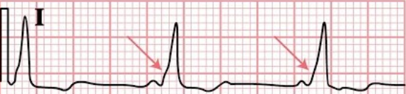
- Vent. Rhythm = Reg
- Rate = 60-100
- P waves = (+), 1 QRS
- PR Interval = .12 - .20 (norm)
- QRS Duration = <.11 (norm)
-
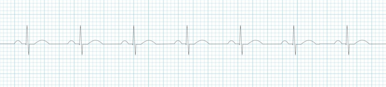
- Vent. Rhythm = Reg
- Rate less than < 60 bpm
- P wave = (+), 1 P wave / QRS
- PR Interval = .12 - .20 (norm)
- QRS less than < .11
-
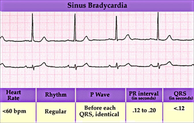
- Ven Rhythm = Reg
- Rate greater than > 100
- P wave = (+), 1 P wave/QRS
- PR Interval =.12 - .20
- QRS less than < .11 (norm)
-
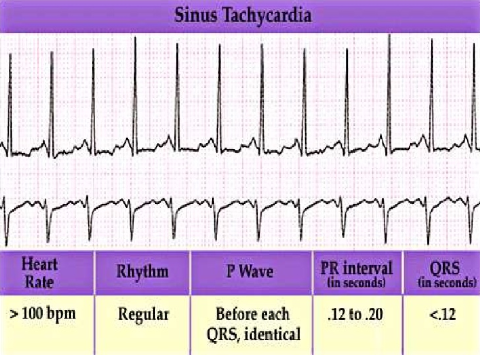
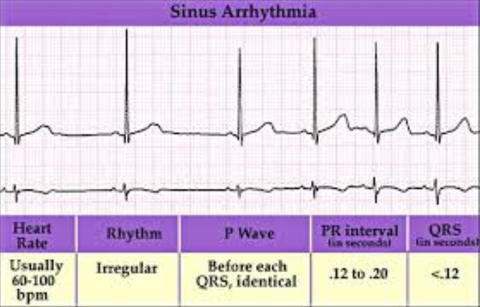
- Ven Rhythm = Irreg
- Rate = 60-100bpm
- P wave = (+), 1 P wave/QRS
- PR Interval =.12 - .20 (norm)
- QRS less than < .11 (norm)
- Reason: Autonomic nervous system causes small changes in HR associated with breathing
- Inspiration - Increased HR
- Expiration - Decreased HR
- Sympathetic Stimulation of SA Node (Increased Pacing) - Activated by Inspiration
- Parasympathetic Stimulation of SA Node (Inhibits Pacing) - Activated by Expiration
- Variability is normal.
- Reduced variability is pathological and indicates increased mortality, especially in heart disease or after infarction.
-
- Ven Rhythm = Reg (Except for Event)
- Rate = Varies
- P wave = (+), 1 P wave/QRS
- PR Interval =.12 - .20 (norm)
- QRS less than < .11 (norm)
- Misc. = Same as distance between 2 P-P waves
-
- Unhealthy SA node temporarily fails for at least 1 cycle
- -Missed beat has no P wave
- -SA node resumes at same rate as before block
-
AV Blocks: Slow / Block Impulse Completely
- 3 Types AV Blocks:
- 1st Degree AV Block: Lengthens delay between Atrial and Ventricular depolarization
- -PR Interval longer than .20 sec (1 big block) (Each cycle is long)
- 2nd Degree AV Block: Allow some Atrial depolarizations to conduct Vent. while others are blocked.
- -Result P wave with no QRS
- -PR interval is longer and longer and Drop
- 2 Types of 2nd Degree AV Blocks: @ AV node & Below AV node
- -
-
- 2nd Degree:
- Type I: Wenkebock
-
- 3rd Degree AV Block or Complete AV Block
-
- Ven Rhythm = Reg (Except for Event)
- Rate = Varies
- P wave = (+), 1 P wave/QRS
- PR Interval =.12 - .20
- QRS less than < .11 (norm)
- Misc. = Not the same as distance between 2 P-P intervals
- # of big boxes between P-P interval after SA arrest is not divisible by # of big boxes between normal P-P interval
-
Premature Atrial Contraction (PAC's)
- Ven Rhythm = Reg (Except for Event)
- Rate = Varies on underlying rhythm
- P wave = Premature, (+), 1 precedes QRS, Differ from sinus P wave
- PR Interval = Normal or Prolonged
- QRS less than < .11 (norm)
- -The Irritable Focus fires early producing a unique P wave.
- -Next P wave is the SA REST
-
Wandering Atrial Pacemaker (WPM)
- Vent. Rhythm = Usually Irreg
- Rate = 60 - 100bpm
- P wave = Size/Shape/Direction may Vary
- PR Interval = Varies
- QRS less than < .11 (norm)
- Misc. = If rate is > 100 then called Multifocal Atrial Tach
- -Wandering = foci PPM wanders from one to another
- -Multifocal Atrial Tach = foci PPM fire at once
-
Multifocal Atrial Tachycardia (MAT)
- Vent Rhythm = Usually Irregular
- Rate = Over 100 bpm
- P waves = P wave shape varies
- PR Interval =
- QRS Complex =
- Misc. = MAT is Tachy but WAP (Wandering Atrial Pacemaker) is not Tachy
- Seen in pt with COPD. Atrial automaticity foci show early signs of parasystole (entrance block)
-
- Rate = 150 -250 bpm
- P wave = P waves differ from Sinus P waves
- PR Interval = Shorter or Longer than Normal
- QRS less than < .11 (norm)
-
AVNRT (AV Nodal Re-entrant Tachy)
- Rate = 150 - 250 bpm
- P wave = P waves often hidden in QRS
- PR Interval = Normal IF seen (.12 - .20)
- QRS less than < .11 (norm)
-
- Ven Rhythm = Reg/Irreg
- Rate = Variable
- P wave = Saw Tooth
- PR Interval = n/a
- QRS less than < .11 (norm)
- Only 1, 2 or 3 series of Flutter waves reach Vent.: Result is Vent. don't depolarize at same rate
- Identifying Atrial Flutter: Invert Tracing or Perform Vagal Maneuver
-
- Ven Rhythm = Reg/Irreg
- Rate = Variable Rate
- P wave = F waves present, No discernible P waves
- PR Interval = n/a
- QRS less than < .11 (norm)
- Misc. = Uncontrolled: >100 bpm & Controlled: <100 bpm
- Cause: Rapid firing of multiple atrial automaticity foci suffering from entrance block pacing rapidly. (the atrial foci are parasystolic so they are all insensitive to overdrive suppression)
- -no single impulse depolarizes atria completely, and only occasional impulse reaches Vent.
- -Result: No P waves just erratic spikes,
- -Result: occasional impulse to Vent. may produce a rapid or slow Vent. rate, but is always Irregular
-
- Ven Rhythm = Reg (Unless a-fib present)
- Rate = 60-100 bpm if sinus is underlying
- P wave = (+), Normal (Unless a-fib)
- PR Interval less than < .12 if P waves are seen
- QRS > .12 usually
- Misc. = If PR interval > .12, then Delta wave may be seen (wide slurred QRS)
-
Premature Junctional Contractions(PJC's)
- Ven Rhythm = Reg with premature beat
- Rate = Usually Normal
- P wave = Occur before, during, or after QRS. If BEFORE then INVERTED
- PR Interval = if present, <.12, if no P wave then n/a
- QRS less than < .11 (norm)
-
- Ven Rhythm =
- Rate =
- P wave =
- PR Interval =.12 - .20
- QRS less than < .11 (norm)
-
- Ven Rhythm =
- Rate =
- P wave =
- PR Interval =.12 - .20
- QRS less than < .11 (norm)
-
- Ven Rhythm =
- Rate =
- P wave =
- PR Interval =.12 - .20
- QRS less than < .11 (norm)
-
- Ven Rhythm =
- Rate =
- P wave =
- PR Interval =.12 - .20
- QRS less than < .11 (norm)
-
Premature Ventricular Contraction (PVC)
- Ven Rhythm =
- Rate =
- P wave =
- PR Interval =.12 - .20
- QRS less than < .11 (norm)
- R on T PVC: extra beat occurs on T wave of previous beat
-
- Watchout may accually be Agonal rhythm
-
- Accelerated Junctional(s) - No P wave (What all have in common)
- Arrhythmia -
- Vent. Tach Know diff Vent. Fib - chaotic and not uniform
- Idoventricular - Slow rate
- Label EKG
Informationen
- Beschreibung
- EKG Rhythms with Rapid Interpretation of EKGs by Dale Dubin, and The Only EKG Book You Will Need by
- Erstellt von
- Elizabeth McKinley
- Aufrufe
- 53
- Bewertung