
Mind map
CN II (OPTIC NERVE)
Map content
CN II (OPTIC NERVE)
AFFERENT NERVE
ORIGIN
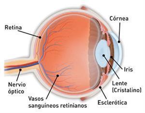
- Its real origin is given by the retina cells of the eyeball.
- ROUTE
- The ganglion cells of the retina
- They create nerve branches that come together to form:
- Optic Disc
- In the posterior part of the eyeball.
- Of the optical disc comes off:
- Optic Nerve
- leaves the eyeball
- leaves the orbital cavity
- Crosses the superior orbital fissure (vertex of the orbit), until it reaches the optic canal where it leaves next to the ophthalmic artery.
- It enters the cranial cavity where it will reach its apparent origin:
- Optic Chiasm
- BRANCHES
- optic tracts
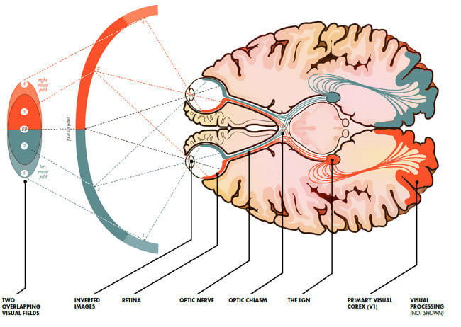
- the fibers project to the lateral geniculate nucleus of the thalamus, located in the depth of the lateral geniculate body. The thalamic neurons project, through the optical radiations, to the primary visual cortex.
- optic tracts
- The anterior cerebral fossa, in front of the sella turcica.
- BRANCHES
- Optic Chiasm
- It crosses the posterior pole of the eyeball (the choroid and the sclerotic layer).
- leaves the orbital cavity
- leaves the eyeball
- Optic Nerve
- Optic Disc
- posterior pole of the eyeball
- They create nerve branches that come together to form:
- The ganglion cells of the retina
ESPECIAL SENSORY:VISION
FUCTION
- It is also responsible for sending the signals
- that the eye perceives to our brain.
- The brain interprets these signals from external stimuli to conform the mental image of what we are seeing.
- that the eye perceives to our brain.
- In the optic chiasm, all the fibers that carry information from the nasal hemirretine pass to the contralateral side, and the fibers of the temporal hemirretina pass ipsilateral, to form the right and left optic tracts.
- This allows the images on each side of the field of both eyes to be transmitted to the appropriate side of the brain.
- The retina receives the inverted image on its walls. The light stimulates the cones and the staffs
- This structures involved
- In receiving and transmitting optical stimuli
- as the optical fibers and neural retina
- In receiving and transmitting optical stimuli
- This structures involved
- It is also responsible for sending the signals
Are surrounded by extensions of the cranial meninges and subarachnoid space
- The meninges extend all the way to the eyeball.
Images and files in the map
Information
- Description
- Mind Map on CN II (OPTIC NERVE), created by Laura Biviana Sánchez on 24/05/2018.
- Created by
- Laura Biviana Sánchez
- Views
- 53
- Rating