
Mind map
Block 9 part 1
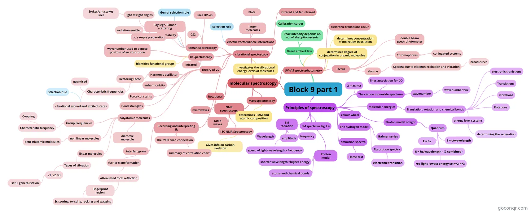
Mind map: Block 9 part 1 spectroscopy
Map content
Block 9 part 1
molecular spectroscopy
- Studies the interaction of electromagnetic radiation with matter to obtain info about molecules.
- NMR spectroscopy
- 13C NMR Spectroscopy
- Gives info on carbon skeleton
- radio waves
- 13C NMR Spectroscopy
- Mass spectroscopy
- determines RMM and atomic composition
- UV-VIS spectrophotometry
- determines degree of conjugation in organic molecules
- determines concentration of molecules in solution
- electronic transitions occur
- UV vis
- Measures the intensity of the light. Quartz, is used below around 300 nm, glass used for organic solvents and cheap plastic cuvettes used when using water or alcohol solvents. Also depending on wavelength - light must be able pass through them.
- alanine
- The wavelength of the peak of the spectrum is called 'wavelength subscript max' This is where it exhibits the maximum absorption. The maxima is wider than for hydrogen which absorbs in very narrow bands at particular wavelengths. Alaninea bosrobs at a wider range of wavelengths. The wide range is due to vibrational energy increase too. IN solution rotation is huindered but tumbling motion occurs broadening the range of the wavelength.
- Spectra due to electron excitation and vibration
- broad curve
- Chromophores
- The part of the molecule responsible for UV vis spectra is called the chromophore. often functional groups but generally conjugated molecules in organic molecules.
- conjugated systems
- CHromophores generally involve conjugated systems in which the electrons are delocalised and electron transitions involve the pi orbitals. Double bonds separated by single bonds form conjugated systems. So penta=1,3-diene will produce an absoption spectra but Penta-1,4- diene will not. Pi electrons can spread across the single bond. This results in a decrease in the separation of energies between pi boning and pi antibonding electronic energy levels so that absorption occurs in the UV vis spectrum. Lower energy spearation means lower frequency which means longer wavelength so penta-1,4-diene peaks at 177 nm but penta-1,3-diene peaks at 224 nm.
- double beam spectrphotometer
- Source beam is split to travel through the reference cell and the sample cell at the same time.
- Rotational
- microwaves
- vibrational spectoscopy
- IR Spectroscopy
- identifies functional groups
- infrared
- wavenumber used to denote positoin of an absorption
- Raman spectroscopy
- Raman spectroscopy is a companion spectroscopy to IR - it finds vibrations that may not appear on the IR spectrum. Some vibrations appear on neither. If a vibrational mode can be observed the molecule is said to be active. Raman spectroscopy gains information from the light that is scattered rather than absorbed revealing info about symetrical vibrations that do not show up on IR spectroscopy.
- uses UV-vis
- Solutions can be used as water is transparent at these frequencies. Solutions can not be used for IR.
- Rayliegh/Raman scattering
- When the frequency of the light is unchanged by the scattering it is called Rayleigh scattering. Typically the intensity of the scattered light is around 10 to the minus 3 of the incident light. Raman scattering is the study of the scattered light when the frequency is changed. It is weaker at about 10 to the minus 6 of the incident light.
- light at right angles
- Chemists study the light that is scattered at right angles to the incident beam in Ramen spectroscopy. Raman spectra consist of the rayleigh line flanked on either side the stokes and antistokes lines. It is the shift in frequency that is important not the frequency it changes to.
- Stokes/antistokes lines
- The shift in frequency happens because the incident light transfers some of its energy to the vibration or rotation of the molecule - it shifts to a lower frequency and is a stokes line. Anti stokes lines shift to a higher frequency and occur when the molecules transfers some of its energy to the photon. Molecules in the ground state absorb energy to give stokes lines and in the excited state they transfer energy to give antistokes lines. The shift in wavenumber corresponds to the virational energy of the molecule. at normal temps there are more molecules at the ground state and the raman spectra shows this.
- Polarisability
- The electric field of radiation causes the electrons in a molecules to oscillate at the frequency of the radiation. The ease with which the electrons can be moved by the radiation is the polarisability of the molecule. Most of the time the electrons emit radiation at the frequency of the exciting radiation - this is the rayleigh radiation. The electron cloud oscillates under the influence of a passing wave (see video). The atoms also vobrate and the interaction of the two is called the Raman effect. As the nucleus stretches the electrons are less constrained by the nucleus and there more easily polarisable. As it is compressed the oppostie is true.
- radiation emitted
- The radiation emitted is at the same frequency of the exciting radiation but the amplitude of the molecule varies producing radiation of two slightly differing frequencies. Detected as two waves, one of higher frequency - (Stokes) and one of lower frequency (Anti stokes)
- Genral selection rule
- The general selection rule for vibrational transitions in Raman spectroscopy is that there must be a change in the polarisability of the molecule during the vibration.
- CS2
- 3 vibrational modes but only the symetrical stretch is Raman active as the electron cloud changes size. The asymetrical stretch and bend do not change size and so are not Raman active. Instead for these two there is a change of dipole moment and they absorb in the IR spectrum. During the symmetric stretch the dipole moment remains at zero.
- no sample preparation
- infrared and far infrared
- investigates the vibrational energy levels of molecules
- electric vector/dipole interactions
- |The electric vector of a EM wave varies in magnitude and changes direction every half cycle. Interaction of the electric vector and the dipole of a molecule causes it to vibrate. This only happens when the molecule and electric vector share the same frequency. CO has a dipole. (C is neg) when it interacts with the wave it is stretched or compressed as + charge experiences a force in direction of of vector stretching it. The - charge moves in opposite direction to vector compressing it.
- selection rule
- The field and the vibrating molecule only interact if there is a change of dipole moment. So homonuclear molecules such as O2 or N2 will not interact as they have no dipole.
- larger molecules
- Polyatomic molecules can vibrate in many more modes. Triatomic molecules like SO2 have 3 vibrational modes, symetric stretch, bend and asymetric stretch. NH3 has 6, some of which have the same energy and so only produce one peak between them . propanone with 10 atoms has 24. IN practice it is too difficult to differentiate them all. FGs have their own distinct vibrations
- Plots
- An IR spectrum is a plot of the extent to which radiation is absorbed by the molecule. Wavenumber is the reciprocal of wavelength and is usually plotted on the horizontal axis. Transmittance is is usually plotted on the vertical axis and is at 100% when no absorption occurs. When bonds within the molecule absorb the radiation this number drops and a peak is observed on the spectrum.
- Theory of VS
- Harmonic oscillator
- The model to describe the regular separation of quantised energy levels of vibrational energy changes.
- Restoring Force
- The harmonic oscillator is the simplest model to depict the vibration of a diatomic molecule. The restoring force causes the molecule to vibrate. When it is stretched it pulls it back and when it is compressed it pushes it away. The restoring force is proportional to the extension or compression of the spring. At equilbrium length the potential energy is zero. When stretched or compressed it changes following the shape of a parabola. Max kinetic energy is reached at the bottom of the parabola.
- Characteristic frequencies
- Frequency depends on the mass of the atom and the stiffness of the bonds so information can be deduced from the frequency using Eqn 2.3. As force constant K increases, stiffness of the bond increases and so does the frequency. Vibrational energies are quantised as molecules will only emit and absorb energies at characteristic frequencies. They are separated by a value of hv (Eqn.2.5) and can only move to an adjacent energy level.
- vibrational ground and excited states
- The general selection rule for vibrational energy changes in IR spectra is 'Change if v =plus or minus 1'. ONly changes to adjacent energy levels are allowed. The tranistion has the same energy separation and therefor the same absorption frequency. So only one absorption line related to vibrational frequencies would be expected for a diatomic molecule. So v=0-v=1 would be the same line as v=1-v=2.
- selection rule
- The selection rule is broken when there is an overtone absorption line at exactly double the wavenumber of the real one. This is a forbidden two level jump and is because the molecule is displaying anharmonicity
- quantised
- anharmonicity
- If a molecule is given sufficient energy it dissociates. As the molecule is raised to a higher energy level the amplitude increases but this caannot continue to infinity, eventually the restorative force gets weaker until no restorative forcee xists and the bond no longer exists. A Morse curve more accurately describes this behavior. It is like the harmonic ocsilator curve when the v number is small but the energy separation levels converge as the number gets higher until the molecule dissociates at the continuum.
- Force constants
- Frequency will decrease with increase in mass in molecules. Light molecules absorb at higher wave numbers than heavier ones at similar bond strengths.
- Bond strengths
- Strongly bonded molecules (eg double or triple bonded) absorb at higher wavenumbers than weakly bonded molecules
- polyatomic molecules
- By considering harmonic and anharmonic models you can get info about force constants and bond energies of diatomic molecules. This can be extended to polyatomic molecules but it is more difficult. You can determine shape - linear or bent.
- non linear molecules
- Each atom can move in any direction. To locate them specify 3 coordinates x,y and z so Each atom has 3 degrees of freedom so a molecule has 3n degrees of freedom where n is the number of atoms in the molecule. To identify the vobrational modes of the non linear molecule you must first eliminate translational motion (3 degrees) and rotational motion (3 degrees). So for a non linear molecule there are 3n-6 degrees of vibrational freedom (or normal modes of vibration). Each of the normal modes is associated with a simple potential energy curve and hense a set of energy levels v=0 etc. The three fundamental modes are symetrical stretch, bend and asymetrical stretch.
- bent triatomic molecules
- SO2 is an example. It has 3N-6 = 3 normal modes of vibration. Symmetric stretch - v1, asymmetric stretch-v2 and bend in bond angle - v3. All three are active in IR and Raman as all three produce achange in dipole moment and polarisabiltiy.
- linear molecules
- Linear molecules behave differently from non- linear molecules because rotation about the molecular axis does not displace the atoms of the molecule. So rotation is described by two degrees of freedom. Therefore 3n-5 degrees gives the number of vibrational modes. The difference occurs in the out of plane motions - there is a bending of the linear molecule CS2 but a rotation of the non-linear one - SO2.
- Types of vibration
- CS2 is linear so it has 3n-5 =4 modes of vibration. symmetric stretch asymmetric stretch and two doubly degenerate bending modes. You will only find three bands on the spectrum because two of the modes have the same energy and are degenerate.
- v1, v2, v3
- To be IR active the dipole moment must change during a vibration. To be Raman active there must be a change in polarisability. THe asymetric stretch and bending vibrations are IR active as the dipole moment changes. The symetric stretch is Raman active because the electron cloud changes size, causing polarisability. Bending modes are generally found at a lower wave number than stretching modes. Often the frequency for the symmetric stretch is less than that of the asymmetric stretch. By convention v1 is the symmetric stretch modes, v2 is the bending modes and v3 is the asymmetric modes. This is true for a linear molecule with xyx. It is different for xyz molecules where there is a change in the dipole moment for both stretching modes as well as the both bending modes. The central atom moves in the symetric stretch mode this time to compensate for the different masses of the two different atoms.
- useful generalisation
- XYX molecules possess a symmetry element that XYZ molecules do not. Molecules with a centre of symmetry will either be active in IR or Raman but not both. If they are active in both then they must be XYZ molecules.
- diatomic molecule
- Can only vibrate in one way - stretching vibration.
- Group Frequencies
- so far the molecules normal modes of vibration have involved all the atoms moving in phase. This is true when all the atoms have a similar mass and similar bond lengths. However if there is a group within the molecule that has a multiple bond it will vibrate independently of the rest of the molecule. The natural frequency will be higher than the rest of the molecule. Also a very light atom H or a very heavey one Iodine can vibrate independently. When the vibrations are independent a group frequency will be observed
- Characteristic frequency
- Often any number of compounds containing a particular functional group such as C=N will, regardless of the rest of the molecule vibrate within a narrow range of frequencies allowing the particular group to be identified. They may also sometimes display a similar intensity.
- Coupling
- Harmonic oscillator
- Recording and interpreting IR
- lighter atoms give higher frequencies. Greater mass, lower frequency. Stronger bonds give higher frequencies. If radiation is applied then the bond absorbs energy. With greater energy there is a greater amplitude. Most bonds absorb in the IR region which corresponds to heat. Each type of bond absorbs at a particular frquency which allows it to be identified. Chemists use units of wavenumbers(cmto the minus 1) which are proportional to frequency.
- interferogram
- Splits the IR beam and it holds information about the intensities of the radiation at all frequencies.
- furrier transformation
- mathematical technique to decode the interferogram information to give the intensity of each frequency of the IR separately to produce a graph of % transmission vs wavenumber. Some frequencies are absorbed more than others depending on what bonds are present in the sample.
- Attenuated total reflection
- The IR is passed through the sample which sits on a crystal of either diamond or germanium. It is passed through at an angle called attenuated total reflection.
- Fingerprint region
- The fingerprint region is below about 1500 cm to the minus 1. (or between 1000-1600 cm-1) It is various bending modes of vibration. It is used to match the spectra of an unknown sample to a database of known samples.
- Scissoring, twisting, rocking and wagging
- It requires less energy to distort a bond than it does to stretch it. so these bending modes are usually observed at lower wavenumbers. two absrptions at around 1400 cm-1 are due to a scissoring mode of CH2 groups and a bending mode of CH3 groups. These modes are sensitive to the nature of the atoms and bonds around them and so difficult to distinguish. Therefore more use for comparison than for identifying particular groups or bonds. The region above 1600 cm-1 is more useful for spotting bonds.
- The 2900 cm-1 connection
- 2900 cm-1 is the most common peak of an organic compound in the IR spec. It is the frequency of the C-H stretch bond. You can now start to build up a correlation chart.
- summary of correlation chart
- Bonds to hydrogen appear at higher wavenumbers. Single bonds to C absorb in the fingerprint region below 1600 cm-1. Double bonds to carbon occur just above the fingerprint region or 1600 cm-1 and are spotted easily. Triple bonds to carbon occur around 2200 cm-1. Aromatic out of plane C-H bends occur in the 600-900 cm-1 region.
- IR Spectroscopy
Principles of spectroscopy
- EM radiation
- Light is an electromagnetic wave comprising oscillating electric and magnetic fields. The electrical and magnetic waves are oscillating at right angle to each other. Both are at right angles to the direction of travel. It is a sinusoidal wave. - like a mathematical function it follws a smooth, repetative oscillation.
- Wavelength
- amplitude
- frequency
- speed of light=wavelength x frequency
- EM spectrum Fig 1.4
- logarithmic scale boundaries are vague and loose
- The hydrogen model
- Balmer series
- The five possible electron transitions of hydrogen. Electrons occupy different energy levels and different electron shells according to the amount of energy they have.
- Absorption spectra
- Absorption spectra are the result of electrons absorbing enery and moving to a higher energy level. It can be used to identify molecules.
- electronic transition
- The electron transition is the amount of energy required for one jump of energy level by the electron. Black absorption lines appear on the spectrum where hydrogen has absorbed energy of particular wavelengths. This energy is absorbed by an electron which then moves to a higher energy level. THe balmer series shows the possible energy levels for hydrogen.
- Balmer series
- colour wheel
- The colour wheel in Fig 1.7 shows the complementary colours. you look at the one opposite.
- emmision spectra
- emmision spectra are the result of electrons moving from excited states to the lower n=2 energy level.
- Flame test
- Saturated solutions of metals salts are sprayed into a flame. The flame excites electrons to higher energy levels. Light is emitted as the electrons return to their ground state. THe light is a characteristic colour. red strontium, orange sodium and green copper.
- Photon model
- Amount of energy carried by a photon is called a quantum. In a ray of light of particular wavelength and frequency there is a stream of photons - each photon carries a particular amount of energy (E) which relates to the frequency of the ray. It is given by E = hv. h is the planck constant 6.63 x 10 to the minus 34 J s to the minus 1. See section 1.4 for further equations, combining this equation with the earlier one.
- shorter wavelength =higher energy
- atoms and chemical bonds
- individual atoms or chemical bonds can absorb or emit light of a particular frequency. THe energy corresponds to the exact energy gap of the transition.
- molecular energies
- Molecules are composed of atoms so the model still holds however electrons in molecules are under the influence of more than one nuclei so the quantised energies are more complex.
- Translation, rotation and chemical bonds
- Translate - move from one region of space to another Rotate - rotate as they do so and the energy associated with rotations is also quantised. Vibrational motion is also quantised - chemical bonds can stretch and bend like springs.
- energy level systems
- Vibrations occur on shorter timescales than rotations which occur on shorter time scales than translations. so these different energies can be treated independently. Etotal=Eelectronic+Evibration+Erotation+Etranslation. Every molecule has a unique energy level system which is not sensitive to temp or pressure or small amounts of impurities. Therefore identity of molecule can be determined.
- determming the seperation
- Determining the seperation between one energy and the next is done by adding some energy to see how it takes. This is equivilent to the seperation energy. It can also be determined by looking at the emmision process.
- Translations
- When a molecules moves in space.
- vibrations
- When the bonds are stretched between atoms in a molecule.
- Rotations
- When the molecules rotates
- electronic tranistions
- When electrons jump from one energy level to another.
- The carbon monoxide spectrum
- lines association for CO
- Lines greater than 10 to the 15 Hz are associated with electronic transitions. Vibrations at infrared and far infrared between 10 to the 14 and 10 to the 11 Hz. Rotational energy changes occur in the far infrared and microwave regions. 10 to the 13 to 10 to the 9 Hz..
- wavenumber
- wavenumber is used to denote the position of an absorption. wavenumber is proportional to frequency and energy and inversely proportional to wavelength. It has units of cm to the minus 1. See 1.5.2 for relevent equations
- wavenumber=v/c
- 2 maxima
- Rotational is superimposed on the vibrational transition on the absorption spectra.
- lines association for CO
- Photon model of light
- Quantum
- The energy carried by a single photon of light. The energy of this photon can be found E=hv where h is the planck constant (6.63 x10 to the minus 34) and v is the frequency
- E = hv
- E = c/wavelength
- E = hc/wavelength - (2 combined)
- h and c are constants so the shorter the wavelength of the light the greater the energy as wavelength is the inverse of energy in this equation.
- red light lowest energy so n=2-n=3
- Quantum
- EM radiation
Beer-Lambert law
- Peak intensity depends on no. of absoption events
- Intensity is converted into absorption using equation in 2.4 Absorbance = log10 (Io/I)
- Calibration curves
- A calibration is a set of operations that establishes, under specific conditions, the relationship between an instrument response and the corresponding values of a standard. (An analyte of known concentration.) Using the beer lambert law a graph of absorbance against concentration of solutions of known concentration produces a calibration curve. Absorbance can be measured for a solution of unknown concentration and the concentration determined from the calibration curve. If path length is 1 cm then the curve will show a linear relationship between absorbance and concentration. The straight line portion of the curve defines the working range of your instrument.
- Peak intensity depends on no. of absoption events
Information
- Description
- degree Chemistry: essential concepts S215 Mind Map on Block 9 part 1, created by vicstevens on 15/03/2015.
- Created by
- vicstevens
- Views
- 4
- Rating