
Mind map
Ischemic Stroke
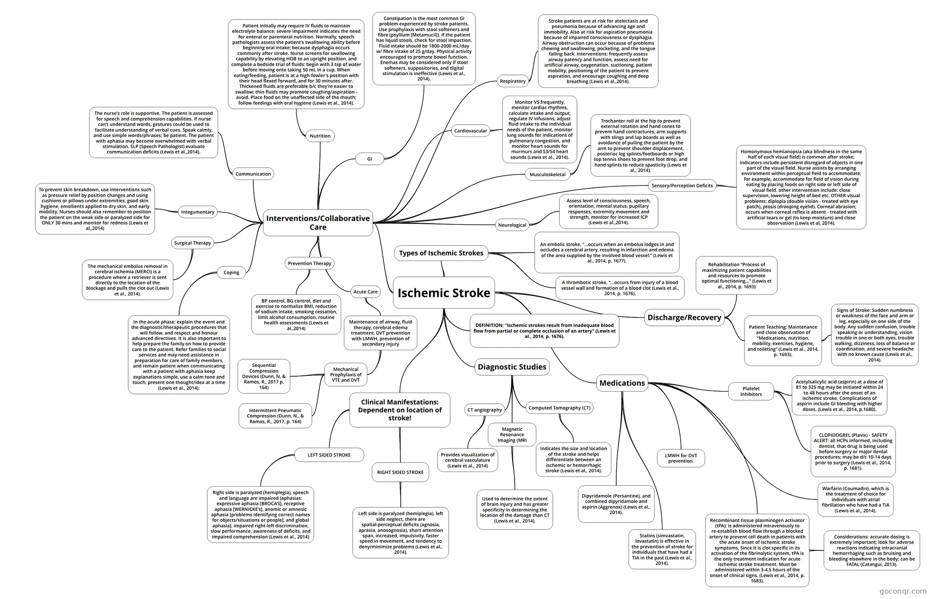
Mind map: Ischemic Stroke
Map content
Ischemic Stroke
DEFINITION: “Ischemic strokes result from inadequate blood flow from partial or complete occlusion of an artery” (Lewis et al., 2014, p. 1676).
Types of Ischemic Strokes
- An embolic stroke, “...occurs when an embolus lodges in and occludes a cerebral artery, resulting in infarction and edema of the area supplied by the involved blood vessel.” (Lewis et al., 2014, p. 1677).
- A thrombotic stroke, “...occurs from injury of a blood vessel wall and formation of a blood clot (Lewis et al., 2014, p. 1676).
Discharge/Recovery
- Rehabilitation “Process of maximizing patient capabilities and resources to promote optimal functioning…” (Lewis et al., 2014, p. 1693)
- Patient Teaching: Maintenance and close observation of “Medications, nutrition, mobility, exercises, hygiene, and toileting” (Lewis et al., 2014, p. 1693).
- Signs of Stroke: Sudden numbness or weakness of the face and arm or leg, especially on one side of the body. Any sudden confusion, trouble speaking or understanding, vision trouble in one or both eyes, trouble walking, dizziness, loss of balance or coordination, and severe headache with no known cause (Lewis et al., 2014).
Medications
- Platelet Inhibitors
- Acetylsalicylic acid (aspirin) at a dose of 81 to 325 mg may be initiated within 24 to 48 hours after the onset of an ischemic stroke. Complications of aspirin include GI bleeding with higher doses. (Lewis et al., 2014, p.1680).
- CLOPIDOGREL (Plavix) - SAFETY ALERT: all HCPs informed, including dentist, that drug is being used before surgery or major dental procedures; may be d/c 10-14 days prior to surgery (Lewis et al., 2014, p. 1681).
- Recombinant tissue plasminogen activator (tPA): is administered intravenously to re-establish blood flow through a blocked artery to prevent cell death in patients with the acute onset of ischemic stroke symptoms. Since it is clot specific in its activation of the fibrinolytic system, tPA is the only treatment indication for acute ischemic stroke treatment. Must be administered within 3-4.5 hours of the onset of clinical signs. (Lewis et al., 2014, p. 1683).
- Considerations: accurate dosing is extremely important; look for adverse reactions indicating intracranial hemorrhaging such as bruising and bleeding elsewhere in the body; can be FATAL (Catangui, 2013).
- Warfarin (Coumadin), which is the treatment of choice for individuals with atrial fibrillation who have had a TIA (Lewis et al., 2014).
- Dipyridamole (Persantine), and combined dipyridamole and aspirin (Aggrenox) (Lewis et al., 2014).
- LMWH for DVT prevention.
- Statins (simvastatin, lovastatin) is effective in the prevention of stroke for individuals that have had a TIA in the past (Lewis et al., 2014).
- Platelet Inhibitors
Diagnostic Studies
- Magnetic Resonance Imaging (MRI
- Used to determine the extent of brain injury and has greater specificity in determining the location of the damage than CT (Lewis et al., 2014).
- CT angiography
- Provides visualization of cerebral vasculature (Lewis et al., 2014)
- Computed Tomography (CT)
- Indicates the size and location of the stroke and helps differentiate between an ischemic or hemorrhagic stroke (Lewis et al., 2014).
- Magnetic Resonance Imaging (MRI
Clinical Manifestations: Dependent on location of stroke!
- LEFT SIDED STROKE
- Right side is paralyzed (hemiplegia), speech and language are impaired (aphasias: expressive aphasia [BROCA’S], receptive aphasia [WERNICKE’s], anomic or amnesic aphasia [problems identifying correct names for objects/situations or people], and global aphasia), impaired right-left discrimination, slow performance, awareness of deficits, and impaired comprehension (Lewis et al., 2014)
- RIGHT SIDED STROKE
- Left side is paralyzed (hemiplegia), left side neglect, there are spatial-perceptual deficits (agnosia, apraxia, anosognosia), short attention span, increased, impulsivity, faster speed in movement, and tendency to deny/minimize problems (Lewis et al., 2014).
- LEFT SIDED STROKE
Interventions/Collaborative Care
- Prevention Therapy
- BP control, BG control, diet and exercise to normalize BMI, reduction of sodium intake, smoking cessation, limit alcohol consumption, routine health assessments (Lewis et al.,2014)
- Acute Care
- Maintenance of airway, fluid therapy, cerebral edema treatment, DVT prevention with LMWH, prevention of secondary injury
- Mechanical Prophylaxis of VTE and DVT
- Sequential Compression Devices (Dunn, N, & Ramos, R., 2017 p. 164)
- Intermittent Pneumatic Compression (Dunn, N., & Ramos, R., 2017, p. 164)
- Mechanical Prophylaxis of VTE and DVT
- Maintenance of airway, fluid therapy, cerebral edema treatment, DVT prevention with LMWH, prevention of secondary injury
- Integumentary
- To prevent skin breakdown, use interventions such as pressure relief by position changes and using cushions or pillows under extremities, good skin hygiene, emollients applied to dry skin, and early mobility. Nurses should also remember to position the patient on the weak side or paralyzed side for ONLY 30 mins and monitor for redness (Lewis et al.,2014)
- Nutrition
- Patient initially may require IV fluids to maintain electrolyte balance; severe impairment indicates the need for enteral or parenteral nutrition. Normally, speech pathologists assess the patient’s swallowing ability before beginning oral intake; because dysphagia occurs commonly after stroke. Nurse screens for swallowing capability by elevating HOB to an upright position, and complete a bedside trial of fluids: begin with 3 tsp of water before moving onto taking 50 mL in a cup. When eating/feeding, patient is at a high-fowler's position with their head flexed forward, and for 30 minutes after. Thickened fluids are preferable b/c they’re easier to swallow; thin fluids may promote coughing/aspiration - avoid. Place food on the unaffected side of the mouth; follow feedings with oral hygiene (Lewis et al., 2014).
- GI
- Constipation is the most common GI problem experienced by stroke patients. Use prophylaxis with stool softeners and fibre (psyllium [Metamucil]). If the patient has liquid stools, check for stool impaction. Fluid intake should be 1800-2000 mL/day w/ fibre intake of 25 g/day. Physical activity encouraged to promote bowel function. Enemas may be considered only if stool softeners, suppositories, and digital stimulation is ineffective (Lewis et al., 2014).
- Cardiovascular
- Monitor VS frequently, monitor cardiac rhythms, calculate intake and output, regulate IV infusions, adjust fluid intake to the individual needs of the patient, monitor lung sounds for indications of pulmonary congestion, and monitor heart sounds for murmurs and S3/S4 heart sounds (Lewis et al., 2014).
- Musculoskeletal
- Trochanter roll at the hip to prevent external rotation and hand cones to prevent hand contractures, arm supports with slings and lap boards as well as avoidance of pulling the patient by the arm to prevent shoulder displacement, posterior leg splints/footboards or high top tennis shoes to prevent foot drop, and hand splints to reduce spasticity (Lewis et al., 2014).
- Neurological
- Assess level of consciousness, speech, orientation, mental status, pupillary responses, extremity movement and strength, monitor for increased ICP (Lewis et al.,2014).
- Communication
- The nurse's role is supportive. The patient is assessed for speech and comprehension capabilities. If nurse can’t understand words, gestures could be used to facilitate understanding of verbal cues. Speak calmly, and use simple words/phrases; be patient. The patient with aphasia may become overwhelmed with verbal stimulation. SLP (Speech Pathologist) evaluate communication deficits (Lewis et al.,2014).
- Coping
- In the acute phase: explain the event and the diagnostic/therapeutic procedures that will follow, and respect and honour advanced directives. It is also important to help prepare the family on how to provide care to the patient. Refer families to social services and may need assistance in preparation for care of family members, and remain patient when communicating with a patient with aphasia keep explanations simple, use a calm tone and touch, present one thought/idea at a time (Lewis et al., 2014).
- Sensory/Perception Deficits
- Homonymous hemianopsia (aka blindness in the same half of each visual field) is common after stroke; indicators include persistent disregard of objects in one part of the visual field. Nurse assists by arranging environment within perceptual field to accommodate; for example, accommodate for field of vision during eating by placing foods on right side or left side of visual field. other intervention include: close supervision, lowering height of bed etc. OTHER visual problems: diplopia (double vision - treated with eye patch), ptosis (drooping eyelid). Corneal abrasion; occurs when corneal reflex is absent - treated with artificial tears or gel (to keep moisture) and close observation (Lewis et al, 2014).
- Respiratory
- Stroke patients are at risk for atelectasis and pneumonia because of advancing age and immobility. Also at risk for aspiration pneumonia because of impaired consciousness or dysphagia. Airway obstruction can occur because of problems chewing and swallowing, pocketing, and the tongue falling back. Interventions: frequently assess airway patency and function, assess need for artificial airway, oxygenation, suctioning, patient mobility, positioning of the patient to prevent aspiration, and encourage coughing and deep breathing (Lewis et al.,2014).
- Surgical Therapy
- The mechanical embolus removal in cerebral ischemia (MERCI) is a procedure where a retriever is sent directly to the location of the blockage and pulls the clot out (Lewis et al., 2014).
- Prevention Therapy
Information
- Description
- Mind Map on Ischemic Stroke, created by Janine Loyola on 07/03/2017.
- Created by
- Janine Loyola
- Views
- 1
- Rating