
Mind map
Spinal Reflexes
Map content
Spinal Reflexes
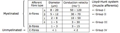
Afferent Fibre Classification
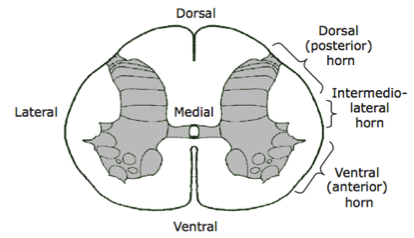
- Spinal Cord and Rexed's Laminae
- Dorsal is at the back (think of animal on all fours)
- Grey matter
- Where cell bodies lie
- White matter
- Where afferent fibres run through
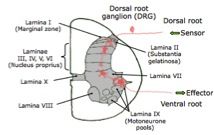
- Spinal cord splits into 10 different laminae
- Motoneurones lie largely in laminae IX
- Dorsal root ganglion (DRG)
- Cell body of the afferent fibre
- Lies outside the spinal cord
- Terminates in lamina I
- Pathways go through the laminae to the efferent pathway
- Erlanger and Gasser system
- Fibres are A, B or C
- Lloyd-Hunt system
- Fibres grouped by I, II, III and IV
- Spinal Cord and Rexed's Laminae
Muscle Spindle Stretch Reflex
- Afferent Innervation
- Single Ia afferent fibres
- Large myelinated A-α fibre
- Signals dynamic and static response
- Primary (annulospiral) endings
- Wrap around the swollen portion of the NBF
- Also activated by tap or vibration of the tendon that the muscle is attached to
- Firing Characteristics
- Constant level of stretch shows a constant level of firing
- Increase in stretch shows an increase in frequency of firing
- Increases further with faster stretching
- After stretch, firing is increased but is consistent
- Group II afferents
- Large, myelinated A-β fibres
- Secondary (flower-spray) endings
- Signals static response only
- Firing Characteristics
- Consistent frequency of firing before and during stretch
- Increased frequency with sustained stretch
- Single Ia afferent fibres
- Efferent Innervation
- Extrafusal fibres are innervated by α-motoneurones
- Intrafusal fibres are innervated by γ-motoneurones
- Ensure the muscle spindle is always in direct contact with the changes occurring in the muscle
- Tension in the spindle is altered to adjust for changes in muscle length due to extrafusal fibre contraction
- Ensure the muscle spindle is always in direct contact with the changes occurring in the muscle
- α- and γ- motoneurones are usually co-activated during voluntary movements
- Iα fibres signal the length of the muscle
- γ-motoneurones cause contraction of the spindle
- Ensures no slackness
- Myotatic Stretch Reflex
- Monosynaptic
- Simplest form of reflex
- Afferent fibres terminate on the motoneurones which innervate the same muscle
- Iα muscle spindle afferents directly synapse onto α-motoneurones in the ventral horn
- No interneurons are used
- Motoneurones synapsed onto can be homonymous muscle (same) or heteronymous (synergistic)
- Glutamate is the neurotransmitter
- e.g. knee Jerk Reflex
- Tapping provides the stimulus that causes the contraction
- e.g. standing upright
- Spindles in the leg detect stretch when leaning forwards or back
- They fire and stimulate α-motoneurones to cause the muscle to contract and bring the body back upright
- Spindles in the leg detect stretch when leaning forwards or back
- Antagonistic inhibition
- Opposing antagonistic muscles about a joint are inhibited/relaxed
- Disynaptic Pathway
- Occurs via Iα interneurons
- Homonymous muscle is stimulated to contract via glutamate, whilst the antagonist muscle is inhibited by glycine
- Monosynaptic
- Key Terms
- Flexors
- Bend limbs into the body about a joint
- Extensors
- Straighten limbs about a joint
- Synergists
- Contract, working together
- Antagonists
- Relax, working against each other
- Flexors
- Muscle Spindles
- Regulate the level of stretch in the skeletal muscles
- Feedback mechanism to the nervous system about the length of the muscle, showing it's state of contraction
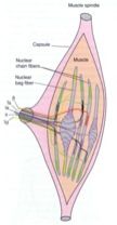
- Made up of intrafusal fibres
- Encapsulated bundles of specialised muscle fibres that make up the intrafusal fibres
- Nuclear bag fibres (NBF)
- Swollen central portion
- Nuclei of the fibres positioned centrally
- Very little sarcoplasm between myofibrils
- Swollen central portion
- Nuclear chain fibres (NCF)
- Thin and evenly shaped
- Nuclei are positioned along the length of the fibre
- Lots of space between myofibrils
- Thin and evenly shaped
- Centre is non-contractile
- Afferent fibres found here
- Sends action potentials to the CNS
- Afferent fibres found here
- Dynamic sensitivity
- Muscle spindles detect the rate at which something is changing
- e.g. length of the muscles
- Muscle spindles detect the rate at which something is changing
- Regulate the level of stretch in the skeletal muscles
- Afferent Innervation
Golgi Tendon Organs (GTOs)
Withdrawal Reflexes
One reflex or many?
Also known as sensorimotor integrations. Organised within the spinal cord into the spinal reflex arc
- Automatic rapid response to a stimulus
- Initiated by a stimulus, which is detected by a sensor, causing the generation of action potentials
- Action potentials are passed along afferent (sensory) neurones to the CNS
- Central processing takes place, causing the release of neurotransmitter
- Neurotransmitter acts upon interneurons, which link afferent and efferent pathways
- Can be short, acting within the spinal cord
- Can send projections to the brain
- Interneurons are not used in monosynaptic reflexes, where the sensory pathway links directly to the motor pathway
- Efferent (motor) pathway is connected to the effector and causes it to create an output
- Neurotransmitter acts upon interneurons, which link afferent and efferent pathways
- Central processing takes place, causing the release of neurotransmitter
- Action potentials are passed along afferent (sensory) neurones to the CNS
- The reflex arc can be modulated by other neurones and propriospinal pathways (interneurones within the spinal cord) as well as the brain
Images and files in the map
Information
- Description
- Undergraduate Systems Neurophysiology Mind Map on Spinal Reflexes, created by Lydia Buckmaster on 22/05/2014.
- Created by
- Lydia Buckmaster
- Views
- 14
- Rating