
Mind map
Visual system 2
Map content
Visual system 2
What does the visual system do?
- Localisation of an object or an event
- Object recognition
- Identifies the direction of motion and can predict where the object will be in the next moment of time
- Colour processing
- Adaptation
Adaptation
- Prevents saturation
- Visual neuron can be either saturated or not sensitive enough
- Therefore, it should be plastic
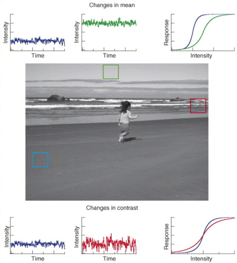
- Desensitise when contrast increases
- Resensitise when contrast increases
- One strategy: increase in gain i.e. overall sensitivity to the stimulus
- Another strategy: hyperpolarisation
- To temporal contrast (modulation of light intensity)
- Recording from ganglion cells
- Responses to increase or decrease in
temporal contrast
- Overall spiking rate gradually decreases after increase in contrast: adaptation. One of the main functions of adaptation is to prevent saturation of visual neurons
- Two types
- Do not confuse facilitation and recovery from depression. Recovery from depression occurs when the stimulus decreases in amplitude while facilitation occurs when the stimulus amplitude increases
- Depression
- Decrease in sensitivity
- Is a result of depletion of a readily releasable pool
- I.e. depression has synaptic origin
- Facilitation
- Increase in sensitivity
- Is a result of circuit mechanisms
- Depression of an inhibitory cells leads to sensitisation in other cells
- Prevents saturation
Visual scenes
- Have different properties
- Different patches of the visual scene differ in average light intensity
- Different patches of the visual scene differ in modulation of light intensity (contrast)
- Because eyes constantly move, the retina receives modulation in light intensity
- Have different properties
Localisation of an object or event
- E.g.
- Orienting reflex (orientation of the head and eyes to focus salient stimulus on the fovea)
- When novel stimulus appears animals tend to turn their heads and eyes in a position suitable for inspection of this stimulus
- Ablation of optic tectum (superior colliculus in lower vertebrates) leads to disappearance of orienting reflex
- When novel stimulus appears animals tend to turn their heads and eyes in a position suitable for inspection of this stimulus
- Smooth pursuit (following moving object)
- Prediction of motion during prey capture
- Saccadic movements during object inspection
- Superior colliculus
- Receives input from ganglion cells, auditory system, somatosensory system
- Integrates information from different sensory modalities
- Main function = regulation of saccadic movements
- Superior colliculus
- Orienting reflex (orientation of the head and eyes to focus salient stimulus on the fovea)
- E.g.
Retinotopic maps
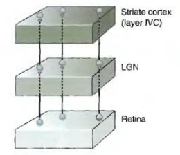
- organisation whereby neighboring cells in the retina feed information to neighboring places in their target structures (LGN, SC, cortex)
- Command neurons
- Regulate eye saccades
- Have retinotopic organisation
- Deeper layers in Superior Colliculus contain neurons that spike before the saccadic movements
- These neurons are organised in maps, similar to retinotopic maps and send projections to layers, regulating eye movements
- Maps of both layers are aligned
- “Foveation hypothesis” states that interaction between these maps initiates orienting reflex
- Interaction between the maps seems to be indirect and foveation hypothesis, therefore, is not valid. Nevertheless, such interaction is a basis of object localisation
- “Foveation hypothesis” states that interaction between these maps initiates orienting reflex
- Maps of both layers are aligned
- These neurons are organised in maps, similar to retinotopic maps and send projections to layers, regulating eye movements
Motion detection and direction selectivity
- Types of motion
- Bar moves in certain direction
- Approaching motion : looming spot
- Direction selectivity is evident in the retina
- Typical ON/OFF cells
- Responds to movements in some directions but not others
- Morphology of DS cells
- Highly asymmetric
- Prefered direction can be guessed from the morphology
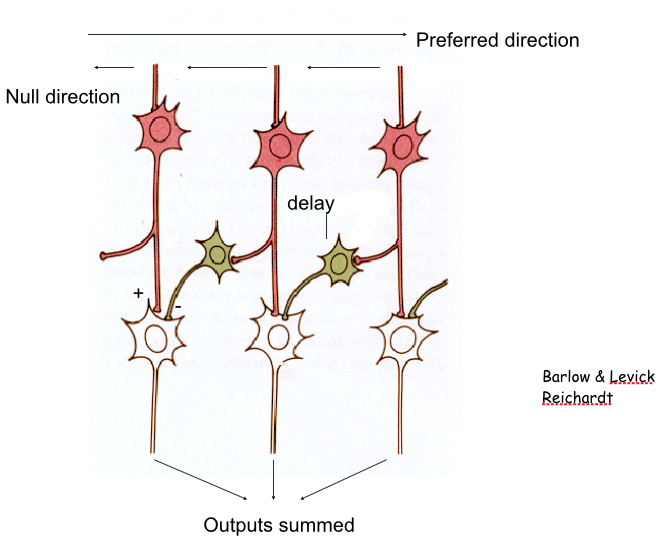
- DS cells receive excitation from bipolar cells and inhibition from amacrine cells
- In preferred direction excitation is larger and Inhibition is smaller and delayed
- In null direction, the excitation is smaller and delayed and inhibition is larger
- In preferred direction excitation is larger and Inhibition is smaller and delayed
- Typical ON/OFF cells
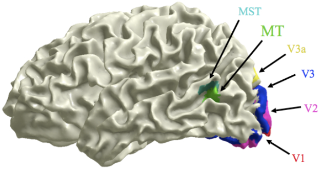
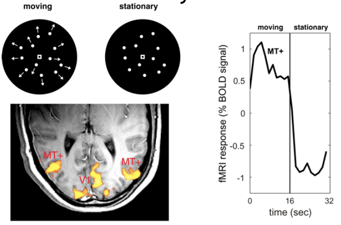
- Motion is analysed by the dorsal pathway in the brain
- Response to moving vs stationary dots
- Why do we need motion perception?
- Patient LM who following a stroke had difficulty perceiving certain forms of motion
- Object recognition and colour perception were intact
- She couldn’t see coffee flowing into the cup and didn’t perceive fluid rising and often lets coffee spill.
- “People were suddenly here or there, but I have not seen them moving... When I'm looking at the car first it seems far away, but then when I want to cross the road suddenly the car is very near”
- Akinetopsia
- Patient LM who following a stroke had difficulty perceiving certain forms of motion
- Types of motion
Processing of colour
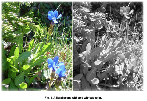
- Why do we need colour vision?
- Helps with object recognition
- In retina
- Three types of cones with different spectral sensitivities
- These types activate different populations of bipolar cells
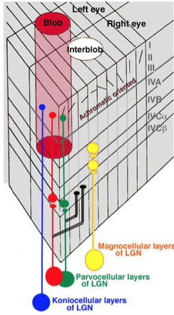
- The output of the retina then projects to V1 cortex via LGN
- Chromatic cells project to specific areas of the cortext called “blobs”
- Three types of cones with different spectral sensitivities
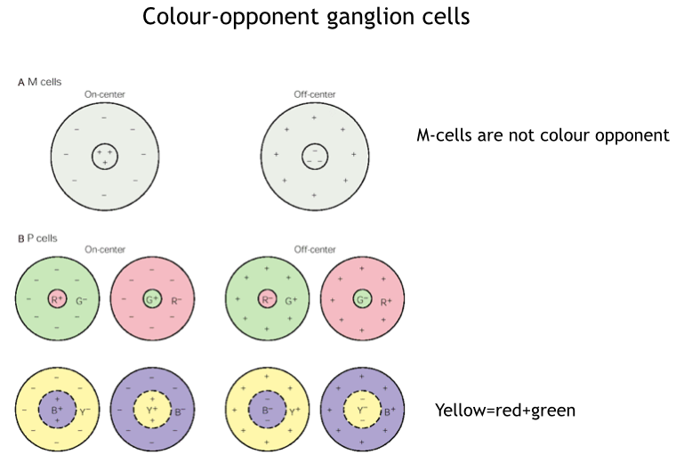
- Colour opponent ganglion cells
- Why do we need colour vision?
Images and files in the map
Information
- Description
- Undergraduate BMS236 Building Nervous Systems (Visual system) Mind Map on Visual system 2, created by Kristi Brogden on 05/08/2014.
- Created by
- Kristi Brogden
- Views
- 16
- Rating