
Mind map
Cranial Bones
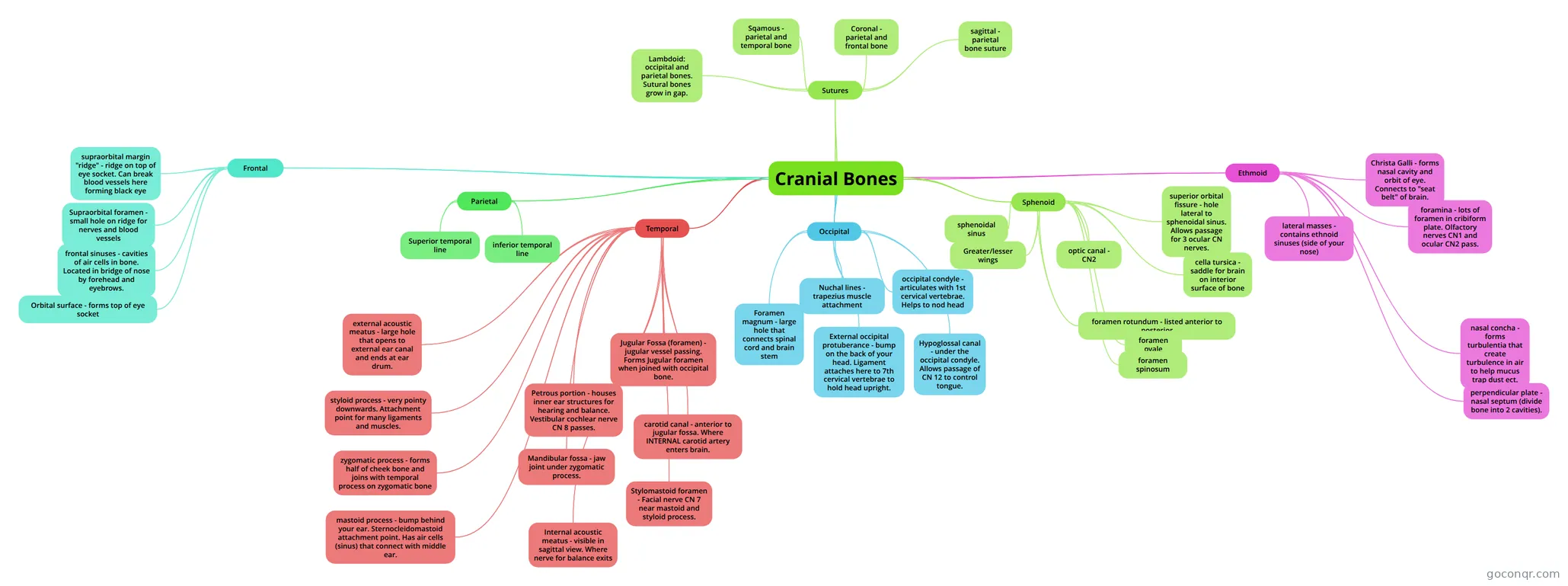
Mind map: cranial bones
Map content
Cranial Bones
Frontal
- supraorbital margin "ridge" - ridge on top of eye socket. Can break blood vessels here forming black eye
- Supraorbital foramen - small hole on ridge for nerves and blood vessels
- Orbital surface - forms top of eye socket
- frontal sinuses - cavities of air cells in bone. Located in bridge of nose by forehead and eyebrows.
Parietal
- Superior temporal line
- inferior temporal line
Temporal
- zygomatic process - forms half of cheek bone and joins with temporal process on zygomatic bone
- mastoid process - bump behind your ear. Sternocleidomastoid attachment point. Has air cells (sinus) that connect with middle ear.
- styloid process - very pointy downwards. Attachment point for many ligaments and muscles.
- external acoustic meatus - large hole that opens to external ear canal and ends at ear drum.
- Internal acoustic meatus - visible in sagittal view. Where nerve for balance exits
- Mandibular fossa - jaw joint under zygomatic process.
- Stylomastoid foramen - Facial nerve CN 7 near mastoid and styloid process.
- Jugular Fossa (foramen) - jugular vessel passing. Forms Jugular foramen when joined with occipital bone.
- carotid canal - anterior to jugular fossa. Where INTERNAL carotid artery enters brain.
- Petrous portion - houses inner ear structures for hearing and balance. Vestibular cochlear nerve CN 8 passes.
Occipital
- Foramen magnum - large hole that connects spinal cord and brain stem
- occipital condyle - articulates with 1st cervical vertebrae. Helps to nod head
- External occipital protuberance - bump on the back of your head. Ligament attaches here to 7th cervical vertebrae to hold head upright.
- Nuchal lines - trapezius muscle attachment
- Hypoglossal canal - under the occipital condyle. Allows passage of CN 12 to control tongue.
Sphenoid
- sphenoidal sinus
- superior orbital fissure - hole lateral to sphenoidal sinus. Allows passage for 3 ocular CN nerves.
- optic canal - CN2
- cella tursica - saddle for brain on interior surface of bone
- foramen rotundum - listed anterior to posterior
- foramen ovale
- foramen spinosum
- Greater/lesser wings
Ethmoid
- Christa Galli - forms nasal cavity and orbit of eye. Connects to "seat belt" of brain.
- foramina - lots of foramen in cribiform plate. Olfactory nerves CN1 and ocular CN2 pass.
- lateral masses - contains ethnoid sinuses (side of your nose)
- nasal concha - forms turbulentia that create turbulence in air to help mucus trap dust ect.
- perpendicular plate - nasal septum (divide bone into 2 cavities).
Sutures
- Lambdoid: occipital and parietal bones. Sutural bones grow in gap.
- Coronal - parietal and frontal bone
- sagittal - parietal bone suture
- Sqamous - parietal and temporal bone
Information
- Description
- Mind Map on Cranial Bones, created by Nikhil Dhall on 16/09/2014.
- Created by
- Nikhil Dhall
- Views
- 1,779
- Rating